1
EXTRADURAL SPINAL MASS LESIONS IN HIV SERO-POSITIVE ADULTS
by
DR SONWABILE GONYA 202500456
Submitted in fulfilment of the academic requirements for the degree of MMED
in the Department of Neurosurgery School of Clinical Medicine
College of Health Sciences University of KwaZulu-Natal
Durban 2017
As the candidate’s supervisor/co-supervisor, I have approved this thesis for submission.
Supervisor: Name: Dr Basil Enicker Date: 20 March 2017
Co-supervisor: Name: Prof Colleen Aldous Date: 20 March 2017
2 DECLARATION
I, Sonwabile Gonya declare that
(i) The research reported in this dissertation, except where otherwise indicated, is my original work.
(ii) This dissertation has not been submitted for any degree or examination at any other university.
(iii) This dissertation does not contain other persons’ data, pictures, graphs or other information, unless specifically acknowledged as being sourced from other persons.
(iv)This dissertation does not contain other persons’ writing, unless specifically acknowledged as being sourced from other researchers. Where other written sources have been quoted, then:
a) Their words have been re-written but the general information attributed to them has been referenced.
b) Where their exact words have been used, their writing has been placed inside quotation marks, and referenced.
(v) Where I have reproduced a publication of which I am an author, co-author or editor, I have indicated in detail which part of the publication was written by myself alone and have fully referenced such publications.
(vi) This dissertation does not contain text, graphics or tables copied and pasted from the Internet, unless specifically acknowledged, and the source being detailed in the dissertation and in the References sections.
Signed: _______________________ Date: 20 March 2017
3
DEDICATION
This thesis is dedicated to my wife, Dr Nonhlanhla Benedicta Shamase, to my mother Victoria Nothando Gonya and in memory of my late father Gerald Vuyisile Gonya.
4
ACKNOWLEDGEMENTS
I would like to acknowledge all resources used for the compilation of this thesis. This includes many people involved in my medical and neurosurgical training. Special appreciation is expressed to Dr Basil Enicker, under whose guidance I completed my neurosurgical training, and supervision of this thesis. It also includes those who granted me permission to train and become a neurosurgeon, Dr E. M. Kiratu, Clinical Head of Neurosurgery, University of KwaZulu-Natal, and the department of health KwaZulu-Natal.
5
Overview of the thesis
The Joint United Nations Programme on HIV (human immuno-deficiency virus) and AIDS (acquired immune deficiency syndrome) (UNAIDS) in 2013 estimated the number of adults and children living with HIV worldwide to be 35 million. The majority of people (68%) living with HIV/AIDS are found in Sub-Saharan Africa. South Africa (SA) is the worst affected, with approximately 5.7 million people living with HIV/AIDS. The prevalence between ages 17 to 49 years is reported at 17%, with an infection rate of 300 000 to 350 000 new cases per year. The province of KwaZulu Natal (KZN) has the highest infection rate in SA.
Individuals who are diagnosed HIV sero-positive often present with neurological disease and in 10 to 20% of patients these are AIDS-defining. The spinal cord can be affected in HIV sero- positive individuals and the commonest pathology is HIV-associated myelopathy. Other conditions that can cause myelopathy are opportunistic infections such as tuberculosis (TB), neoplasms and vascular lesions. The extradural space in the spine is where some of these pathologies can be located. However, these have been infrequently reported in the literature.
Purpose of the study
The purpose of the study was to determine the common histo-pathology diagnoses of extradural spinal mass lesions (EDSMLs) in HIV sero-positive patients treated in the Department of Neurosurgery (DoN) at Inkosi Albert Luthuli Central hospital (IALCH), Durban, SA during January 2003 to December 2014.
It was a retrospective, observational, chart review of all HIV sero-positive adult patients who presented to the DoN at IALCH, and were diagnosed with extradural spinal mass lesions, using magnetic imaging resonance (MRI). These patients then underwent surgical management; with the histo-pathology report used to confirm the diagnosis.
6 Variables investigated were demographics, CD4 count, clinical presentation, diagnostic investigations which included MRI of the spine, concomitant AIDS related disease of the central nervous system and other organs, surgical management and final histo-pathology report, to ascertain the common EDSMLs in this cohort of patients at our institution.
A total of 38 patients were treated in our institution over a 12-year period and the histo- pathology diagnoses were lymphomas [17; 45%], lymphoproliferative disease [8; 21%], Tuberculosis [4; 10%], Epstein-Barr virus associated myoid tumours [3; 8%], pyogenic abscesses [2; 5%], Rosai Dorfmans disease [1; 3%], schwannoma [1, 3%], metastatic ovarian carcinoma [1, 3%], and metastatic prostate carcinoma [1. 3%].
EDSMLs in HIV sero-positive patients are under reported both locally and internationally. The majority of studies have focused on HIV-associated intracranial mass lesions. Bhigjee et al found toxoplasmosis to be the commonest intracranial lesion in HIV sero-positive patients, whilst Modi et al and Smego et al found tuberculosis (TB) to be the commonest cause of HIV- associated focal brain lesions. These studies on intracranial mass lesions have helped to formulate guidelines which assist physicians managing this group of patients to administer a stepwise approach to treatment.
It is our belief that this study will help clinicians working in areas affected by the HIV/AIDS pandemic to familiarize themselves with various potential pathological entities affecting the spine in HIV sero-positive adult population. The study has offered insight into this disease entity and will add to the body of knowledge in the field of neurosurgery and HIV/AIDS.
Understanding these entities is crucial for initiation of appropriate diagnostic tests and in formulating protocols that offer best management practices, which will assist medical professionals to maximize patient recovery, especially in under-resourced environments with an overburdened health system.
7
Table of Contents
Title……….….1
Declaration ... 2
Dedication ... 3
Acknowledgements ... 4
Overview of the thesis ... 5
Purpose of the study……….….…………...5
Chapter 1 ... 8
Introduction ... 8
Research question………... 8
Literature review…….………...……….….…9
Rational for the study…………....……….14
Chapter 2: Manuscript... …15
Abstract……….……….……....16
Introduction…...18
Methods and material……….……19
Results………...…...20
Discussion………...……...23
Conclusion………...…..36
Limitations………..…...38
Conflict of interest……….…....38
Funding………..38
References……….…….39
Appendix 1: ... 41
Appendix 2: The Guidelines for Authorship for the Journal selected for submission of the manuscript ... 62
Ethical approval………...63
Appendix 4: Data collection tools... 66
Appendix 5: Raw data ... 67
8
Chapter 1
1.1 Introduction
The Joint United Nations Programme on human immunodeficiency virus (HIV) and acquired immune deficiency syndrome (AIDS) (UNAIDS) estimated in 2013 the total population living with HIV worldwide to be 35 million [1]. The majority of people (68%) living with HIV/AIDS are found in Sub-Saharan Africa [1, 2]. South Africa (SA) is worst affected, with approximately 6.4 million people (12.2% of population) living with HIV/AIDS and 1.2 million more than in 2008 (5.2 million or 10.6%) [2, 3, 4].
It is reported that the prevalence of HIV infection in the SA adult population between the ages of 14 to 49 years is 17%, with 469, 000 new infections a year [4, 5]. Geographical differences were found by locality type and also by provinces. Rural informal area residents had significantly higher HIV prevalence than did urban formal area residents. The Province of KwaZulu-Natal (KZN) has the highest infection rate in SA [6]. Black Africans were less likely to live in urban formal areas than other racial groups in South Africa. Urban informal areas are generally under- resourced and lack some basic necessities such as formal housing, water, sanitation, and access to preventative health services. The survey found significantly higher HIV prevalence in people who lived in urban informal areas than those in urban formal areas [3, 6].
Individuals who are diagnosed HIV sero-positive often present with neurological disease and in 10 to 20% of patients these are AIDS-defining [7]. The spinal cord can be affected in HIV sero- positive individuals and the commonest pathology is HIV-associated myelopathy [8]. Other conditions that can cause myelopathy are opportunistic infections such as TB, neoplasms and vascular lesions [8]. The extradural space is where some of these pathologies can be located;
however, this has been under reported in the literature.
9 1.2 Research question
What are the common extradural spinal mass lesions in HIV sero-positive adult patients managed at Inkosi Albert Luthuli Central Hospital over a 12-year period?
1.3. Literature review
The HIV epidemic has put a lot of strain in the already over-burdened health system of SA over the years. The first death from AIDS in SA occurred in 1982, with HIV/AIDS being reported as one of the leading causes of death in the country [5].
The introduction of the anti-retroviral therapy (ART) program has gone a long way in reducing the mortality from HIV/AIDS, with more than 1.9 million people receiving ART in SA [6, 9]. In KZN the ART program has increased life expectancy from 49.2 in 2003 to 60.5 years in 2011 [9].
Individuals who are HIV sero-positive with low CD4 counts are at a high risk of developing HIV- associated mass lesions, especially of the spine. These mass lesions can be either infectious or neoplastic.
The infectious lesions can be bacterial (acute pyogenic), chronic granulomatous infections such as TB or fungal infections. Bacterial infections can cause extradural empyema or abscess, the responsible organism commonly being Staphylococcus aureus [10]. The source of infection can be either from haematogenous spread (e.g from respiratory infections) or from direct extension (e.g psoas abscess).
In SA, approximately 60% of HIV sero-positive patients are co-infected with TB and it can manifest as an extradural mass lesion and the organism responsible in majority of cases is Mycobacterium Tuberculosis [11].
10 In the spine, TB can also cause destruction of the vertebral body and result in spinal deformity a condition called Pott’s disease [12, 13]. Pulmonary TB should be excluded in these patients.
Extradural lesions caused by fungal infections are rare, when they do occur are due to organisms such as Aspergillus species and Cryptococcus neoformans [13].
Neoplasms of the spine account for approximately 10% of central nervous system (CNS) tumours [14]. The commonest extradural tumours of the spine are metastatic lesions and majority (70%) occurred in the thoracic spine [14]. These tumours cause destruction of the vertebral body and compress the spinal cord and nerve roots. Metastatic tumours are commonly from the lung, breast, prostate, multiple myeloma and sarcoma [14].
Lymphomas are typically found in the extradural space; they can be primary or metastatic and are considered AIDS-defining. The risk of lymphoma in HIV sero-positive individuals is reported to be 10-fold higher when compared to HIV negative individuals, especially if they have a high viral load (> 100 000 copies/mm3), low CD4 count (< 50 cells/mm3) and are not on ART [15]. However, lymphomas can occur at any CD4 count level.
These tumours are caused by combination of immune dysregulation, loss of T cell immunity against viruses namely Epstein-Barr virus (EBV) and Human herpes virus 8 (HHV8), and lastly from direct oncogenic effect of HIV [15]. Infection with HIV is associated with certain types of lymphomas namely high grade B-cell lymphomas (non-Hodgkin’s) and diffuse large B-cell ([1, 2, 15]. It is reported that more than 90% of patients diagnosed with high grade B-cell lymphoma (HGBCL) are infected with HIV [16].
Other tumours that can manifest in HIV sero-positive individuals are the EBV-associated smooth muscles tumours. These tumours are usually locally invasive and mainly arise from the smooth muscle especially in areas with rich vascular supply [15, 16].
11 Thurnher et al in a study of 55 patients with AIDS and neurological symptoms related to the spine and spinal cord found 58% of patients had extradural lesions and five (9%) had a combination of extradural and intradural (extramedullary) lesions. These lesions were lymphomas (38%), bone marrow changes related to chronic anemia (35%), empyema due to Staphylococcus aureus (18%), TB mass lesions (12%) and metastatic tumours (3%) [10].
These lesions cause symptoms by compression, invasion, obstruction of flow of cerebrospinal fluid (CSF), destruction of the myelinated tracts and anterior horn cells [10]. There are also vascular changes which cause symptoms following occlusion and thrombosis of extradural veins [10]. Symptoms and signs are related to site of the lesion and its growth rate.
Patients often present with local pain, due to stretching of the periosteum by the tumour or inflammatory process [11]. Pain can also be caused by vertebral body collapse and worsened by movement indicating spinal instability [12]. Nerve root compression results in radicular pain.
Patients may complain of constitutional symptoms such as weight loss and night sweats.
Neurological deficits can range from paraplegia or paraparesis, spasticity, sensory loss, abnormal plantar reflexes, urinary bladder and sexual dysfunction [13].
When treating HIV sero-positive patients with back pain and neurological deficits a series of investigations should be performed. Plain x-rays are inexpensive and they demonstrate bony abnormalities such as destruction of pedicles and vertebral collapse often associated with spinal cord compression.
Computerized tomography (CT) scan of the spinal column is also recommended to assess bony quality, destruction and instability [14]. Magnetic resonance imaging (MRI) of the spine is the investigation of choice for diagnosing mass lesions [14, 23]. Sagittal and axial T1-weighted images with and without intravenous contrast, along with T2-weighted images are obtained.
12 MRIs are non-invasive and show better definition of the mass lesion, soft tissue and neural elements. MRI is not ideal for assessing bone quality [14]. CT myelogram is used less frequently because it is time consuming and invasive requiring a lumbar puncture to inject contrast required to demonstrate neural tissue, and when MRI is contra-indicated [14].
The mainstay of treatment of patients with compressive symptoms from extradural lesion is surgical decompression via a laminectomy. At this stage assessment is made whether the mass can be removed totally or sub-totally and tissue obtained is sent for histology. Microscopy, culture and sensitivity (MCS) is sent in addition, if an inflammatory lesion is discovered intra- operatively. In some instances, the lesion affects the entire length of the spinal cord, thus limiting surgical option to a biopsy.
It is of outmost importance to consider the overall condition of these patients when offering surgical treatment as most of these patients especially those with AIDS might not be fit for a general anaesthesia due to co-morbid disease. Patient with infective lesions are put on appropriate antibiotics based on MCS results. Anti-TB therapy is commenced when TB is confirmed by either MCS or histo-pathology results. Therapy is given for a minimum of 12 months [16].
In patients with metastatic disease assessment is made as to whether the primary disease is under control. The goals of treatment in patients with metastatic tumours are preservation and, occasionally, recovery of normal neural function, local tumour control, spinal stability and pain relief. Palliation is a reasonable goal in the management of patients with advanced systemic malignancies and spinal metastases [17]. Once neural function is lost, its return is unlikely [17].
Patient with lymphomas will require adjuvant chemotherapy in combination with ART as lymphomas are considered AIDS-defining [18].
13 The meta-analyses by Specht et al and Leoffler et al showed a lack of an overall survival benefit in patients receiving combined modality treatment (radiotherapy and chemotherapy), and highlighted the impact of radiation-induced cardiovascular complications and secondary neoplasia [19, 20, 21]. Seam et al in their study concurred that combined modality treatment is not superior to chemotherapy alone, and that the widespread use of radiation therapy in all stages of disease seems unjustified [21].
The collaboration of observational research Europe (Cohere) study group evaluated survival and prognosis of 847 patients with non-Hogkin’s lymphoma associated with HIV [22]. The proportion of histological subtypes in the study were Burkitt’s lymphoma (10%), and lymphoma with large B-cell (8%) and 82% were classified as non-specified or other. The median age at time of diagnosis was 41.2 years and 82% of patients were males. The median CD4 count was 114 cells/mm3, median viral load was 181 000 copies/mm3 and 43% were ART naïve.
Lymphoma and HIV infection were diagnosed in 13% of patients, and only 66% survived more than a year [22].
Those patients with EBV-associated smooth muscle tumours benefit from complete tumour resection and ART [23]. They often respond poorly to adjuvant radiotherapy and or chemotherapy [23, 24].
Spinal epidural abscess is a rare but potentially devastating disease. Patients with a spinal epidural abscess, when treated early, the outcome is usually good [25]. Early clinical and radiological detection is therefore critical in patient management. Nathoo et al reported that symptoms are non-specific, with fever and back pain as frequent presentations. It is important to have a high index of suspicion to make the diagnosis early in the course of the illness.
Staphylococcus aureus was the most common pathogen from specimens sent for MCS [25].
14 Khanna et al. analyzed the predictive value of the symptoms and outcomes of patients with spinal epidural abscess [26]. The duration of pain or radiculopathy, location of abscess, and degree of granulation tissue did not appear to affect the outcome of the disease if the condition was treated in a timeously [26]. They concluded that surgery provides a good outcome if bowel and bladder dysfunction, paresis, paraplegia are present for fewer than 72 hours, when the degree of thecal sac compression is less than 50%, and patients are younger than 60 years of age.
However, several studies have shown poor neurological outcome and recovery in patients with paraplegia for more than 12 hours before surgery. Although the neurological outcome depends on location, patients with epidural abscess in the cervical and thoracic spine have poor prognosis.
The neurological outcome is related to severity of the neurological deficit [26, 27].
1.4 Rationale for the study
There have been numerous studies focusing on HIV-associated intracranial mass lesions [28, 29].
The data on EDSMLs in HIV sero-positive individuals is under reported. Studies from African and Central American countries describe either TB or toxoplasmosis as the two most frequently diagnosed lesions in the brain [29].
Bhigjee et al in KZN, SA found toxoplasmosis to be the commonest intracranial lesion in HIV sero-positive patients and advocated for antiretroviral roll-out in addition to treatment with co- trimoxazole [7]. Modi et al and Smego et al in contrast found TB to be the main cause of HIV- associated focal brain lesions in Johannesburg, SA [29, 30]. They recommended HIV-positive patients who present with focal brain lesions (FBL) to be treated initially with medication specific to the infection that is endemic to that population [29, 30].
Our study will offer insight into this disease entity and add to the body of knowledge. This will help in formulating protocols that offer best management practices for medical professionals who manage these patients.
15
Chapter 2: Manuscript
Extradural spinal mass lesions in HIV sero-positive adults Sonwabile Gonya1*, Basil Enicker1, Colleen Aldous2.
1Department of Neurosurgery, University of Kwa-Zulu Natal, Nelson R. Mandela School of medicine, Durban, South Africa.
2School of Clinical Medicine, University of Kwa-Zulu Natal, Nelson R Mandela School of medicine, Durban, South Africa.
*Correspondence: [email protected] Department of Neurosurgery
University of Kwa-Zulu Natal
Nelson R. Mandela School of Medicine Durban, South Africa.
Telephone: +27 31240 2439 Fax: +27 31240 1132
Email addresses: [email protected]
SG [email protected] BE [email protected] CA [email protected]
16
ABSTRACT
Background: Extradural spinal mass lesions (EDSMLs) in HIV sero-positive patients are under reported, with most reports focusing on intracranial mass lesions. These lesions result in severe neurological deficits.
Aims: To determine the common histo-pathology diagnoses of EDSMLs in HIV sero-positive adult patients managed at Inkosi Albert Luthuli Central hospital, Durban, South Africa, over a 12-year period.
Methods: Retrospective, observational, chart review of all HIV sero-positive patients who were diagnosed with EDSML on MRI and underwent surgical treatment during January 2003 to December 2014. The data collected was analyzed for demographics, clinical presentation, anatomical level of the mass lesion on MRI, histo-pathology reports, CD4 cell count, concomitant AIDS defining illness, management, length of hospital stay, complications and in- hospital mortality.
Results: A total of 38 patients were treated during this period. The mean age was 33±13 years (range 20-64). There were 24 (63%) males and 14 (37%) females (M: F 1.7:1). Twenty-nine (76%) patients were paraparetic, of which 32 (84%) had associated bladder dysfunction. The mean CD4 count was 191± 119 cells/mm3 (range 33 - 497). Twenty-nine (47%) patients were on anti-retroviral therapy. The lesions were located in the cervical [6; 16%], thoracic [27; 71 %] and lumbar [5; 13%] spine. The histo-pathological diagnoses were lymphoma [17; 45%], lymphoproliferative disease [8; 21], Tuberculosis [4; 10%], Epstein Barr virus associated myoid tumours [3; 8%], pyogenic abscesses [2; 5%], Rosai-Dorfman disease [1; 3%], schwannoma [1;
3%], metastatic ovarian carcinoma [1; 3%], and metastatic prostate carcinoma [1; 3%]. Two (5%) patients died during the admission period. Only one (3%) patient showed neurological improvement following treatment.
17 Conclusion: It is of outmost importance for clinicians managing patients with HIV/AIDS to be aware of pathology affecting the spine in this group of patients, as this will help in formulating protocols aimed at improving neurological function and outcomes.
Keywords: AIDS, Extradural spinal mass lesion, HIV, Laminectomy, Lymphoma, Tuberculosis
18 Introduction
The Joint United Nations Programme on HIV and AIDS (UNAIDS) in 2013 estimated the number of adults and children living with HIV worldwide to be 35 million [1]. The majority of people (68%) living with HIV/AIDS are found in the Sub-Saharan Africa [1, 2]. South Africa (SA) is the worst affected, with approximately 5.7 million people living with HIV/AIDS [2, 3, 4].
The prevalence between ages 17 to 49 years is reported at 17%, with an infection rate of 300 000 to 350 000 per year [5]. The province of KwaZulu-Natal (KZN) has the highest infection rate in SA [6].
Individuals who are diagnosed HIV sero-positive often present with neurological disease and in 10 to 20% of patients these are AIDS-defining [7]. The spinal cord can be affected in HIV sero- positive individuals and the commonest pathology is HIV-associated myelopathy [8]. The extradural space is where some of these pathologies can be located. However, this has been under reported in the literature.
Spinal cord lesions in HIV sero-positive patients result in debilitating neurological deficits, which are often non-reversible [7, 8]. These impose a serious burden on an already stretch health care system in SA and in particular KZN.
The purpose of this study was to determine the common histo-pathological diagnoses, demographics, clinical presentation, management and outcome of EDSMLs in HIV sero-positive patients managed at the Department of Neurosurgery (DoN) at Inkosi Albert Luthuli Central hospital (IALCH), Province of KZN, SA over a 12-year period. The DoN provides the only public sector neurosurgery service in the Province of KZN.
19 Methods and materials
The study was a retrospective, observational, chart review of all patients managed in the DoN at IALCH during January 2003 to December 2014. Inclusion criterion involved consecutive HIV sero-positive adult patients with a diagnosis of EDSMLs made on magnetic resonance imaging (MRI). We excluded patients < 18 years of age and those with intradural and intramedullary spinal mass lesions.
The Data was collated from the medical records kept in a pass-word protected hospital information system (Sorian TM), and exported into Excel® (Microsoft Inc., WA, USA).
The variables examined included demographics, neurological deficits, CD4 cell count, concomitant AIDS-defining illnesses. We also examined the anatomical location of the lesions on MRI scans and their reports, surgical management, histo-pathology reports to determine the diagnosis, length of hospital stay, improvement of neurological deficits, complications and mortality.
The data was analyzed using the statistical package for social sciences (SPSS version 21).
Descriptive statistics such as frequencies, proportions, mean, standard deviation and percentages were used to summarize normally distributed data.
The study was granted ethical approval by the biomedical research ethics committee (BREC) of the University of KwaZulu-Natal (REF: BE002/15).
Results
A total of 41 HIV sero-positive patients with EDSMLs were treated during this study period.
Only 38 patients met the inclusion criteria, as these were patients ≥ 18 years of age.
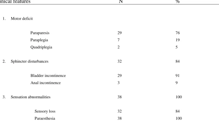
20 The mean age was 33 ±13 years (range 20 - 64), interquartile range (IQR) of 24 - 38. There were 24 (63%) males and 14 (37%) females (M: F 1.7:1). The clinical symptoms and related neurological deficits are shown in table 1.
Table 1:Clinical features of the 38 HIV sero-positive patients diagnosed with EDSMLs
Clinical features N %
1. Motor deficit
Paraparesis Paraplegia Quadriplegia
2. Sphincter disturbances
Bladder incontinence Anal incontinence
3. Sensation abnormalities
Sensory loss Paraesthesia
29 7 2
32
29 3
38
32 38
76 19 5
84
91 9
100
84 100
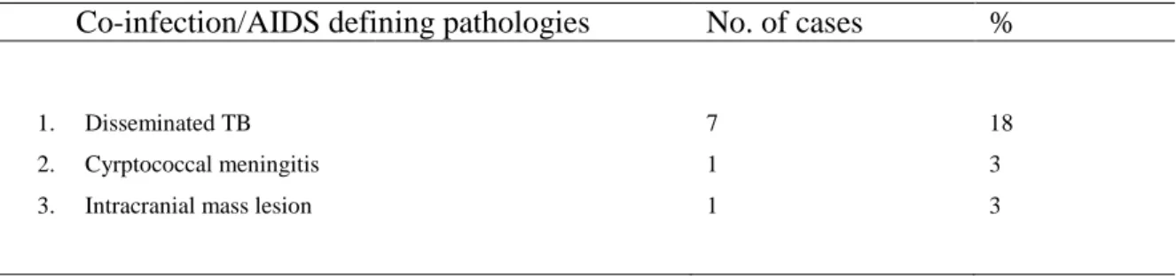
The mean duration of symptoms at presentation was 28±9 days. The mean CD4 count was 191±119 cells/mm3, (IQR 236 - 109). Sixteen (42%) patients were on anti-retroviral therapy (ARVs), and while the rest were ARV naïve. Nine (24 %) patients had concomitant AIDS defining pathologies as shown in table 2. The anatomical location of the EDSMLs on MRI is shown in table 3.
21 Table 2: Concomitant AIDS defining pathologies found in the 38 HIV sero-positive patients with EDSMLs
Co-infection/AIDS defining pathologies No. of cases %
1. Disseminated TB 7 18
2. Cyrptococcal meningitis 3. Intracranial mass lesion
1 1
3 3
Table 3: Anatomical location of EDSMLs on MRI in the 38 HIV sero positive patients
Location No. of cases %
1. Cervical 2. Thoracic 3. Lumbar
6 27 5
16 71 13
All patients underwent surgical intervention which included laminectomy to relieve compression of neural elements (3, 7.9%) and laminectomy and biopsy (35, 92.1%) when complete surgical removal was not possible, especially those patients with extensive and a long segment spinal cord compression. Two patients (5.2%) had spinal fusion for spinal instability, and these were patients diagnosed with spinal TB and metastasis. The histo-pathological diagnoses are shown in table 4.
22 Table 4: Histo-pathology diagnoses of the 38 HIV sero-positive patients with EDSMLs
Abbreviations: TB – Tuberculosis, EBV – Epstein-Barr virus, MTs – myoid tumours
The mean length of hospital stay was 16 days (range 2 - 74). Complications occurred in 4 [10.4%] patients and these included wound sepsis [1, 2.6%], renal failure, [1, 2.6%] pneumonia [1, 2.6%] and pressure sores [1, 2.6%]. The patient with wound sepsis required surgical debridement in the operating theatre under general anaesthesia.
Diagnosis No. of cases %
1. Lymphoma
i. High grade B-cell lymphoma ii. Diffuse large B-cell lymphoma
iii. Plasmacytic non-Hodgkin’s B-cell lymphoma
17 45%
11 65%
5 29%
1 6%
2. Lymphoproliferative disease
i. Fibro-fatty ii. Granulation iii. Inflammatory
3. TB
8 21%
3 37.5%
3 37.5%
2 25%
4 10%
4. EBV associated MTs 5. Pyogenic abscess 6. Metastasis 7. Rosai Dorfmans 8. Schwannoma
3 8%
2 5%
2 5%
1 3%
1 3%
23 Two (5.2%) patients died during the admission period, one patient had a diffuse large non- Hodgkin’s B-cell lymphoma with CD20 immuno-positivity and the other patient had an epidural abscess. The cause of death in the first patient was secondary to complications of renal failure, while the second patient developed septicaemia following wound sepsis.
Patients with EDSMLs due to lymphoma were referred with a haematologist for further adjuvant therapy. Those with EBV-associated MT, Rosai-Dorfman disease and metastases were referred to an oncologist for chemotherapy and radiotherapy. Mean follow up period was 2months and on follow up the majority did not show any improvement, only one (3%) patient had neurological improvement following excision of a schwannoma.
4. Discussion
The HIV epidemic has put a lot of strain in an already over-burdened health system of SA over the years. The introduction of the anti-retroviral therapy (ART) program has reduced mortality from HIV/AIDS, with a reported 1.9 million people on ARVs in South Africa [9]. In KZN the ART program has increased the life expectancy from 49.3 years in 2003 to 60 years in 2011 [6, 9].
HIV sero-positive patients with low CD4 counts are at high risk of developing HIV associated mass lesions, especially of the spine. These mass lesions can either be infectious and/or neoplastic. Our study showed a male predominance, which is interesting because in SA HIV infection has a high prevalence amongst the female population [5, 6, 9]. Majority of patients who developed EDSMLs had a low CD4 count, and were not on ART. The latter point underscores the importance of early initiation of ART in order to reduce sequelae of AIDS related mass lesions and medical illnesses.
24 4.1 Clinical presentation
All patients presented with neurological deficits and these were secondary to compression and invasion of neural tissue, obstruction of cerebrospinal fluid (CSF) flow, destruction of the myelinated tracts and anterior horn cells [10]. The lesions affect the vascular structures resulting in occlusion and thrombosis of extradural veins [10, 19].
The symptoms of pain are often due to stretching of periosteum by tumour or inflammatory process and nerve root compression. Thurnher et al found the neurological symptoms and signs of patients with EDSMLs to be progressive limb weakness (60%), incontinence (21%), back pain (19%), numbness (14%), burning (14%), sensory deficit (8%), paraesthesia (8%), cauda equina syndrome (8%), sexual dysfunction (2%) and radiculopathy (2%) [10]. Bilsky et al reported that in patients with extradural tumours neurological symptoms started with back pain, radiculopathy, myelopathy, proprioceptive sensory loss, and lastly painless urinary dysfunction [11].
4.2 Neuro-imaging
MRI of the spine with sagittal and axial pre and post contrast T1 and T2 weighted images was the investigation of choice. MRI is the gold standard for evaluating disc space infection and osteomyelitis of the spine. It is most effective for demonstrating the extension of disease into soft tissues, and neural compression. MRI with contrast can also be used to assess the response to treatment and regression of the disease [10, 19].
The thoracic spine is most commonly affected by extradural metastatic tumours, followed by lumbar and rarely cervical region [13]. These findings are mirrored in our study. The relative length of the thoracic spine may explain this selectivity [14]. CT scans of the spine are better in showing the quality of bone, especially in patients with metastatic bone lesions when compared to MRI [15].
25 4.3 Surgery
Principles of management included, medical work up, exclusion of concomitant pathologies, fitness for general anaesthesia and surgical decompression of neural structures via a laminectomy, removal of a mass lesion and stabilizing of the spine with instrumental fusion when necessary [16]. However, in some cases the lesions involved an extensive area of the spine and only a biopsy could be obtained in these cases.
4.4 Lymphoma
Our study showed that lymphoma was the most common pathology. Carbone et al [17] reported that these tumors are caused by a combination of immune dysregulation, loss of T-cell immunity against viruses namely Epstein Barr virus, Human herpes virus 8 and direct oncogenic effects of HIV. HIV infection is associated with certain types of lymphomas namely high grade B-cell lymphoma (> 90 %) and diffuse large B-cell lymphoma.
In our study, high grade B-cell lymphoma (CD20 immuno-positive) was reported in over 60% of patients, diffuse large B-cell lymphoma in 29%, and plasmacytic non-Hogkin’s B cell lymphoma in 6%. One case (9%) of high grade B-cell lymphoma favoured an immunoglobulin heavy chain (IGH) MYC translocation identification and confirmed a Burkitt’s lymphoma. All these patients had a low CD4 count.
MRI characteristics of lymphomas are T1W isointense and usually have a solid and homogenous enhancement on T1W post gadolinium MRI. These patients were started on ART and then referred to the hematologist for further management.
26
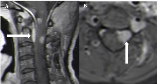
Figure 1: Cervico-thoracic T1W sagittal (A) and axial (B) post contrast MRI showing a lymphoma (arrow) in a 38-year- old male patient, causing spinal cord compression.
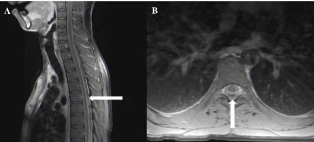
4.5 Lymphoproliferative disease
Eight patients (21%) were diagnosed with lymphoproliferative disease. Histological findings were: Chronic inflammation comprising of lymphocytes and plasma cells, with no granulomatous inflammation or fungi in 2 patients (25%). Three patients (37.5%) had a granulation tissue with mixed cell infiltrate, an acute inflammatory exudate, and the Acid-Fast Bacillus (AFB) staining and fungi were negative. Three patients (37.5%) had fibro-fatty tissue with focal mild peri-vascular chronic inflammation and no evidence of TB or malignancy.
Govender et al in a study of 39 HIV sero-positive patients reported histological features of chronic non-specific infection, and negative cultures for fungal studies in 8 patients (20.5%) [19].
A B
27
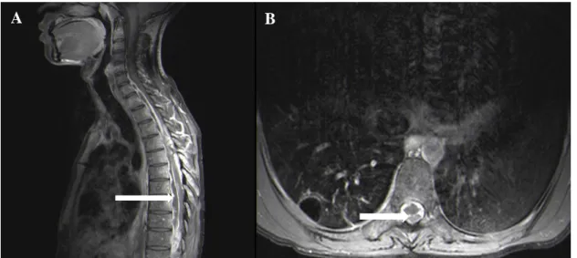
Figure 2: Thoracic T1W sagittal (A) and axial (B) post contrast MRI showing a lymphoproliferative disease (arrow) in a 28-year-old male patient, causing spinal cord compression.
4.6 Tuberculosis
In our study 18% of patients had disseminated TB; spinal TB was confirmed in 4 (10.5%) patients. TB in the extradural spine can mimic other lesions. Infections, eosinophilic granuloma and metastatic disease can be confused with spinal TB with dire consequences if not recognised and appropriately managed [18].
The innate immune system eradicates 85% of the disease, and 10-15% of the incompletely sterilised residual bacteria lay dormant [19]. In a few patients, progressive active disease ensues.
HIV affects the cellular immunity; it reduces the host’s reaction to TB. This manifests as poor control of the initial infection or inability to control the residual TB foci with reactivation of disease [19].
In SA, approximately 60% of HIV sero-positive patients are co-infected with TB and it can manifest as an extradural mass lesion and the organism responsible in majority of cases is Mycobacterium tuberculosis [19]. In the spine, TB can cause destruction of the vertebral body and result in spinal deformity “Pott’s disease”.
A B
28 The medical management of pulmonary TB is 2 months’ intensive phase with four drugs followed by a continuation phase of two drugs for 4 months. In spine TB, the duration is for a minimum of 9 months. If the patient was on medication pre-operatively, the 9 months is calculated from the date of surgery irrespective of the pre-operative medication period. At 9 months, clinical, radiological and erythrocyte sedimentation rate (ESR) is assessed and frequently the duration is extended to 12 months if there are any concerns. Clinically there should be resolution of axial pain, the ESR should be normal and the X-rays confirm bony healing. Once the drugs are stopped the patient is reviewed 3 months later to confirm the ESR remains normal. The ESR is less useful in the HIV patient and may well be overridden by the other criteria [20].
Anti-TB treatment is recommended for minimum of 12 months and compliance is essential to prevent multidrug resistance, especially in the immunocompromised patients [19, 20, 21]. The British Thoracic Society recommends 12 months’ duration for Rifampicin and Isoniazid for spinal TB and CNS involvement [21].
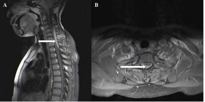
Indications for surgery are neurologic deficit, spinal deformity with instability, severe or progressive kyphosis, retro-pulsed bone fragments in the canal, large abscess causing respiratory embarrassment, and poor response to medical therapy [19, 20, 21]. MRI scan is most useful in delineating epidural TB and evaluating the extent of spinal cord compression and nerve root compromise (fig 3).
29
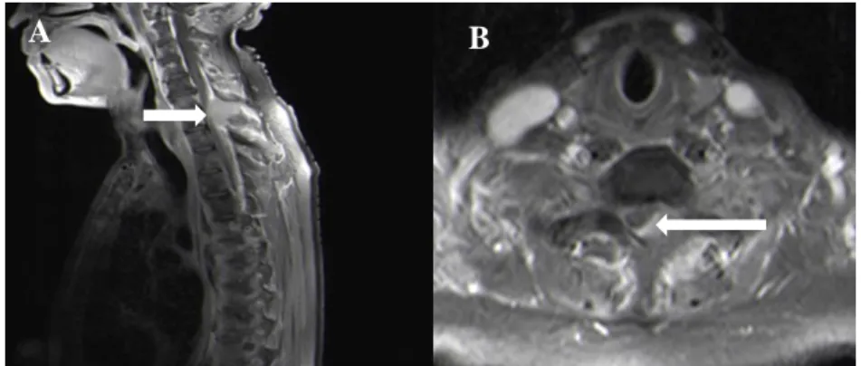
Figure 3: T1W post contrast sagittal (A) and axial (B) MRI showing a showing extradural TB (arrow) involving the long segment of the spine resulting in spinal cord compression in a 30-year-old female.
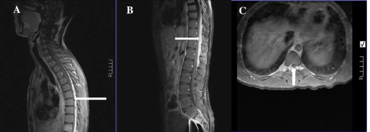
4.7 Smooth muscle tumours (Leiomyosarcoma)
Myoid tumours (MTs) in patients with AIDS have been reported with increasing frequency over the last two decades. They have been documented often, but not always, in association with Epstein–Barr virus (EBV) infection. MT can occur in unusual anatomical locations, are considered independent primary tumour rather than metastasis. These EBV-associated MTs (EBV-positive MTs) are also typified by multi-focality; phenotypic heterogeneity and intra- tumoral T cell lymphocytic infiltration [22]. There is a recent expansion of the spectrum of EBV- positive MTs to include EBV-positive myopericytomas. Globally, AIDS related smooth muscle tumour (SMTs) have been documented up to after 18 years the diagnosis of AIDS, the MTs served as the sentinel clue of HIV infection and AIDS in 44% of patients [23].
A recent global review of 35 adult AIDS patients with EBV positive SMTs confirmed that 68.6%
were male [24]. These tumours are solid, enhancing paravertebral masses with spinal canal extension on MRI (fig 4).
In this cohort 8% of patients (two females and one male) had SMTs – 2 were diagnosed with leiomyosarcoma and one was diagnosed with EBV associated tumour with myopericytic and leiomyomatous components.
A B
30 Both female patients had CD4 count more than 150 cells/mm3, while in the male patient it was unknown. After the surgical decompression, these patients were started on ART and then referred to an oncologist for radiotherapy.
Figure 4: T1W (pre and post gadolinium) sagittal (A and B) and axial (C) MRI showing an invasive paravertebral cervical mass diagnosed as EBV associated MT extending into the spinal canal resulting in spinal cord compression, and causing expansion of the neural foramen.
4.8 Epidural abscess
Spinal epidural abscesses originate from a distant focus such as a skin infection, pharyngitis, or dental abscess and are also associated with discitis or vertebral osteomyelitis [25, 26].
Predisposing condition include AIDS, diabetes mellitus, trauma, and intravenous drug abuse [27].
The diagnosis of epidural abscess is made promptly because delay in treatment can result in irreversible neurologic damage or death [27]. The progression of neurologic dysfunction varies from a few hours to several months. Leukocytosis may be the only abnormal laboratory finding and should be suspected in patients presenting with fever and back pain [27].
A B
31 The diagnostic study of choice is MRI with gadolinium contrast as shown in figure 5. Surgical decompression remains the mainstay of treatment for a spinal epidural abscess in patients with neurological deficit. Non-surgical treatment is indicated in patients who present with minimal neurologic deficit or are poor surgical patients [27].
Figure 5: T1W post contrast thoracic MRI (A and B) showing a T1 – T7 long level extradural pyogenic abscess (see arrow) in a 23-year-old male patient.
Khanna et al. [25] analyzed the predictive value of the symptoms and outcomes of patients with spinal epidural abscess. The duration of pain or radiculopathy, location of abscess, and degree of granulation tissue did not appear to affect the outcome of the disease if the condition was treated timeously.
They concluded that surgery provides a good outcome if bowel and bladder dysfunction, paresis, paraplegia are present for fewer than 72 hours, when degree of thecal sac compression is less than 50%, and when patients are younger than 60 years of age [25, 31].
Several studies have shown no improvement in the neurological outcome or recovery in patients who were paraplegic for more than 12 hours before surgery [19, 25, 26, 27].
A B
32 In our practice, empyaemas were drained via a laminectomy followed by intravenous antibiotics based on microscopy, culture and sensitivity for 6 weeks.
4.9 Metastatic lesions
The most common extradural tumours of the spine are metastatic lesions and majority occur in the thoracic spine [13, 29, 30]. De Leon et al in their series reported that the metastases were commonly from lung (14.6 %), breast (13.9 %), prostate (12.6 %), and multiple myeloma (1.3
%), [13, 14]. These metastatic lesions may cause destruction of the vertebral body and compression of the spinal cord and nerve roots.
Metastases to the spinal column usually occur through haematogenous (main route), lymphatic and CSF spread (rare) or direct invasion by contiguity from other organs. Symptomatic lesions are in 70% of cases located in the thoracic spine, 20% in the lumbosacral region, 10% in the cervical region and are multiple in 17-30% of patients [29, 30]. In our study, metastatic lesions (6%) were from ovarian and prostate tumours (figure 6). The CT scan allowed an accurate demonstration of bone destruction, [fig 7]. These patients were referred to the oncologist for adjuvant therapy.
Figure 6: T1W (post gadolinium) sagittal (A) and axial (B) MRI of the thoracic spine showing a metastatic ovarian carcinoma (arrow) in a 30-year-old female causing spinal cord compression.
A B
33
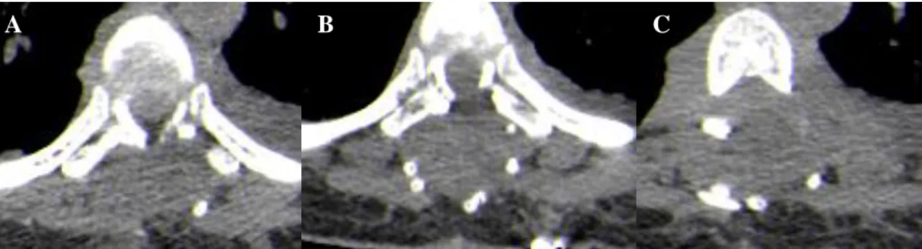
Figure 7 (a): Axial (A, B, and C) views of CT scan of the thoracic spine in a 30-year-old female with metastatic ovarian carcinoma showing extensive destruction of the posterior elements of the spine.
The tumours that usually respond to hormonal therapy are originating from breast and prostate cancer. The response to this modality is related to the presence of receptors [31]. The indications for radiotherapy in spinal metastases are: tumours with high radio-sensitivity, patients with minimal or no neurological deficit, unable to tolerate the surgical procedure, with involvement of multiple vertebral levels (> 2), with pain resistant to other treatment modalities, Karnofksy score
< 70 and with life expectancy less than 3 months are also candidates for radiotherapy [32].
4.10 Rosai-Dorfman disease
Rosai-Dorfman disease (RDD) is a rare disease and was only found in one patient in our series.
This condition is characterized by idiopathic proliferative disease that involves phagocytic histiocytes. It is chronic and self-limiting with a male predominance and occurs in young adults [33]. It is associated with immune dysregulation and viral infection. The most frequent presentation is cervical lymphadenopathy, fever, general malaise, and extradural spinal lesions typically present with spinal cord compression resulting in paraplegia [33, 34].
On MRI post contrast T1W images RDD appears homogenous and iso-intense (fig 8). Gross total resection is the preferred management of CNS RDD to relieve neurologic symptoms and for histo-pathological diagnosis [35].
A A
B C
34 If subtotal resection was performed and neurological symptoms resolve, a conservative approach is adopted, however with persistence of symptoms following surgery adjuvant therapy is considered [34]. Majority (58%) of CNS RDD will remain stable after surgical resection, radiation and has low recurrence rate [34]. Our patient was referred to the oncologist following surgical resection for adjuvant therapy.
Figure 8: T1W (post gadolinium MRI) sagittal A, B and axial C of the thoracic spine showing a Rosai-dorfman disease extending from T2 to L2, a well circumscribed homogenous enhancing extradural mass with spinal cord compression in a 23-year-old male.
4.12 Mortality and morbidity
Mortality rate in this study was 5.3%. Only one patient showed neurological improvement after excision of an EDSML, and was diagnosed with a schwannoma, which is not surprising as complete excision of these lesions are associated with a good outcome. McCormick et al in a series of 11 patients where complete resection was achieved, 92% of patients had neurological improvement [35, 36%].
AIDS related lymphomas are associated with high mortality rates, only (66%) of patients survive more than a year [37, 38]. The median overall survival for patients with diffusely large B-cell lymphomas (DLCCL) was 6 months in the pre-ART era [37].
However, for Burkitt’s lymphoma it remains unchanged at 6 months even with addition of ART to chemotherapy [39]. Severe immunosuppression is the highest risk factor for death [39].
A B C
35 Raviglione at al reported a mortality rate in HIV sero-positive TB patients to be 13% [40]. A Korean study of 116 patients with spinal TB showed favourable outcome in 81% of patients, while 19% had an unfavourable outcome [40]. Govender et al reported the mortality rate for spinal abscess to be 7.7% [19].
The prognosis with respect to survival in patients with extradural spinal metastasis essentially depends on the biology of the primary tumour: 2-year survival rates for spinal tumour are 9%
(lung) to 44% (breast or prostate) cancer [36].
5. Conclusion
EDSMLs in HIV sero-positive adult patients result in non-reversible neurological deficits in our practice. MRI of the spine is the investigation of choice in delineating these lesions; however, the role of a thorough neurological examination should not be overlooked. It is important for clinicians managing patients with HIV/AIDS to be aware of pathology affecting the spine in this group of patients, as this helps in formulating protocols aimed at improving neurological function and outcomes.
Rehabilitation and long term follow-up remains a challenge for the overburdened health care system in the KZN, as most of these patients have long-term neurological deficits which are worsened by other medical illnesses. The emphasis should be on multidisciplinary team approach with the purpose of early voluntary counseling and testing for HIV, early initiation of ARVs, treatment of associated medical illness and routine screening of malignancies in patients at risk.
36 Limitations of the study
The study was retrospective in nature and there was no long-term follow-up of patients. It is a single center study, however, the data emanated from the only public neurosurgical unit in the province of KwaZulu-Natal.
Conflict of interest
Authors had no conflict of interest when preparing this manuscript and research was not funded externally.
Funding sources
This research did not receive any specific grant from funding agencies in the public, commercial, or not-for-profit sectors.
37 References
1. Wiggill TM, Mayne ES, Willem P. Challenges in lymphoma diagnosis in HIV positive patients in the South African setting. Transfusion and Apheresis Science. 2013; 49: 157–
162.
2. Giarelli E, Jacobs LA. Journal of the Association of Nurses in Aids Care. 2001; 12(6):
52-67.
3. Census 2013 Statistical release – PO302/Statistics South Africa, Pretoria. Statistics South Africa. 2013.
4. Human Sciences Research Council. South African National HIV Prevalence, Incidence and Behaviour Survey. 2012.
5. Black S, Wallace M, Middelkoop K, Roobertze D, Bennie T, Wood R et al. Improving HIV testing amongst adolescents through an integrated Youth Centre rewards program:
Insights from South Africa. Children and Youth Services Review. 2014; 45: 98-105.
6. Bor J, Herbst A.J, Newell ML et al. Increases in adult life expectancy in rural South Africa: valuing the scale-up of HIV treatment. Science. 2013; 339 (6122): 961-965.
7. Bhigjee AI, Naidoo K, Patel VB, Govender D. Intracranial mass lesions in HIV-positive patients – the KwaZulu-Natal experience. S Afr Med J. 1999; 89:1284-1288.
8. Chong J, Di Rocco A, Tagliati M, Danisi F, Simpson DM, Atlas SW. MR Findings in AIDS-Associated Myelopathy. Am J Neuroradiol. 1999; 20: 1412–1416.
9. Adam MA, Johnson LF. Estimation of adult antiretroviral treatment coverage in South Africa. S Afr Med J 2009; 99 (9) :661–7.
10. Thurnher MM, Post MJD, Jinkins JR. MRI infections and neoplasms of the spine and spinal cord in 55 patients with AIDS. Neuroradiology. 2000; 42: 551-563.
11. Bilsky MH, Lis E, Raizer J, Henry Lee, Boland P. The diagnosis and treatment of metastatic spinal tumour. The Oncologist. 1999;4: 459 – 469.
38 12. Van Rie A, Westreich D, Sanne I. Tuberculosis in patients receiving antiretroviral treatment: incidence, risk factors, and prevention strategies. J Acquir Immune Defic Syndr 2010.
13. De Leon MEM, Schnell S, Rozental JM. Tumours of the spine and spinal cord. Seminars in Oncology Nursing. 1998; 14:43-52.
14. EpelbaumR, Haim N, Ben Shaker M, Ben Arie Y, Feinsod M, Cohen Y. Non-Hogkin’s lymphoma presenting with spinal epidural involvement. Cancer 1986;58: 21-20 – 2124.
15. Fuchs B, Boos N. Primary spine tumours. [ed] Max Aebi Norbert Boos. Heidelberg:
Springer-Verlag, 2008. pp. 951 – 76.
16. Gallien S, Zuber B, Polivka M. Multifocal Epstein–Barr virus associated smooth muscle tumour in adults with AIDS: case report and review of the literature. Oncology 2008; 74:
167–176.
17. Carbone A, Gloghini A. AIDS-related lymphomas: from pathogenesis to pathology. Br J Haematol 2005; 130(5):662–70.
18. Zondagh I, Dunn RN. Spinal tuberculosis: Diagnostic biopsy in mandatory. SAMJ May 2008;98(5);360-2.
19. Govender S, Parbhoo AH, Kumar KPS, Annamalai K. Anterior spinal decompression in HIV-positive patients with tuberculosis A prospective study. J Bone Joint Surg. 2001; 83:
864 – 7.
20 Dunn RN. Medical management of spinal tuberculosis. SA Orthopaedic journal. Autumn 2010; P:37 – 41.
22. Wiggill TM, Mantuna H, Willem P, Perner Y, Stevens WS. Changing pattern of lymphoma subgroups at a tertiary academic complex in a high-prevalence HIV setting: A South African perspective. J Acquir Immune Defic Syndr 2011; 56 (5):460–6.
23. McLoughlin LC, Nord KS, Joshi VV, DiCarlo FJ, Kane MJ. Disseminated leiomyosarcoma in a child with acquired immune deficiency syndrome. Cancer 1991; 67:
2618–2621.
39 24. Pratistadevi K Ramdial, Yetish Sing, Julian Deonarain, Jalaludin I Vaubell, et al. Extra-
uterine myoid tumours in patients with acquired immunodeficiency syndrome: a clinic- pathological re-appraisal: Histo-pathology. 2011; 59:1122–1134.
25. Martin RJ, Yuan HA. Neurosurgical care of spinal epidural, subdural, and intramedullary abscesses and arachnoiditis. Orthop Clin North Am 1996; 27: 125-36.
26. Khanna RK, Malik GM, Rock JP, Rosenblum ML. Spinal epidural abscess: evaluation of factors influencing outcome. Neurosurgery 1996; 39:958-64.
27. Corr P, Nathoo N, Landers AT. Spinal epidural abscess. South African Medical journal.
2000; 90: (8): 779 – 780.
28. Smego RA, Orlovic D, Wadula J, Modi G. A diagnostic and therapeuric algorithm for CNS mass lesions in HIV/AIDS. S Afr J Epidemiol Infect. 2000; 15:7-13.
29. Quraishi NA, Gokaslan ZL, Boriani S. The surgical management of metastatic epidural compression of the spinal cord. J Bone Joint Surg Br. 2010;92(8):1054-60.
30. Ghogwala Z, Mansfield FL, Borges LF: Spinal radiation before surgical decompression adversely affect outcome for symptomatic metastatic spinal cord compression. Spine 2001; 26(7): 818 -824.
31. Bohlius J, Schidlin K, Costagliola D, Fatkenheuer G, May M, Caro Murillo AM, Mocroft A, Bonnet F, Clifford G, Touloumi G, Miro JM, Chene G, Lundgren J, Egger M, Prognosis of HIV-associated non-Hogkin lymphoma in patients starting combination anti-retroviral therapy. AIDS, 2009, 23915):2029 – 2037.
32. Mounier N, Spina M, Gabarre J, Raphael M, Rizzardini G. AIDS-related non-Hodgkin’s lymphoma. Blood. 2006; 107: 3832- 40.
33. Rosai J, Dorfman RF. Sinus histiocytosis with massive lymphadenopathy: a newly recognized benign clinic-pathological entity. Arch Pathol. 1969; 87:63-70.
34. Kital R, Kazufumi S, Kubota T, et al. Meningeal sinus histiocytosis mimicking lymphoplasmacyte-rich meningioma. J Neurosurg. 1996; 84:1051-1054.
40 35. McCormick PC. Surgical management of Dumbbell tumours of the cervical spine.
Journal of Neurosurgery. 1996; Vol. 38:2 p 294 – 300.
36. Miura T, Nakamura K, Tanaka H, Kawaguchi H, Takeshita K, Kurokawa T. Resection of cervical spinal neurinoma including affected nerve root: recovery of neurological deficit in 15 cases. Acta Orthop Scand.1998; 69: 280–282.
37. Conant MA. Management of human immunodeficiency virus-associated malignancies.
Recent Results Cancer Res. 1995; 139: 423 – 432.
38. Lim ST, Karim R. AIDS-related Burkitt’s lymphoma versus diffuse large-cell lymphoma in the pre-highly active antiretroviral therapy (HAART) and HAART eras. J Clin Oncol 2005; 23: 4430-8.
39. Park DW, Sohn JW, Kim EH, Cho DJ, Lee JH, KIM KT et al. Outcome and management of spinal tuberculosis according to the severity of disease. Spine 2007; 32 (4): E130 - 135 40. Raviglione MC, Narain JP, Kochi A. HIV-associated tuberculosis in developing countries: Clinical features, diagnosis and treatment. World Health Organ 1992; 70 (4):
515-526.
- 41 -
Appendix 1:
STUDY PROTOCOL
Extradural Mass Lesions of the Spine in HIV Sero-positive Adults Primary investigator: Dr. Sonwabile Gonya1
Supervisor: Dr. Basil Enicker2 Co-supervisor: Prof. Colleen Aldous3
1 Department of Neurosurgery,
2 Department of Neurosurgery,
3 School of Medicine, College of Health Sciences University of Kwa-Zulu Natal
Nelson R. Mandela School of Medicine Private Bag X03, Mayville, 4058 Durban
South Africa
- 42 - Contents
A. List of abbreviations and acronyms 24
B. Definition of terms 26
C. Introduction 27
D. Research question 28
E. Study aim 28
F. Study objectives 28
G. Literature review 29 - 33
H. Rationale for the study 33
I. References 34 - 35
J. Study methodology 36
K. Study population 36
L. Study sampling 36
M. Sample size 36
N. Inclusion criteria 37
O. Exclusion criteria 37
P. Data collection methods and tools 37
Q. Statistical analysis 38
R. Study location 38
S. Study period 38
T. Limitations of the study 38
U. Ethical approval 39
V. Ethical considerations 39
- 43 -
W. Annexure A (data collection sheet) 40 - 41
- 44 - List of abbreviations and acronyms
AIDS Acquired immunodeficiency syndrome ART Antiretroviral treatment
CSF Cerebrospinal fluid
CT Computerized tomography EDSML Extradural spinal mass lesion HIV Human immunodeficiency virus IALCH Inkosi Albert Luthuli Central Hospital KZN KwaZulu-Natal
MCS Microscopy culture and sensitivity MRI Magnetic resonance imaging
NSOPD Neurosurgery out-patient department TB Tuberculosis
- 45 - Definition of terms
Dura mater: The outermost membrane of the meninges that surround the brain and spinal cord
Extradural mass: Mass that arises outside the dura mater from bones of the spinal column or paraspinal tissue.
Laminectomy: Surgical procedure involving removal of spinous process and lamina of the vertebrae to gain access to the spinal cord
Myelopathy: Functional disturbance and/or pathological change of the spinal cord Radicular pain: Pain occurring at the distribution of the nerve root
- 46 - Introduction
The majority of people (68%) in the world living with HIV are found in Sub-Saharan Africa [1, 2]. South Africa is worst affected, with approximately 5.7 million people (10.9% of population) living with HIV/AIDS [2, 3, 4].
It is reported that the prevalence of HIV infection in South African adults between the ages of 14 to 49 years is 17%, with 300 000 to 350 000 new infections a year [5]. The Province of KwaZulu-Natal, has the highest infection rate in South Africa [6].
Individuals who are diagnosed HIV sero-positive often present with neurological disease and in 10 to 20% of patients these are AIDS-defining [7]. The spinal cord is often affected in HIV sero- positive individuals and the commonest pathology is HIV-associated myelopathy [8]. Other conditions that can cause myelopathy are opportunistic infections such as TB, neoplasms and vascular lesions [8]. The extradural space is where some of these pathologies can be located.
Research question
What is the histo-pathology of extradural mass lesions of the spine that affect HIV-sero-positive adult patients?
- 47 - Study aim
To determine the histo-pathological findings of extradural spinal mass lesions (EDSML) in HIV sero-positive patients treated in the Department of Neurosurgery at Inkosi Albert Luthuli central hospital (IALCH), Durban, South Africa from 01 January 2003 to 31 August 2014.
Objectives of the study
I. To determine the demographic profile of HIV sero-positive patients presenting with extradural spinal mass lesions (EDSML) to the Department of Neurosurgery at IALCH.
II. To determine their clinical features and neurological deficits at presentation.
III. To determine the CD4 count of these patients on admission.
IV. To determine results of neuro-radiology investigations.
V. To determine the presence of opportunistic co- infections in other organ systems.
VI. To determine surgical management and morbidity.
VII. To determine final histo-pathology reports.
VIII. To determine length of hospital, stay and treatment outcomes.
IX. To determine the mortality rate at follow-up.
- 48 - Literature review
The HIV epidemic has put a lot of strain in the already over-burdened health system of South Africa over the years. The first death from AIDS in South Africa occurred in 1982, with HIV/AIDS being reported as one of the leading cause of death in the country [2].
The introduction of the ART program has gone a long way in reducing the mortality from HIV/AIDS, with reported 1.9 million people receiving ART in South Africa [9]. In KwaZulu- Natal, the ART program has increased life expectancy from 49.2 in 2003 to 60.5 years in 2011 [6].
Individuals who are HIV sero-positive with low CD4 counts are at a high risk of developing HIV- associated mass lesions, especially of the spine. These mass lesions can be either infectious or neoplastic.
The infectious lesions can be bacterial (acute pyogenic), chronic granulomatous (TB) or fungal.
Bacterial infections can cause extradural empyema or abscess, the responsible organism commonly being Staphylococcus aureus [10]. The source of infection can be either from haematogenous spread (e.g from respiratory infections) or from direct extension (e.g psoas abscess).
- 49 - In South Africa approximately 60% of HIV sero-positive patients are co-infected with TB and it can manifest as an extradural mass lesion and the organism responsible in majority of cases is Mycobacterium tuberculosis [11]. In the spine TB can also cause destruction of the vertebral
body and result in spinal deformity a condition called Pott’s disease [12,13]. Pulmonary TB has to be excluded in these patients. Extradural lesions caused by fungal infections are rare, when they do occur are due to organisms such as Aspergillus species and Cryptococcus neoformans [13].
Neoplasms of the spine account for approximately 10% of CNS tumours [14]. The commonest extradural tumours of the spine are metastatic lesions and majorities (70%) are found in the thoracic spine [14]. These tumours cause destruction of the vertebral body (70%) and compress the spinal cord and nerve roots. Metastatic tumours are commonly from the lung, breast, prostate, multiple myeloma and sarcoma [14].
Lymphomas are typically found in the extradural space; they can be primary or metastatic and are considered AIDS-defining [1]. The risk of lymphoma in HIV sero-positive individuals is reported to be 100 higher when compared to HIV negative individuals, especially if they have a high viral load (> 100 000 copies/mm3), low CD4 count (< 50 cells/mm3) and are not on ART [15]. However, lymphomas can occur at any CD4 count level.
These tumours are caused by combination of immune dysregulation, loss of T cell immunity against viruses namely Epstein-Barr virus (EBV) and Human herpes virus 8 (HHV8), and lastly from direct oncogenic effect of HIV [15].