THE UTILITY OF BASIC FIBROBLAST GROWTH FACTOR AS A NON-INVASIVE BIOMARKER OF FOCAL SEGMENTAL GLOMERULOSCLEROSIS IN HIV POSITIVE AND NEGATIVE
CHILDREN
By
Nokwanda Zamahlubi Gumede
Submitted in partial fulfillment for the degree of MASTER OF MEDICAL SCIENCE
in the
Discipline of Clinical Medicine Nelson R Mandela School of Medicine
College of Health Sciences University of KwaZulu-Natal
Durban, South Africa
2018
i PREFACE
This study represents original work by the author and has not been submitted in any other form to another University. Where use was made of the work of others, it has been duly acknowledged in the text.
The research described in this dissertation was carried out in the Optics & Imaging Centre, Doris Duke Medical Research Institute, College of Health Sciences, University of KwaZulu- Natal, Durban, South Africa under the supervision of Professors Thajasvarie Naicker and Rajendra Bhimma.
Nokwanda Gumede Thajasvarie Naicker (co-supervisor)
Rajendra Bhimma (supervisor)
ii DECLARATION
I, Nokwanda Zamahlubi Cele declare that:
(i) The research reported in this dissertation, except where otherwise indicated is my original work.
(ii) This dissertation has not been submitted for any degree or examination at any other university.
(iii) This dissertation does not contain other person’s data, pictures, graphs or other information, unless specifically acknowledged as being sourced from other persons.
(iv) This dissertation does not contain other persons writing, unless specifically acknowledged as being sourced from other researchers. Where other sources have been quoted, then:
a) Their words have been rewritten but the general information attributed by them has been referenced.
b) Where their exact words have been used their writing had been placed inside quotation marks and referenced.
(v) Where I have reproduced a publication of which I am an author, co-author, I have indicated in detail which part of the publication was actually written by myself alone and have fully referenced such publications.
(vi) This dissertation does not contain text, graphics, or tables copied and pasted from the internet, unless specifically acknowledged and the source being detailed in the dissertation and the reference sections.
Signed: __________________ Date: 03/08/03
iii DEDICATION
To my grandmother, Lindiwe Cele - You have always been my pillar of strength. You are the woman behind all my achievements and you the reason why giving up has never been an option. Thank you for your support and words of wisdom. I hope this dissertation will be an inspiration to my children as well as my grandchildren.
To my mother and father - Bongiwe and Mlaleni Yengwa; if it was not for you I will not be where I am today. Thank you for your understanding, patience and for making me realize my potential.
Family and Friends - My deepest gratitude and special thanks to my loved ones, for all their prayers and continued support.
To God - Above all, I thank the living God Almighty for granting me this opportunity and strength to successfully complete this research project. I could never have done this without the faith I have in you.
“Therefore I tell you, whatever you ask for in prayer, believe that you have received it, and it will be yours.”
(Mark 11:24)
iv ACKNOWLEDGEMENTS
Special gratitude to my supervisors Professors T Naicker and R Bhimma for their support, patience, valuable and constructive advice.
To all my lab members, thank you for your technical assistance and for creating a warm working atmosphere.
My deepest gratitude and special thanks to my loved ones, grandmother, my mother and my friends for all their prayers and continued support.
Lastly, thank you to the National Research Foundation (South Africa) and College of Health Sciences for financial support.
Above all, I thank the living God Almighty for granting me this opportunity and strength to successfully complete this research project.
v TABLE OF CONTENTS
PREFACE ... i
DEDICATION ... iii
ACKNOWLEDGEMENTS ... iv
LIST OF ABBREVIATIONS ... vii
LIST OF TABLES ... viii
LIST OF FIGURES ... ix
ABSTRACT ... x
CHAPTER ONE ... 1
BACKGROUND AND LITERATURE REVIEW ... 2
1.1 Basic fibroblast growth factor ... 2
1.1.1 Molecules that mediate the function of FGF ... 3
1.1.2 Interaction with heparin or heparan sulfate ... 3
1.1.3 FGF in functional and structural kidney damage ... 4
1.1.4 Progression of kidney disease ... 5
1.2 Focal segmental glomerulosclerosis ... 6
1.2.1 Pathogenesis of focal segmental glomerulosclerosis ... 8
1.2.2 Therapy ... 10
1.2.3 Adjunctive therapies for FSGS ... 11
1.2.4 Aims and Objectives ... 13
Aim ... 13
Objectives ... 13
CHAPTER TWO ... 14
Submitted Manuscript ... 15
Abstract ... 17
Introduction ... 19
Method and Materials ... 21
vi
Results ... 24
Discussion ... 26
Conclusion ... 29
Acknowledgement ... 30
Funding ... 30
References ... 31
CHAPTER THREE ... 38
SYNTHESIS ... 39
Conclusion ... 43
CHAPTER FOUR ... 44
References ... 45
vii LIST OF ABBREVIATIONS
HIV - Human Immunodefiency Virus HIVAN - HIV-associated Nephropathy FSGS - Focal segmental glomerulosclerosis bFGF - Basic fibroblast growth factor HSPG - Heparin sulfate proteoglycans
viii LIST OF TABLES
Chapter 2 –Manuscript
Table 1 ... Error! Bookmark not defined.
ix
LIST OF FIGURES
Chapter 1
Figure 1 ... Error! Bookmark not defined.
Chapter 2
Figure 1 ... Error! Bookmark not defined.
Figure 2 ... Error! Bookmark not defined.
Figure 3 ... Error! Bookmark not defined.
Figure 4 ... Error! Bookmark not defined.
Figure 5 ... Error! Bookmark not defined.
Figure 6 ... Error! Bookmark not defined.
Figure 7 ... Error! Bookmark not defined.
x ABSTRACT
Background: The human immunodeficiency virus (HIV) infection can lead to the development of HIVAN with the majority of patients progressing to end-stage kidney disease. Importantly, individuals of African ancestry are more at risk of developing HIVAN than their European descent counterparts. Early diagnosis and immediate nephrology referral are key steps in management because this enable predialysis education, allows for implementation of preventive measures that delay or even halt progression of HIVAN to end- stage kidney disease, as well as decrease morbidity and mortality. Currently, the diagnosis of HIVAN requires a kidney biopsy. Due to the development of genomics, epigenetics, transcriptomics, proteomics, and metabolomics, the introduction of novel techniques will allow for the identification of novel biomarkers in kidney diseases. Previous studies have recognized basic fibroblast growth factor (bFGF) as a biomarker for HIVAN, since significant levels of bFGF low affinity receptors have been previously found in the kidneys from HIV infected children. bFGF is an angiogenic growth factor that is involved in kidney growth and the pathogenesis of kidney diseases.
The aim of this study was to investigate the use of basic fibroblast growth factor (bFGF) as a non-invasive biomarker for the diagnosis of primary FSGS and HIVAN
Method: The study group consisted of 31 children; HIVAN (n=11) and idiopathic FSGS (n=20). The control group consisted of 40 children with no kidney disease; HIV positive (n=20) and HIV negative (n=20). Serum samples were stored at -800C and were analysed for bFGF using the Bio-Plex ProTM Human Cytokine. Statistical analysis was performed using GraphPad Prism version 5.
xi Results: The concentration of bFGF was higher in the HIVAN (mean= 9.0 ng/ml; 95% CI:
10.18 – 7.18) vs idiopathic FSGS (mean= 7.0 ng/ml; 95% CI: 8.16 – 6.59) (Mann-Whitney U= 66.5; p= 0.0685). There was a significant elevation of serum bFGF Mean Flourescence Intensity (MFI) in children with HIVAN when compared to controls [HIV positive (mean= 7.0 ng/ml; 95% CI: 7.56 – 6.38) (Mann-Whitney U= 58.8 ; p= 0.004) and negative controls (mean= 6.5 ng/ml; 95% CI: 7.00-6.06) (Mann-Whitney U= 43.5 p= 0.029)]. There was no statistically significant differences in serum bFGF MFI between patients with HIVAN vs.
idiopathic FSGS and between idiopathic FSGS vs. controls.
Conclusion: This study demonstrated statistically significant difference between bFGF levels in children with HIVAN and controls, although it failed to distinguish statistically significant differences in bFGF levels between HIVAN and idiopathic FSGS.
1
CHAPTER ONE
2 BACKGROUND AND LITERATURE REVIEW
1.1 Basic fibroblast growth factor
The fibroblast growth factor (FGF) family consists of 18 secreted ligands. These secreted ligands can be categorized into two subfamilies; the canonical FGFs (FGF1-10, 16-18 and 20), and the hormone-like FGFs (FGF 19, 21 and 23) (Burgess and Maciag, 1989). FGFs are classified as paracrine, intracrine and endocrine FGFs based on their mode of action (Itoh et al., 2015) . Paracrine and endocrine FGFs are secreted signaling molecules acting through cell-surface FGF receptors (FGFRs) (Ornitz and Itoh, 2015). In comparison, several endocrine FGFs, require α-Klotho or β-Klotho as a cofactor for binding to FGFRs (Ornitz and Itoh, 2015).
The phenotypes of endocrine FGF knockout mice demonstrate that FGF are involved in phosphate and vitamin D metabolism (Itoh et al., 2015). There is evidence to suggest that endocrine FGF have a pathophysiological role in genetic and metabolic diseases. Abnormal levels of endocrine FGF may serve as a potential risk factor for metabolic diseases but also are useful biomarkers for detection of metabolic diseases (Itoh et al., 2015). These discoveries provide new perceptions into the physiological and pathophysiological roles of endocrine FGFs and their role as potential biomakers and therapeutic targets for metabolic diseases.
FGF2, also known as basic fibroblast growth factor (bFGF) and FGF-β, is a growth factor and signaling protein encoded by the FGF2 gene (Dionne et al., 1990). It is synthesized primarily as a 155 amino acid polypeptide, resulting in an 18 kDa protein (Kim, 1998). Basic FGF as well as their receptors control certain key behaviours such as migration, differentiation, survival and proliferation (Brooks et al., 2012). Furthermore, bFGF
3 specifically promotes fibroblast proliferation and up-regulates the expression of proliferation- associated genes (Strutz et al., 2000).
1.1.1 Molecules that mediate the function of FGF
The activated FGFs transfer their signals to cellular targets via four FGF receptors (FGFRs) (Ornitz and Itoh, 2016). A number of alternative splicing occur in different intracellular and extracellular encoding regions (Ornitz and Itoh, 2015). Furthermore, alternative splicing within the third immunoglobulin-like domain of FGFRs 1-3 generating b and c alternates are mainly important ligand binding specificity determinants. Remarkably, FGF1 is the only ligand that is capable of activating all FGFRs regardless of alternative splicing. It has been demonstrated by ligand binding specificity assays that the FGF7 subfamily specifically binds and stimulates a splice variant (Ornitz et al., 1996). Uniquely, the FGF9 subfamily also stimulates FGFR 3b. As for the canonical FGF subfamilies (FGF 1, 4, 7, 8, 9) heparan sulfate (HS) acts as the primary endogenous co-factor for the activation of the receptor as well as its ligand binding. Lastly, intracellular the FGFs subfamily specifically bind and mediate Nar channels without activating FGFRs (Ornitz and Itoh, 2016).
Often FGF signals reciprocally and directionally through epithelial mesenchymal boundaries (Olsen et al., 2003). The functionality of these signaling pathways need exceedingly tight mediation of FGF activity as well as receptor specificity. Directional signaling mediates the patterning and outgrowth of a given compartment during development. Hence, differential expression of the receptors' alternate splice forms can inhibit or limit autocrine signaling (Olsen et al., 2003).
1.1.2 Interaction with heparin or heparan sulfate
One of the important features of FGF is its interaction with heparan sulfate proteoglycan (HSPG) (Ornitz and Itoh, 2016). These interactions are responsible for the stabilization of
4 FGFs to inhibit proteolysis and thermal denaturation; thereby strictly preventing their release and diffusion into interstitial spaces (Virag et al., 2007). Interaction between HS and FGFs lead to the formation of higher order oligomers and dimers (Nusayr et al., 2013). A study by (Schreiber et al., 1985) demonstrated that HS increases the half-life and affinity of the FGF- FGFR complex and that HS can bridge FGF 2 including the FGFR by attaching to a groove created by the heparan-binding sites of both the receptor and ligand.
1.1.3 FGF in functional and structural kidney damage
Recent studies have established the intrinsic capacity of the kidney to undergo repair subsequent to acute damage via re-expression or repair proteins (Villanueva et al., 2014).
Initiation with bFGF could speed up this process. Nonetheless, there is not sufficient evidence that supports whether bFGF can induce this phenomenon in damaged kidney cells (Villanueva et al., 2014).
bFGF is a protein that is released for the induction of cellular aggregation (Karavanova et al., 1996). The core effect of bFGF includes maintaining WT-1 synthesis, a transcription factor that is responsible for inducing the conversion of mesenchymal cells into metanephric tissue (Karavanova et al., 1996). Furthermore, bFGF is able to induce expression of repair proteins and speed up the process of repairing tissue when it is exogenously added or conversely, renal injury could be sustained when it is inhibited (Villanueva et al., 2008). Moreover, it has been established that bFGF stimulates the proliferation rate of kidney cells (Dudley et al., 1995).
Previous studies have recognized bFGF as a biomarker for renal disease, particularly in focal segmental glomerulosclerosis (FSGS), where they demonstrated significant levels of bFGF low affinity receptors in the kidney of HIV infected children (Ray et al., 1999). These studies
5 highlighted that bFGF in circulation is subsequently harboured in the kidney bound to HSPG (Klagsbrun and Baird, 1991).
1.1.4 Progression of kidney disease
In both animal models and in humans with FSGS, glomerulosclerosis is categorized by scarring of small sections of individual glomeruli as well as tubular structure (D'Agati et al., 2011). The glomeruli show loss of capillaries leading to localized areas of progressive scarring and cell proliferation that ultimately result in breaking down of the capillary bed (Klahr and Morrissey, 2000). Tubules, mainly those bound to scarred glomeruli, are shrunken and in most cases, are delimited by inflammatory cells (Mullins et al., 2016) . This is followed by activation and proliferation of matrix-producing fibroblasts, mainly bFGF which synthesize substantial amounts of extracellular matrix deposited in an organized manner (Klahr and Morrissey, 2000). Several studies have established that the differential expression of certain bFGFs is one of the key reasons for scar development (Klahr and Morrissey, 2000).
Thus, bFGF may play an important role in the scarring process pathognomic to FSGS.
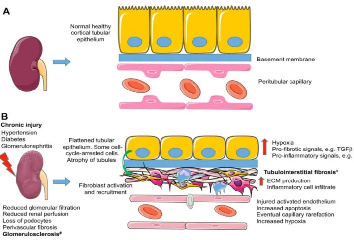
6
Figure 1: The pathophysiological processes linked to FSGS. (A) A healthy, normal kidney (left), and
an enlarged view of the tubule its associated vasculature (right). (B) A chronically diseased kidney, displaying the progression of factors that lead to tubule-interstitial fibrosis (Mullins et al., 2016).
1.2 Focal segmental glomerulosclerosis
FSGS is one of the most common forms of steroid resistant nephrotic syndrome and is categorized histologically by obliteration of glomerular capillaries, mesangial sclerosis, foam cells, hyalinosis and adhesion between the Bowman’s capsule and glomerular tuft (Rood et al., 2012). It is a worldwide public health issue due to its increasing incidence and prevalence. Nephrotic patients with FSGS have a poor prognosis with 50% progressing to end stage renal disease (ESRD) over 3 to 8 years (Korbet, 2000).
7 FSGS may be primary or secondary to various causes including infections such as Human Immunodeficiency virus type I (HIV-1) (Kim et al., 2016). Over the past 20 years there has been a significant increase in the incidence of FSGS, particularly in adult patients where there has been a 2-3 fold rise in the rate of diagnosis of FSGS (Kiffel et al., 2011). A similar increase has been documented in children (Kiffel et al., 2011). Research has shown that majority of steroid resistant nephrotic syndrome is due to increasing levels of angiotensin II, which together with other growth factors, also up regulate the expression of basic fibroblast growth factors (bFGF) (Klahr and Morrissey, 2000). Hence, the development and progression of FSGS is determined by the action of these growth factors. FGFs are involved in the regulation of the balance between proliferation and cell growth versus apoptosis, necrosis and cell death as well as the balance of degradation against matrix accumulation (Fogo, 2015).
Hence, glomerulosclerosis may result due to an increase in bFGF expression leading to the accumulation of extracellular matrix which contribute to renal malfunction (Fogo, 2015).
In patients with FSGS, bFGF is up regulated (Strutz et al., 2000). This is supported by a study done on HIV positive children where elevated levels of bFGF were observed and this was explained by the fact that renal HSPG functions as a sink, trapping bFGF as well as other growth factors (Strutz et al., 2000).
Globally, the incidence of FSGS has significantly increased over the past 30 years across all ethnic groups (Kiffel et al., 2011). It is one of the most significant clinical disorders that causes acquired chronic kidney disease (CKD) in both adults and children (Kiffel et al., 2011). FSGS has been the subject of intensive basic medico-clinical studies because of the significant morbidity arising from this condition. It was initially diagnosed on kidney biopsy specimens taken from patients suffering from steroid resistant nephrotic syndrome (Kiffel et al., 2011). There is not significance data on the epidemiology of FSGS in many parts of Africa due to the unavailability of renal registries. In Africa, glomerular diseases have been
8 reported to account for a large proportion of patients. Most renal biopsies reported from Africa originates from studies in North Africa (Okpechi et al., 2016). Of the 12,093 biopsies that were reported, 70% were from studies carried out in North Africa and these countries had more papers published after the year 2000 compared to countries from sub-Sahara Africa (Okpechi et al., 2016).
Attempts have been made to establish a histological classification of renal pathology so as to predict pharmacological response and treatment. In most of the pathological scoring systems used worldwide, the scarring associated with FSGS is characterised into five categories;
collapsing, perihilar, not otherwise specified (NOS), hypercellular, and tip lesions (D'Agati, 2003). The intensity of mesangial cellularity can differ and there is continuing debate whether the severity of this observation has prognostic implications (Stokes et al., 2006). The collapsing variant, which is subtype of FSGS, has constantly been proven to be resistant to treatment and runs a severe to moderately aggressive course (Kiffel et al., 2011).
Ultrastructural studies display occasional immune deposits, segmental scarring, and fusion of the podocyte foot process (Markowitz et al., 2003). Reticular inclusion bodies are observed on various secondary types of FSGS such as HIV nephropathy. Foot process effacement is often more severe and prevalent in primary FSGS than in secondary FSGS (Kiffel et al., 2011). Immunofluorescence findings often display deposition of IgM and complement factor C3 in sclerotic portions but this is reflected as a non-specific finding (Jennette and Hipp, 1985).
1.2.1 Pathogenesis of focal segmental glomerulosclerosis
Podocyte dysfunction is currently one of the central features in the pathogenesis of FSGS (Barisoni et al., 2009). Podocyte dysfunction could be due to an inherent abnormality within the podocyte or an exogenous element that stimulates podocyte damage. Loss of these non-
9 proliferative terminally differentiated cells is also critical in the progression of FSGS (Kiffel et al., 2011). In vivo experimental models that categorise podocyte damage and loss, showed that podocytopenia is largely related with the histological pattern of damage (Wharram et al., 2005). In experiment models that were performed it was observed that, if ≤20% podocytes are lost, resident glomerular epithelial stem cells transit from a position adjacent to the Bowman’s space into the glomerulus to recover injured podocytes that were damaged by apoptosis and necrosis. However, if the loss is between 20-40%, FSGS lesions result, whereas if loss of podocyte is >40%, then there is global sclerosis of the glomerulus (Kiffel et al., 2011).
Transformed expression of various signalling molecules are linked with podocyte damage and the progression of glomerulosclerosis in animal models of FSGS (Kaufman et al., 2010).
These include Sidekick-1 and Notch proteins which play integral roles in the morphology of the glomerulus and normal nephrogenesis (Kaufman et al., 2010). Upcoming genomic studies may assist in determining the role of these as well as other molecules in the broad spectrum of disease severity that occur in FSGS patients.
One of the interesting improvements that have been discovered in the past decade in understanding FSGS is the association between genetic mutations and several structural proteins of the podocyte (Kaufman et al., 2010). The first FSGS genetic studies revealed the occurrence of glomerulopathy in humans with altered genes for podocin included NPHS1, NPHS2, WT-1, LAMB2, CD2AP, TRPC6, ACTN4 and INF2 (Chen and Liapis, 2015).
Furthermore, seven other genes have been demonstrated in FSGS family pedigrees which include; IFN2, CD2AP, α-actinin -4, laminin β2, Wilms tumor-1, phospholipase C epsilon (Kiffel et al., 2011). In addition, nephron protein mutations, which were initially demonstrated in connection with congenital nephrotic syndrome, can also result in steroid- resistant nephrotic syndrome in adults and children (Philippe et al., 2008). Generally, there
10 are two forms of mutations, autosomal dominant disease with onset in adulthood or in late adolescence and an autosomal recessive pattern of inheritance seen mainly in children (Philippe et al., 2008). The incidence of sporadic FSGS cases related with each mutation differs from 3-20% depending on the patient cohort being tested. Current studies with genotype-phenotype correlations have simplified certain mutations that have an effect on the clinical course of disease in FSGS patients (Caridi et al., 2009). For instance, the NPHS2 gene in podocin are related with onset of disease earlier in childhood and rapid progression to end stage kidney disease (Caridi et al., 2005).
1.2.2 Therapy
There is dearth of clinical information describing the outcome of randomized trials (RCT) in glomerular and nephrological diseases to guide treatment of FSGS (Leaf et al., 2010). All FSGS patients profit from the use of antiproteinuric agents that antagonize the renin- angiotensin II-aldosterone axis(Kiffel et al., 2011). This therapy comprises inhibition of renin by specific renin antagonists, aldosterone antagonists, angiotensin converting enzyme inhibitors, and lastly angiotensin receptor blockers(Mercier et al., 2014). There is emerging recognition of the role of T-cells in the pathogenesis of the disease, and the successive focus on immunosuppressive agents as possible treatments may be misguided and delay the search for therapeutic agents targeting T-cells (Meyrier, 2009). This is supported by current data that cyclosporine may be responsible for reducing proteinuria in FSGS animal models by acting specifically on the podocyte and maintaining the actin cytoskeleton instead of aiming at immunological mechanisms, specifically inhibition of nuclear factor of triggered T-cell (NFA7) (Meyrier, 2009).
About 25% of FSGS patients have total or partial remission of proteinuria after a course of corticosteroids (Meyrier, 2009). One exception to this typical grim outcome is the feedback
11 from a single center signifying that almost 80% (18/23) of children suffering from FSGS may accomplish a thorough or partial remission over a mean continuation period of 46 ± 5 months in response to extended treatment with pulses doses of methylprednisolone (Mendoza et al., 1990). Using oral dexamethasone pulses, Kopp et al (unpublished data) tried to duplicate this in adults; however the outcomes were not as encouraging as reported in children by (Kiffel et al., 2011). Hence, steroids are not consistently optimal therapy in FSGS patients.
Furthermore, the major side effects of prolonged use of steroids significantly restricts its use in patients suffering from bone disease, hypertension, or those that are overweight (Meyrier, 2009).
The effectiveness of rituximab to encourage a remission in 25-40% of patients with steroid- resistant FSGS suggests that B-cells rather than T-cells may be possible immunosuppressive therapy targets (Kiffel et al., 2011). Additionally, other therapies that could be utilized include, mycophenolate mofetil (MMF) which is one of the most commonly used immunosuppressive drugs used either alone or in combination with other immunosuppressive drugs (e.g. corticosteroids and/or calcineurin inhibitors) for the prevention of organ rejection after solid organ transplantation as well as in the therapy of autoimmune and neoplastic diseases (Oellerich et al., 2000, Allison and Eugui, 2000) .
1.2.3 Adjunctive therapies for FSGS
Several adjunctive therapies have been proposed in an attempt to reduce the progression of tissue injury in FSGS. These include angiotensin I-converting enzyme inhibitors, renin- angiotensin-aldosterone, angiotensin receptor blockers (ARBs) and statins. However, these therapies have not displayed significant positive improvements specifically in tubular function and glomerular filtration rates that could prevent the final phase of renal disease
12 (Wolf et al., 2003). Hence, research for new promising and alternative treatments is one of the main concerns in nephrology.
Statins are well known to decrease cardiovascular morbidity and mortality in patients with and without recognized cardiovascular disease as well as in several high-risk populations (Shepherd et al., 1995). Nonetheless, definite evidence for developed cardiovascular outcomes with statin therapy for renal disease is not yet available (Agarwal, 2007).
The overall conclusion established from various FSGS researchers suggests that suitable pharmacological treatments for the development of renal fibrosis may require an approach that is multi-pharmacological (Kiffel et al., 2011). More research is required to understand the developmental role of bFGF and other FGFs as these will provide further insight as to whether a single or multiple FGFs interact during the progression of FSGS. The aim of this study is to investigate bFGF as an non-invasive biomaker to renal biopsy in children with primary FSGS and HIVAN compared to controls.
13 1.2.4 Aims and Objectives
Aim
The aim of this study was to investigate the use of basic fibroblast growth factor (bFGF) as a non-invasive biomarker for the diagnosis of primary FSGS and HIVAN.
Objectives
1. To compare the levels of bFGF in children with biopsy proven FSGS to healthy controls.
2. To measure and compare the levels of bFGF in FSGS according to HIV status.
3. To compare the levels of bFGF in FSGS and HIVAN to HIV positive children with no kidney disease and healthy controls.
14
CHAPTER TWO
15 Submitted Manuscript
16 The utility of basic fibroblast growth factor as a non-invasive biomarker of focal segmental glomerulosclerosis in HIV positive and negative children
Authors: aNokwanda Gumede, bThajasvarie Naicker and aRajendra Bhimma
Affiliations: aClinical Medicine, College of Health Sciences, University of KwaZulu-Natal, Durban, KwaZulu-Natal, South Africa, Department of Pediatrics & Child Health, Nelson R.
Mandela School of Medicine, University of KwaZulu-Natal, Durban, KwaZulu-Natal, South Africa
*Corresponding author:
Nokwanda Gumede Pediatrics & Child Health, College of Health Sciences, University of KwaZulu-Natal, Durban, South Africa.
Postal address: Private Bag 7
Congella
KwaZulu-Natal
4013
South Africa
Email address: [email protected];[email protected]; [email protected] Total word count:
Word count excluding abstract and references:
Number of figures: 7 Number of tables: 1
17 Abstract
Background: The human immunodeficiency virus (HIV) infection can lead to the development of HIVAN with the majority of patients progressing to end-stage kidney disease. Importantly, individuals of African ancestry are more at risk of developing HIVAN than their European descent counterparts. Early diagnosis and immediate nephrology referral are key steps in management because this enable predialysis education, allows for implementation of preventive measures that delay or even halt progression of HIVAN to end- stage kidney disease, as well as decrease morbidity and mortality. Currently, the diagnosis of HIVAN requires a kidney biopsy. Due to the development of genomics, epigenetics, transcriptomics, proteomics, and metabolomics, the introduction of novel techniques will allow for the identification of novel biomarkers in kidney diseases. Previous studies have recognized basic fibroblast growth factor (bFGF) as a biomarker for HIVAN, since significant levels of bFGF low affinity receptors have been previously found in the kidneys from HIV infected children. bFGF is an angiogenic growth factor that is involved in kidney growth and the pathogenesis of kidney diseases.
The aim of this study was to investigate the use of basic fibroblast growth factors (bFGF) as a non-invasive biomarker for the diagnosis of FSGS in HIV positive (HIVAN) and HIV negative children (primary FSGS).
Method: The study group consisted of 31 children; HIVAN (n=11) and idiopathic FSGS (n=20). The control group consisted of 40 children with no kidney disease; HIV positive (n=20) and HIV negative (n=20). Serum samples were stored at -800C and were analysed for bFGF using the Bio-Plex ProTM Human Cytokine. Statistical analysis was performed using GraphPad Prism version 5.
18 Results: The concentration of bFGF was higher in the HIVAN (mean= 9.0 ng/ml; 95% CI:
10.18 – 7.18) vs idiopathic FSGS (mean= 7.0 ng/ml; 95% CI: 8.16 – 6.59) (Mann-Whitney U= 66.5; p= 0.0685). There was a significant elevation of serum bFGF Mean Flourescence Intensity (MFI) in children with HIVAN when compared to controls [HIV positive (mean= 7.0 ng/ml; 95% CI: 7.56 – 6.38) (Mann-Whitney U= 58.8 ; p= 0.004) and negative controls (mean= 6.5 ng/ml; 95% CI: 7.00-6.06) (Mann-Whitney U= 43.5 p= 0.029)]. There was no statistically significant differences in serum bFGF MFI between patients with HIVAN vs.
idiopathic FSGS and between idiopathic FSGS vs. controls.
Conclusion: This study demonstrated statistically significant difference between bFGF levels in children with HIVAN and controls, although it failed to distinguish statistically significant differences in bFGF levels between HIVAN and idiopathic FSGS.
Keywords: HIV, HIVAN, bFGF, FSGS
Running title: FGF levels in HIV associated nephropathy
19 Introduction
Human immunodeficiency virus (HIV) infected antiretroviral therapy naïve children display a high viral load during the late phase of infection, placing them at risk of developing several types of kidney diseases, including HIV associated nephropathy (HIVAN) [1]. However children on antiretroviral therapy, kidney disease is usually uncommon [2]. The spectrum of kidney disease that occurs with HIV infection includes acute kidney injury, disorders of tubular function, thrombotic microangiopathies, kidney injury secondary to drug use and various forms of chronic glomerular diseases including HIVAN and HIV associated immune complex disorders [3] . The classical findings of HIVAN include: persistent proteinuria usually accompanied by varying degrees of haematuria, urinary sediment with urinary microcysts, azotemia, normal to large kidneys on ultrasound images, normal blood pressure, and focal segmental glomerulosclerosis (FSGS) on kidney biopsy findings [4,2]. During HIV infection, FSGS is an important comorbidity [5].
FSGS is a primary cause of nephrotic syndrome in children, and if untreated, has a poor prognosis [6]. Nonetheless, the pattern of segmental and focal sclerosis is not specific to disease with the primary podocyte lesions, and several other diseases e.g. HIVAN, demonstrate light microscopic lesion patterns such as overlying cell hyperplasia as well as glomerular tuft injuries similar to primary FSGS [7].
In the pre-antiretroviral therapy era, HIVAN was characterized by rapid progression to end- stage kidney disease leading to the need for dialysis [8]. Highly active antiretroviral therapy (HAART) has improved the natural course of this disease, increasing the importance of early diagnosis and allied suitable care [8]. Robust biomarkers may assist in diagnosing and monitoring kidney disease commencing from initial stages of the disease [9]. Previous studies have recognized bFGF as a biomarker for FSGS, since significant levels of bFGF low affinity receptors have been previously found in the kidney of HIV infected children [10]. bFGF is an
20 angiogenic vascular growth factor, that is essential for cellular aggregation induction [11]. It is involved in the stimulation of epithelial condensation, apoptosis inhibition as well as maintenance of WT-1 synthesis [12]. Furthermore, bFGF is mainly stored as an inactive pool within the extracellular matrix and vessel wall, and is absent in the circulation unless it is secreted during tissue injury and angiogenesis [13]. Elevated levels of bFGF may result in the progression of FSGS, and unrestrained elevation of bFGF in the kidney extracellular matrix can generate the fibrotic lesions and tubulointerstial proliferative distinctive to HIVAN [13]. However, the processes that regulate secretion and behaviour of bFGF in glomeruli and renal tubules are as yet not clearly understood [14].
Based on the findings that elevated bFGF may result in the progression of FSGS and plays a role in the generation of fibrotic lesions and tubulintersitial proliferation distinct to HIVAN, our aim was to determine if bFGF can be used as a non invasive biomarker for the detection of HIVAN and idiopathic FSGS.
21 Method and Materials
Ethical consideration
This study was performed on blood samples collected from Black African children with idiopathic FSGS and HIVAN aged between 1-16 years. Ethical approval was received from the University of KwaZulu-Natal Biomedical Research Ethics Committee (BREC 220/17) and permission to perform the project was given by the Hospital managers at Inkosi Albert Luthuli Central Hospital and King Edward VIII Hospital. Furthermore, written informed consent was obtained from the children’s parents or legal guardian prior to collection of blood samples. All identifies were removed and following collection, each blood sample was assigned a study identity number to maintain anonymity. All data collected from the hospital records on a Microsoft Excel spreadsheet using Windows (version 7)® was protected by a password only known by the researchers.
Diagnosis of HIVAN
The diagnosis of HIVAN was made following confirmation of HIV-1 infection and presence of persistent proteinura ≥1+ on urinary dipstick examination (on at least 3 separate occassions in non-febrile children) with one or more of the following: (i) presence of enlarged echogenic kidneys by renal ultrasound; (ii) abnormal urinary sediment; (iii) microcystic tubular dilation, a childhood variant of HIVAN in the absence of significant podocyte lesions; and (iv) histological finding of FSGS [23].
Creatinine measurement
Serum samples were analysed for creatinine using the modified Jaffe kinetic method on the Siemens Advia 1800 analyser (Siemens Healthcare Diagnostics, Tarrytown, USA), The revised Schwartz equation was used to estimate GFR using the equation [24].
22 Revised Schwartz GFR
= 36.5 x height in cm creatinine in (umol/l)
The study population
Black South African children aged between 1-16 years from KwaZulu-Natal were recruited.
The study population included children with biopsy proven HIVAN (n = 11) and idiopathic FSGS (n =20). A control group (n= 40) was further stratified based on the HIV status i.e., HIV positive children with no kidney disease (n = 20) and HIV negative children with no kidney disease (n = 20).
Inclusion criteria for HIVAN group
Black South African children with HIV-associated biopsy-proven nephropathy (FSGS) aged between 1-16 years old who gave written in formed consent for participation in the study.
Exclusion criteria for HIV Associated FSGS (HIV positive) group
HIV negative Black South African with histological forms of HIV related nephropathy other than FSGS, children <1 year old, failure to obtain written informed consent for participation in the study, absence of kidney biopsy or inadequate tissue for histology.
Inclusion criteria for idiopathic FSGS (HIV Negative) group
Black South African children with biopsy-proven idiopathic FSGS aged between 1-16 years old with written informed consent for participation in the study.
Exclusion criteria for idiopathic FSGS (HIV Negative) group
23 HIV negative Black South African children between 1-16 years old with idiopathic nephrotic syndrome having histological forms of nephropathy other than FSGG, children <1 year old, failure to obtain written informed consent for participation in the study, or inadequate tissue for histology.
Inclusion criteria for control group
HIV negative and positive Black South African children between the ages of 1-16 years with no kidney disease.
Exclusion criteria for control group
Black South African children with FSGS or other kidney diseases and children below the age of 1 year or over 16 years.
Bioplex Immunoassay
Serum from stored samples were analysed for bFGF using the quantitative BioPlex ProTM Human Cytokine. Briefly, samples as well as the standards were diluted using the Bioplex sample diluent HB and standard diluent HB, respectively. Coupled beads were added into each well of the 96 well assay plate, and a sealing tape was used to protect the beads from light. Using 100 µl of assay buffer, the plate was washed and left to incubate at 850 rpm at room temperature (RT). With 10 min left in the incubation, the detection antibodies were vortexed and added to each well, followed by a second incubation at 850 rmp at RT. To complete the reaction an addition of fluorescent conjugate streptavidin-phycoerythrin (SAPE) was added to the wells prior to incubation of the plate at RT. After the final wash, beads were resuspended in 125 µl assay buffer.
24 Analysis
Samples were analysed on Bio-plex MAGPIXTM Multiplex system (Bio-Rad laboratories Inc., USA) and the data was obtained using the Bio-Plex ManagerTM version 4.1 software. A standard curve was generated using the known concentration (ng/ml) of each analyte by plotting the median fluorescent intensity (MFI) signal against concentration39. These standards were used to interpolate the concentration of the unknown samples. Intra-plate variability were determined with CV ˂20% and ( ) between 70- 130% (r=0.8, p=0.05). The data was imported into an Excel spreadsheet for statistical analysis.
Statistical analysis
Data was entered into SPSS version 24 (Statistical Packages for the Social Sciences) and GraphPad Prism version 5 (GraphPad software version 5, San Diego, Califonia, USA) for analysis. A p value <0.05 was considered as statistically significant. A descriptive statistical analysis of the data (means, standard deviations, ranges, frequencies and percentages, etc.) were initially conducted prior to inferential analysis. The independent samples T-Test test and Anova were used to determine if high levels of bFGFs are associated with the development of FSGS. The Chi-square test of association was used to assess any associations between categorical variables.
Results
Patient demographics and Clinical Characteristics
The overall study population was inclusive of 71 children, 31 of whom had a histopathological pattern of FSGS. A summary of the patients’ demographics are outlined in
25 Table 1. The mean age for idiopathic FSGS and HIVAN was (9 ± 3.11 years) and (10 ± 3.62 years) respectively.
To evaluate associations with the variability, bFGF concentration was compared with urea, cholesterol, eGFR, CD4, age, weight and creatinine. Non-significant statistical correlations were demonstrated, therefore signifying that these demographic data had no observable effect on the bFGF MFI in children.
The Fluorescence Intensity (FI) of bFGF
The serum FI of bFGF is displayed in figures 1 – 7.
There was a significant elevation of serum bFGF MFI in children with HIVAN when compared to the HIV positive (mean= 7.0 ng/ml; 95% CI: 7.56 – 6.38) (Mann-Whitney U=
58.8 ; p= 0.004) and HIV negative (mean= 6.5 ng/ml; 95% CI: 7.00-6.06) (Mann-Whitney U= 43.5 p= 0.029) control groups. There was no statistically significant difference between children with idiopathic FSGS vs positive and negative controls.
26 Discussion
Several studies have confirmed the presence of HIVAN in children who are most likely to develop nephrotic syndrome in association with FSGS [10]. This study reports a significant elevation of serum bFGF MFI in children with HIVAN when compared to the HIV positive and HIV negative control groups (p = 0.0043). Similarly a study by Liu et al. detected elevated levels of bFGF in HIVAN children [13]. HIVAN is associated with an increased expression of renal HSPGs that may facilitate the accumulation of bFGF in the kidney and the progression to end-stage kidney disease. Additionally, renal tubular epithelial cells harvested from the serum of children with HIVAN produce and release high levels of bFGF, as well as a FGF-binding protein, that facilitates the release of several members of the FGF’s family, including bFGF [13]. These findings may explain the elevated levels of serum bFGF in children with HIVAN. However, our study demonstrated no significant difference in paediatric bFGF between HIVAN versus idiopathic FSGS.
Furthermore, we noted higher albumin levels in HIVAN children compared to primary FSGS children. These results corroborate findings that suggest increased levels of albumin result from improved appetite and general well-being as well as decreased loss of protein in the urine. Proteinuria is a marker of HIV nephropathy and has been correlated with decreased renal function and progression to ESRD [14]. It must be noted that in our study all HIV infected children were on HAART treatment. HAART therapy and angiotensin-converting enzyme antagonists are highly effective in reducing proteinuria and protecting renal function [15].
In our study high levels of cholesterol and creatinine together with reduced eGFR were found in children with idiopathic FSGS compared to children with HIVAN. These parameters further support the view that children with HIV-associated FSGS treated with HAART may achieve full remission and delay or prevent progression to end-stage kidney disease. Such
27 findings are supported by a study on 152 biopsy proven HIVAN patients treated with HAART that were found to have better renal survival compared with patients who did not receive HAART. Hence, HIVAN should be considered as an indication to initiate HAART [16].
In our study, the bioactive protein, bFGF was significantly lower in HIV negative and positive controls compared to HIVAN children. The possible reason for the significantly raised bFGF in children with HIVAN compared to HIV positive and negative controls includes stimulation of cell growth by bFGF [17] [18]. High levels of bFGF may lead to the development of FSGS [13]. bFGF can increase the attachment of HIV-infected mononuclear cells to renal epithelial cells [19], enhance the expression of hypoxia-inducible genes in these cells [14], induce renal microcysts [20], and/or cause FSGS [21]. Excessive accumulation of bFGF in the renal extracellular matrix can induce tubulointerstitial proliferative and fibrotic lesions typical of HIVAN [13]. However, the mechanisms that control the release and activity of bFGF in renal tubules and glomeruli are not clearly described.
This study was based on previous studies displaying an advanced accumulation of heparin- binding growth factors in association with the occurrence of the renal microcytic tubular lesions that are biomarkers for children with HIVAN [18]. Another study that was done based on this approach that revealed significant differences in the levels of bFGF between HIVAN and the control group, with bFGF showing elevation. In support of the latter finding, our study also reports similar results [22].
In our study, no significant difference was observed between idiopathic FSGS vs. HIV positive and negative controls as well as between children having idiopathic FSGS vs.
HIVAN. This lack of significant differences in bFGF levels between HIVAN study group and controls may be also be attributed to the small sample size that was used in our study or that
28 our patients had established disease and were on treatment, with arrested or markedly attenuated distal tubular cell injury.
Our study and that of Wyatt [19], demonstrate that bFGF can be used as a potential candidate renal biomarker for HIVAN. However, further studies in a larger cohort of children are necessary to validate these data. It is also essential to examine other new biomarkers for HIVAN as an approach to making a more conclusive diagnosis of HIVAN [24].
There are several other limitations to our study. Samples were collected and stored at -800C to prevent protein degradation until further use. It is likely that during the storage periods, there was a decrease of the serum proteins due to protein degradation. This could have led to the non-significant differences in serum levels of bFGF. Moreover, this study only included a homogenous group of Black African children and may therefore not be applicable to other population groups. Children recruited for the study were on treatment and this could have had an impact on the levels of serum levels of bFGF studied.
From the observations of the study the serum profile of bFGF was the highest in HIVAN, followed by idiopathic FSGS, and HIV positive controls, with lowest level found in HIV negative controls. The significant difference between bFGS in children with HIVAN compared to controls suggests that bFGF may be a useful biomarker in distinguishing HIVAN from HIV positive children with no kidney disease. In HIV-infected children, one can minimize the risk of developing kidney disease by beginning antiretroviral therapy early in the course of the disease and continue with the treatment. bFGF could be used as an adjunctive marker for the detection of kidney disease in HIV infected children although this study did not address the sensitivity or specificity of bFGF against proteinuria or microalbuminuria or other biomarkers for the early detection of HIVAN.
29 Conclusion
This study demonstrated statistically significant difference between bFGF levels in children with HIVAN and controls, although it failed to distinguish statistically significant differences in bFGF levels between HIVAN and idiopathic FSGS. Further studies with a larger samples size are required to assess the role of bFGF as a potential biomarker for the detection of HIVAN in children.
30 Acknowledgement
We appreciatively acknowledge the patients that consented to participate in this study, to King Edward VIII Hospitals and Inkosi Albert Luthuli nurses for their assistance during the sample collection. We would also like to thank the Optics and Imaging Centre at DDMRI of Nelson R Mandela School of Medicine in South Africa, for the use of the Bioplex equipment.
Funding
The authors would like to thank the College of Health Science UKZN and National Research Foundation, for financial support.
31 References
1. SOLER-GARCÍA, Á. A., RAKHMANINA, N. Y., MATTISON, P. C. & RAY, P. E. 2009. A urinary biomarker profile for children with HIV-associated renal diseases. Kidney international, 76, 207-214.
2. KALAYJIAN, R. C. 2010. The treatment of HIV-associated nephropathy. Advances in chronic kidney disease, 17, 59-71.
3. BHIMMA, R., PURSWANI, M. U. & KALA, U. 2013. Kidney disease in children and adolescents with perinatal HIV‐1 infection. Journal of the International AIDS Society, 16.
4. WYATT, C. M. & KLOTMAN, P. E. 2009. HIV-associated nephropathy. Genetic Diseases of the Kidney. Elsevier.
5. HUSAIN, N. E., AHMED, M. H., ALMOBARAK, A. O., NOOR, S. K., ELMADHOUN, W.
M., AWADALLA, H., WOODWARD, C. L. & MITAL, D. 2018. HIV-Associated Nephropathy in Africa: Pathology, Clinical Presentation and Strategy for Prevention. Journal of clinical medicine research, 10, 1.
6. ROY, S. & STAPLETON, F. B. 1987. Focal segmental glomerulosclerosis in children:
comparison of nonedematous and edematous patients. Pediatric Nephrology, 1, 281-285.
7. KIFFEL, J., RAHIMZADA, Y. & TRACHTMAN, H. 2011. Focal segmental glomerulosclerosis and chronic kidney disease in pediatric patients. Advances in chronic kidney disease, 18, 332-338.
8. RÖLING, J., SCHMID, H., FISCHEREDER, M., DRAENERT, R. & GOEBEL, F. 2006.
HIV-Associated Renal Diseases and Highly Active Antiretroviral Therapy—Induced Nephropathy. Clinical Infectious Diseases, 42, 1488-1495.
9. SONI, S. S., RONCO, C., KATZ, N. & CRUZ, D. N. 2009. Early diagnosis of acute kidney injury: the promise of novel biomarkers. Blood purification, 28, 165-174.
10. RAY, P. E., LIU, X.-H., XU, L. & RAKUSAN, T. 1999. Basic fibroblast growth factor in HIV-associated hemolytic uremic syndrome. Pediatric Nephrology, 13, 586-593.
11. KOPP, J. B. 2000. BMP receptors in kidney. Kidney international, 58, 2237-2238.
12. KOPP, J. B., SMITH, M. W., NELSON, G. W., JOHNSON, R. C., FREEDMAN, B. I., BOWDEN, D. W., OLEKSYK, T., MCKENZIE, L. M., KAJIYAMA, H. & AHUJA, T. S.
2008. MYH9 is a major-effect risk gene for focal segmental glomerulosclerosis. Nature genetics, 40, 1175.
13. LIU, X.-H., AIGNER, A., WELLSTEIN, A. & RAY, P. E. 2001. Up-regulation of a
fibroblast growth factor binding protein in children with renal diseases. Kidney international, 59, 1717-1728.
32 14. VILLANUEVA, S., CONTRERAS, F., TAPIA, A., CARREÑO, J. E., VERGARA, C., EWERTZ, E., CESPEDES, C., IRARRAZABAL, C., SANDOVAL, M. & VELARDE, V.
2013. Basic fibroblast growth factor reduces functional and structural damage in chronic kidney disease. American Journal of Physiology-Renal Physiology, 306, F430-F441.
15. FREDRICK, F., FRANCIS, J. M., RUGGAJO, P. J. & MARO, E. E. 2016. Renal abnormalities among HIV infected children at Muhimbili National Hospital (MNH)—Dar es Salaam, Tanzania. BMC nephrology, 17, 30.
16. LU, T.-C. & ROSS, M. 2005. HIV-associated nephropathy: a brief review. The Mount Sinai journal of medicine, New York, 72, 193-199
17. OLAWUMI, H. & OLATUNJI, P. 2006. The value of serum albumin in pretreatment assessment and monitoring of therapy in HIV/AIDS patients. HIV medicine, 7, 351-355.
18. ATTA, M. G., GALLANT, J. E., RAHMAN, M. H., NAGAJOTHI, N., RACUSEN, L. C., SCHEEL, P. J. & FINE, D. M. 2006. Antiretroviral therapy in the treatment of HIV- associated nephropathy. Nephrology Dialysis Transplantation, 21, 2809-2813.
19. WYATT, C. M. 2017. Kidney disease and HIV infection. Topics in antiviral medicine, 25, 13.
20. KLAHR, S. & MORRISSEY, J. J. 2000. The role of vasoactive compounds, growth factors and cytokines in the progression of renal disease. Kidney International, 57, S7-S14.
21. TANG, P., JEREBTSOVA, M., PRZYGODZKI, R. & RAY, P. E. 2005. Fibroblast growth factor-2 increases the renal recruitment and attachment of HIV-infected mononuclear cells to renal tubular epithelial cells. Pediatric Nephrology, 20, 1708-1716.
22. LI, Z., JEREBTSOVA, M., LIU, X.-H., TANG, P. & RAY, P. E. 2006. Novel cystogenic role of basic fibroblast growth factor in developing rodent kidneys. American Journal of Physiology-Renal Physiology, 291, F289-F296.
23. RAMSURAN, D., BHIMMA, R., RAMDIAL, P. K., NAICKER, E., ADHIKARI, M., DEONARAIN, J., SING, Y. & NAICKER, T. 2012. The spectrum of HIV-related nephropathy in children. Pediatric nephrology, 27, 821-827.
24. SCHWARTZ, G. J., MUNOZ, A., SCHNEIDER, M. F., MAK, R. H., KASKEL, F., WARADY, B. A. &
FURTH, S. L. 2009. New equations to estimate GFR in children with CKD. Journal of the American Society of Nephrology, 20, 629-637.
33
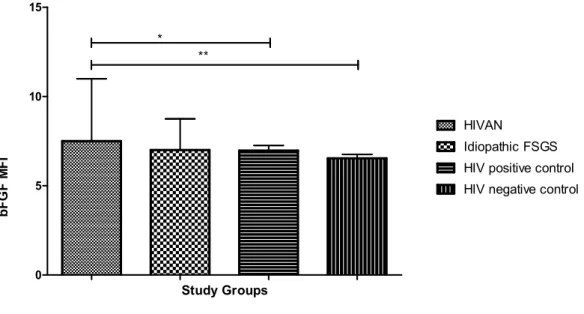
0 5 10 15
HIVAN
Idiopathic FSGS HIV positive control HIV negative control
bFGF MFI
*
*
**
Study Groups
Figure 1: The FMI (median and interquartile) of bFGF in HIVAN, idiopathic FSGS, HIV positive
and HIV negative control groups. Serum MFI of bFGF was statistically different between the study groups, p = 0.003.
0 5 10 15
HIVAN
Idiopathic FSGS
bFGF MFI
Study Groups p > 0.05
34 Figure 2: Serum MFI between HIVAN and idiopathic FSGS groups. Result are presented as median and interquartile range. Serum MFI between the two groups are not significantly different, p = 0.068.
0 5 10 15
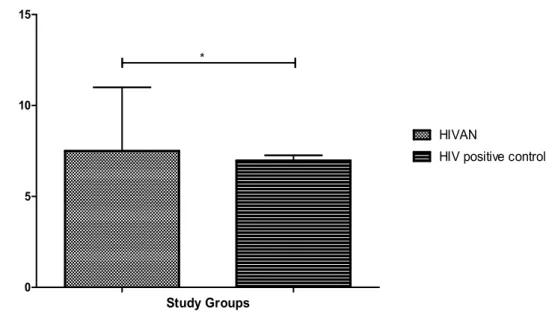
HIVAN
HIV positive control
bFGF FMI
*
Study Groups
Figure 3: Serum MFI between HIVAN and HIV positive control group. Result are presented as median and interquartile range. Serum MFI between the two groups was significantly different, p = 0.004.
0 5 10 15
HIVAN
HIV negative control
bFGF MFI
**
Study Groups
35 Figure 4: Serum MFI between HIVAN and HIV negative control group. Result are presented as median and interquartile range. Serum MFI between the two groups was significantly different, p = 0.028.
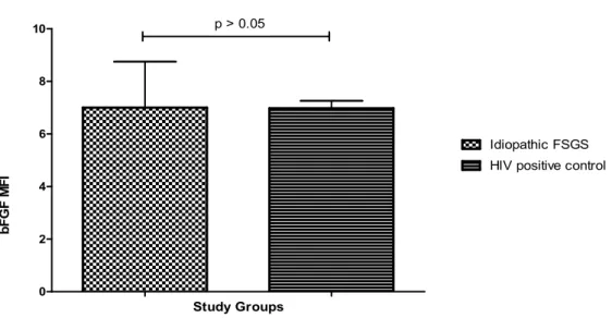
0 2 4 6 8 10
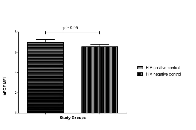
Idiopathic FSGS HIV positive control
bFGF MFI
p > 0.05
Study Groups
Figure 5: Serum MFI between Idiopathic FSGS and HIV positive control group. Result are presented
as median and interquartile range. Serum MFI between the Idiopathic FSGS and HIV positive group was observed to be not significantly different, p = 0.702.
0 2 4 6 8 10
Idiopathic FSGS HIV negative control
bFGF MFI
P > 0.05
Study Groups
36 Figure 6: Serum MFI between Idiopathic FSGS and HIV negative control group. Result are presented
as median and interquartile range. No statistical significant was observed between the two groups, p = 0.181.
0 2 4 6 8
HIV positive control HIV negative control
bFGF MFI
p > 0.05
Study Groups
Figure 7: Serum MFI between HIV positive and HIV negative children control groups. Result are presented as median and interquartile range. Serum MFI are not significantly different between the HIV positive and HIV negative control groups, p = 0.318.
37 Table 1: Clinical and laboratory demographics of patients, expressed as mean ± standard deviation
HIVAN - Human immunodeficiency virus associated nephropathy FSGS - Focal segmental glomerulosclerosis
HIVAN n = 14
Primary FSGS n = 20
HIV Positive Control
n = 20
HIV Negative Control
n = 20
Age (years) 10 ± 3.62 9 ± 3.11 11 ± 3.25 7 ± 3.87
Weight (kg) 31.13 ± 11.57 29.04 ± 10.33 37.25 ± 13.73 18.20 ± 10.55
Creatinine (µ/mol)
82.83 ± 62.36 45.32 ± 15.44 40.10 ± 9.25 29.14 ± 9.25
Protein:creatinine ratio
3.07 ± 2.04 4.22 ± 4.21 - 1.85 ± 0.67
eGFR (ml/ min /1.73m2)
119.98±57.84 125.09±102.53 207.13±58.02 218.00±122.01
Urea blood (mmol/l)
5.30 ± 4.26 7.64 ± 7.02 5.34 ± 9.81 2.71 ± 1.58
Albumin (g/l) 29.91 ± 14.76 28.96 ± 7.96 30.77 ± 10.94 37.30 ± 9.25 Cholesterol
(mmol/l)
4.82 ± 2.36 8.21 ± 4.80 3.75 ± 0.68 -
CD4 count 820.20±642.10 900.60±620.00
38
CHAPTER THREE
39 SYNTHESIS
Human Immunodefiency Virus (HIV) is a disease that raises serious concerns nationwide, with a prevalence of about 8% (Olawumi and Olatunji, 2006). In HIV infected patients, nephropathy is a common outcome (Röling et al., 2006). Direct influence of HIV has a substantial role in the development of HIV-associated nephropathy (HIVAN) (Röling et al., 2006). HIVAN is commonly related with rapid progression to end-stage kidney disease (ESKD), occurring in persons who are newly diagnosed with advanced HIV infection (Wyatt et al., 2007). It has a discrete histology demonstrating a collapsing arrangement of focal segmental glomerulosclerosis (FSGS) (Wyatt et al., 2007). HIVAN pathogenesis involves local HIV infection of the kidney, with the virus causing injury to the glomerular and tubular epithelial cells (Wyatt et al., 2007). FSGS accounts for nearly 20% of incidence of the nephrotic syndrome in children, with Black children showing a unique susceptibility (D'Agati et al., 2011).
Kidney biopsy is the only conclusive approach of establishing a diagnosis (Soler-García et al., 2009), but given the complication of performing this procedure in HIV-infected children, biomarkers should be investigated. Basic fibroblast growth factor (bFGF) is a growth factor and signaling protein encoded by the FGF2 gene (Dionne et al., 1990). It is an angiogenic growth factor that participates in kidney injury and in the pathogenesis of kidney disease (Liu et al., 2001).
This study describes the role of bFGF as a potential biomarker in the detection of FSGS in HIV positive and negative children. We noted high levels of bFGF in children with HIVAN compared to HIV negative and HIV positive controls (with no kidney disease). These findings when compared with previously published studies of HIVAN in children suggest that bFGF may have a significant clinical role for the detection of FSGS in HIV infected children. Current studies have displayed the crucial role of biologically active bFGF in the
40 pathogenesis and development of kidney disease. Liu et al., found high levels of bFGF in the kidney and circulation of HIV-transgenic mice; HIV infected children with renal disease (Liu et al., 2001), which was also demonstrated in our study. These findings suggest that bFGF is released into the systemic circulation and renal interstitium by injured endothelial and renal epithelial cells, and that it may play a role in the pathogenesis of hemolytic-uremic syndrome and HIV nephropathy (Liu et al., 2001).
The present study observed an increase of bFGF in HIVAN affected children in contrast to the levels of bFGF in controls. In healthy persons, bFGF is identified in glomerular Bowman’s capsule and the wall of blood vessels, with minimal staining covering the renal extracellular matrix (Ray et al., 1999). Over the years, several studies have supported this notion such as the one done by Soler-Garcia et al that showed levels of bFGF were higher in patients with HIVAN in contrast to those without kidney disease (Soler-García et al., 2009).
Furthermore, comparable outcomes were observed in HIV-Tg 26 mice correlating to these findings (Soler-García et al., 2009). Nonetheless, in HIVAN kidneys, bFGF was excessively high in the renal glomeruli (Soler-García et al., 2009). Rall et al discovered high circulating and renal tissue levels of bFGF in children suffering from acute stages of renal disease (Rall et al., 1985). Consequently, these findings further upkeeps the theory that bFGF released by injured glomerular endothelial cells amass in the HIVAN kidneys leading to renal cell death.
Despite the knowledge that has been accumulated, up until now there is still insufficient data to support how bFGF becomes solubilized and hence activated in the human kidney (Liu et al., 2001). There is a possibility that bFGF solubilisation occurs from the digestion of the glycosaminoglycan portion of the cells attachment molecule by heparanases, released by blood monoclear cells which in turn activate bFGF (Moscatelli, 1992). Another possibility could be digestion of the protein backbone HPSG by proteases thus releasing bFGF from the immobilized state resulting to its activation (Yayon et al., 1991). However, at present it is