HUMAN T CELL LYMPHOTROPHIC VIRUS 1 ASSOCIATED INFECTIVE DERMATITIS IN KWAZULU NATAL
SOUTH AFRICA
DR CAROL HLELA
MB Ch B (NATAL), FC DERM (SA), MSc GHS FACULTY OF CLINICAL SCIENCES
UNIVERSITY OF KWAZULU NATAL 2008
SUBMITTED IN PARTIAL FULFILLMENT OF THE DEGREE OF MASTER OF MEDICINE IN
DERMATOLOGY
Supervisor Dr A Mosam
DECLARATION
I hereby declare that this is my original work and has not previously been submitted to this or any other university.
Carol Hlela
CONGRESS PRESENTATIONS
1. Hlela C, Mosam A, Dlova NC, Aboobaker J, Bhigjee A. HTLVl associated infective dermatitis in KwaZulu Natal, South Africa. Galderma Fellowship Feedback. Dermatology Congress of the Dermatological Society in South Africa, April 2005, Sun City , Mpumalanga.
2. Hlela C, Mosam A, Ramdial PK , Bhigjee A, Dlova NC, Aboobaker J. HTLVl associated infective dermatitis in KZN, South Africa. 11 Prague Dermatology Symposium, Czech Republic, 15-17 September 2005.
3. Hlela C, Mosam A, Ramdial PK , Bhigjee A. HTLVl associated infective dermatitits in KwaZulu Natal, South Africa, Congress of the Dermatological Society of South Africa. April 2006, Durban.
AWARDS
1. GALDERMA FELLOWSHIP AWARD - R35 000
Awarded to registrars conducting research in their respective fields. A stipend to assist young researchers in starting up a research project. This is a national award administered by the Dermatological Society of South Africa. Judges are heads of Dermatology Departments countrywide.
2. YOUNG DERMATOLOGIST TRAVEL AWARD - $ 1000
Awarded to junior consultants within 5 years of registering as a specialist. This international award is administered by the International Dermatological Society. The award of $ 1000 is paid towards travel expenses for attendance and presentation in an international dermatology congress. In addition congress registration and
accommodation is paid by the Society.
PUBLICATIONS
Alcantara LCJ , de Oliveira T, Gordon M, Pybus O, Mascarenhas RE, Goncalves M, Seixas MO, Hlela C, Cassol S and Galvao-Castro B (2006). Tracing the origin of Brazilian HTLVl in as determined by analysis of host and viral genes, AIDS, 20 (5):
780-782.
ACKNOWLEDGEMENTS
I owe a huge thanks to my supervisor Dr Anisa Mosam for her ceaseless support. A special thanks to Prof PK Ramdial, from the Anatomical Pathology Department, IALCH, for her tireless efforts working on the histology sections and for her scholarly input into the study. Natalie Graham from Africa Centre Laboratory and Dr Liuz Alcarantra, from Brazil for their contribution in extracting DNA, performing PCR. I also acknowledge Ms Tonya Esterhuizen, from the Statistics Department, Nelson R Mandela School of
Medicine, for her invaluable insights and the analysis of the results. I wish to acknowledge Ms Cheryl Baxter for formatting this document. Prof A Bhigjee, from Neurology Department, IALCH, for his scholarly contribution in the initial stages of the project and the analysis of some of the patients. Drs Ncoza Dlova and Nilesh Morar from the Department of Dermatology, UKZN, who were pioneers in the commencement of this work, I thank them for their support. Dr Victoria Mubuiwa for her invaluable contribution earlier on the study and Mr Sphamandle Mkhize for his technical support.
TABLE OF CONTENTS
DECLARATION 1 CONGRESS PRESENTATIONS 2
AWARDS 3 PUBLICATIONS 3 ACKNOWLEDGEMENTS 4
TABLE OF CONTENTS 5 LIST OF FIGURES 7 LIST OF TABLES 8 LIST OF ABBREVIATIONS 9
ABSTRACT 10 CHAPTER 1 14 INTRODUCTION AND LITERATURE REVIEW 14
1.1 Background 15 1.2 Virological Characteristics 18
1.3 Epidemiology 19 1.3.1 Seroprevalence 19 1.4 Geographical Subtypes 20 1.5 Clinical Features of HAID 21
1.6 Pathogenesis 22 1.7 Pathology 24 1.8 Disease Transmission 25
1.9 Modes of Detection 25 1.10 HTLV-I associated diseases 26
1.10.1 Adult T cell leukaemia / lymphoma (ATLL) 27 1.10.2 HTLV-I Associated Myelopathy / Tropical Spastic Paraparesis (HAM/TSP) 27
1.10.3 HTLV-I Associated Uveitis 27
1.11 Complications 28 1.12 Natural History 29 1.13 Treatment 30 1.14 HIV / HTLV-I co-infection 30
1.14.1 Historical Review 30 1.14.2 Structural Differences 30 1.14.3 Local Experience 32
CHAPTER 2 34 2.1. Aims 34 2.2. Objectives 34 2.3 Methods 34 2.3.1 Clinical examination 35
2.3.2 Laboratory investigation 35 2.3.3 Histological Investigations 37
CHAPTER 3 38 RESULTS 38 3.1 Demography 38
3.3 Dermatological examination 41
3.4 Complications 43 3.5 Microscopy results 44 3.6 Bloods Results 45 3.7 Virology 48 3.9 Data for the family members 54
CHAPTER 4 56 DISCUSSION 56 Demography 56 4.2 Dermatological Examination 57
4.3 Complications 58 4.4 Microbiology 59 4.5 Blood results 60 4.6 Genotyping 62 4.7 Data for the family members 62
4.8 Histopathology 63 4.9 Study Limitations 63
CHAPTER 5 65 CONCLUSION 65 REFERENCES 68
APPENDK: Raw Data Spreadsheet, Data Sheet
LIST OF FIGURES FIGURES
Figure 1: HTLV-I structure
Figure 2a and 2b: Clinical picture of a relapse in a patient with HAID Figure 3:A schematic map HTLV-I/II genome in comparison with HIV genome
Figure 4: A histogram of patient age in axis and number in the y axis Figure 5: Gender distribution between adults and children with HAID Figure 6a and 6b: Nasal discharge and crusting
Figure 7: Picture of a patient demonstrating flexural involvement Figure 8: A generalised papular eruption in HAID
Figure 9: Anatomical sites of involved in the HAID patients Figure 10a and 10b: Pictures demonstrating corneal opacities affecting some patients with HAID
Figure 11: A histogram showing the distribution of miscroscopy results
Figure 12: Schematic diagram of protein electrophoresis result Figure 13: Superficial and deep perivascular dermatitis (SVD-PVD) Figure 14: SVD-PVD and Lichenoid Inflammatory Dermatitis (LID) Figure 15: Interface dermatitis
Figure 16a and 16b: Seborrhoeic dermatitis Figure 17a and 17b: Chronic folliculitis
Figure 18: Histological characteristics of HAID
PAGE 20 23 32
40 41 42 43 43 44 45
46
48 51 52 52 53 54 55
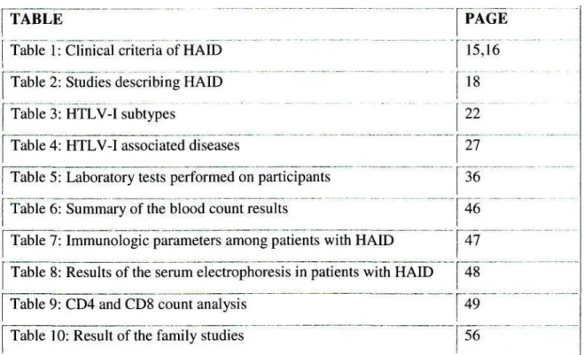
LIST OF TABLES '"TABLE
fable lfChnicai criteria of HAID Table 2: Studies describing HAID Table 3: HTLV-I subtypes
Table 4: HTLV-I associated diseases
Table 5: Laboratory tests performed on participants Table 6: Summary of the blood count results
Table 7: Immunologic parameters among patients with HAID Table 8: Results of the serum electrophoresis in patients with HAID fable 9: CD4 and CD8 count analysis
Table 10: Result of the family studies
LIST OF ABBREVIATIONS
AIDS Acquired Immune Deficiency Syndrome ATLL Adult T cell leukaemia / lymphoma AD Atopic dermatitis
BHS Beta Haemolytic Streptococcus CF Chronic folliculitis
ESR Erythrocyte sedimentation ratio ELISA Enzyme linked immunosorbent assay FBC Full blood count
HAID Human T cell lymphotropic virus type 1 associated infective dermatitis HAM Human T cell lymphotropic virus type 1 associated myelopathy HIV Human Immunodeficiency Virus
HIV- VL Human immunodeficiency virus viral load HLA Human leucocyte antigen
HTLV-I Human T cell lymphotropic virus type 1 HTLV-IT Human T cell lymphotropic virus type 2 KEH King Edward VIII Hospital
KZN KwaZulu-Natal LCV Leucocytoclastic vasculitis LD Lichenoid dermatitis LTR Long tandem repeat MF Mycosis Fungoides PCR Polymerase Chain Reaction PTCL Peripheral T cell lymphoma S.aureus Staphylococcus aureus Seb derm Seborrhoeic dermatitis
SD/ PVD Superficial and deep perivascular dermatitis SPEP Serum Protein Electrophoresis
TSP Tropical spastic paraparesis WCC White cell count
WB Western Blot
ABSTRACT
Background
Human T cell Lymphotropic Virus Type I (HTLV-I) associated infective dermatitis, first described by Sweet in Jamaican children, is a pattern of eczema characterized by
exudation, crusting around the nostrils, ears and scalp with eventual appearance of a generalized fine papular rash. More recently LeGranade and co-workers have proposed major and minor criteria in establishing the diagnosis of HTLV-I associated infective dermatitis (HAID).
HTLV-I has been aetiologically linked to Adult T cell leukaemia/lymphoma (ATLL) and tropical spastic paraparesis (TSP). HAID is not only a marker of childhood infection with HTLV-I but may be a harbinger of more serious HTLV-I associated diseases later on in life such as ATLL or TSP. The pathogenesis of HAID is poorly understood so are the histopathological features of this entity. The effects of co-infection with human immunodeficiency virus- 1 (HIV-1) are inconclusive.
HAID is described in Sub Saharan Africa, Senegal but no data is published on this entity in Southern Africa, characterizing the clinical, laboratory features and the histopathology of this entity.
Aims and Objectives
1) To describe the clinical and histological features of HTLV-I associated infective dermatitis in KZN, South Africa
2) To determine the virological characteristics of HTLV-I in KZN, South Africa 3) To assess for HTLV-I / HIV co-infection
Methods
This was a prospective study of all patients with HAID who presented to King Edward VIII hospital (KEH), outpatient department over a period of 42 months. These were patients who fulfilled the clinical criteria of HAID. Enrolled patients were subjected to a
seropositive patients. Their clinical examination included dermatological, neurological and pathological examination. A blood count, immunoglobulin levels, serum protein electrophoresis measuring albumin levels and globulin fractions were measured. For bacteriological assessment skin swabs were taken from the affected sites with stool samples examined for parasites, ova and cysts.
The HIV-1 status together with HIV-1 viral load were determined on those enrolled. The CD4 count, CD8 counts and CD4/CD8 ratio were also calculated. Skin biopsies were taken for histological examination. PCR for HTLV subtyping was performed on a subset of the cohort.
Results Demography
Of the 60 patients recruited, 33 fulfilled criteria for HAID. The majority of patients fell between age categories of 6 to lOyears. The male to female ratio was 1:1. There were more females in the adult group than there were within the childhood group. All of the patients in our cohort were African.
Clinical features
The lesions were erythematous, scaly, exudative, and crusted in all cases. The distribution of lesions was as follows: scalp (77.4%), retroauricular areas (71%), the axilla (65%) and paranasal areas (58%) were the sites more commonly affected. Nasal crusting was not a significant feature in this series.
Bacteriology
Culture was positive for Staphylococcus aureus (S. aureus) in 90%, with streptococcal group of organisms found in 68% of the skin swabs taken from the lesional skin.
Haematological
Our patients were mildly anaemic as has been shown in previous studies. They had a mean Hb of 11.5g/dl. In 12 of the 14 patients tested, the erythrocyte sedimentation rate (ESR) was elevated. Serum protein electrophoresis and levels of Immunoglobulin A, G and M were raised. The mean CD4 count in the entire group was elevated at 1730 cells/fil, CD8 was 1299 cells/ul
Histopathology
The major histological findings were as follows: 38% demonstrated a superficial and deep perivascular inflammatory infiltrate, 28% had a superficial and deep perivascular inflammatory infiltrate together with a lichenoid dermatitis, 12.9% had features of superficial and deep inflammatory infiltrate with an interface dermatitis, 6.4% revealed features of seborrhoeic dermatitis.
Genotyping
Our patients were infected with the strains belonging to the Cosmopolitan, A Subtype (HTLV-Ia).
Complications
Complications were low in this series with the commonest being scabies in 6(18.1%), corneal opacities in 3(8.6%), 2(6 %) with HAM/TSP. No parasitic worm infestations were isolated.
HIV/HTLV-I co-infection
Of the 33 patients, 9 (30 %) were co-infected with HIV. The mean viral load in this group was 52 000 copies/ml. Their mean CD4 count was also elevated at 1505cells/^il with a CD8 of 1704 cells/Mi and a CD4/CD8 ratio of 1.15.
Discussion
Thirty three of the 60 patients enrolled met the diagnosis for HAID according to the established criteria. The mean age in this series was 17 years (range: 8 months-46 years) however; almost a third (30.3%) were children under 12 years, reinforcing the entity as a childhood infective condition.
There was an equal male female distribution in the childhood group and a female predominance in the adult group.
Clinically patients presented with infected erythematous, scaly lesions mainly on the scalp, neck and post- auricular area. The clinical features were in keeping with other series worldwide. The complication rate was low in our cohort.
S. aureus was the predominant organism in both anterior nares and lesional skin. The most common histological pattern was superficial and deep perivascular inflammatory infiltrate. The subtype in our series was the Cosmopolitan Subtype A (HTLV-Ia) as opposed to subtype B in Japan. We share with Brazil a common subtype.
A subset of our patients (30%) was co-infected with HIV. The CD4 cell count in this subgroup was lower than the entire group but this was not statistically significant. The histological patterns found in this subgroup infected with HIV were similar to the rest of the group except for a more intense eosinophilic infiltrate in these skin biopsy specimens.
Conclusion
HTLV-I associated infective dermatitis is distinct entity which affects the African population of KwaZulu Natal, South Africa. It is predominantly a disease of childhood with an equal female to male ratio in children. The clinical features are an exudative, erythematous scaly rash most commonly found involving the scalp, axillae, paranasal and retroauricular areas. HTLV-I positivity is essential for the diagnosis; the Cosmopolitan Subtype A is commonest in South Africa. The commonest histological pattern is a superficial and deep perivascular infiltrate in 38%. A subset, 30%, was co-infected with HIV.
CHAPTER 1
INTRODUCTION AND LITERATURE REVIEW
Human T cell lymphotropic virus type I (HTLV-I) associated infective dermatitis is a chronic and severe form of childhood dermatitis characterized by an exudative, infective dermatitis involving mainly the scalp, neck and ears. Other symptoms include a
generalized papular rash, nasal discharge and crusting of the nostrils. The majority of described patients with this condition originate from Jamaica.
The disease has also been reported in several HTLV-I endemic populations including Japan, Trinidad, Brazil and Columbia. Diagnosis is based on specific clinical and
laboratory criteria, Table 1. The average onset is 2 years and 60% of patients are female.
The incidence and prevalence are undefined, as is the pathogenesis. The skin manifestation becomes less severe with age.2 Long-term clinical studies have
demonstrated that the rash is often followed by the development of HAM/TSP or ATLL.3
Table 1. Clinical criteria of HAW4
MAJOR
1. Eczema of scalp, axillae and groin external ear and retro-auricular areas, eyelid margins, paranasal skin and/or neck
2. Chronic watery nasal discharge without other signs of rhinitis and/or crusting of the anterior nares
3. Chronic relapsing dermatitis with prompt response to appropriate therapy but prompt recurrence on withdrawal of use of antibiotic
4. Usual onset in early childhood
5. Human T cell lymphotropic virus type 1 antibody seropositivity
Table 1: Clinical Criteria of HAW4 (Ctd) MINOR
1. Positive cultures for Staphylococcal aureus and/or Beta-haemolytic streptococci from the skin or anterior nares
2. Generalized fine papular rash (in most severe cases)
3. Generalized lymphadenopathy with dermatopathic lymphadenitis 4. Anaemia
5. Elevated erythrocyte sedimentation rate 6. Hyperimmunoglobulinaemia (IgD and IgE)
7. Elevated CD4 count, CD8 count, and CD4/CD8 ratio
* Of the major criteria, 4 are required for the diagnosis with mandatory inclusion of 1, 2 and 5; to fulfil criteria 1, involvement of at least 2 sites are required.
1.1 Background
Recognition of this condition dates back to 1966 when RD Sweet brought the world's attention to a unique pattern of eczema in a group of children and adults that seemed to vary from the patterns seen in Europeans.5 He recognized that 17 of 28 patients who had been diagnosed with eczema were Jamaican. He noted that this peculiar type of eczema started at the age of 2 years. The lesions were infected from the time of onset and were located on nostrils, ears and spread to the rest of the face, the scalp and around the neck.
He documented that some of these children developed a generalized fine papular eruption and that some patients came from the same family.5 He observed that the eruption cleared rapidly when treated with an antibiotic and steroid therapy and that the condition tended to relapse when these patients returned home after discharge on withdrawal of antibiotics.
Recognising that this pattern defied the tidy classification of eczema he was accustomed to, he named it "A pattern of eczema in Jamaica".5
The following year Margaret Walshe documented a study of 40 Jamaican children, 25 of whom suffered from the condition which had been described as "infective dermatitis" by Sweet.6 She amplified Sweet's original description, established criteria for diagnosis and
documented bacteriological findings. She also documented a high incidence of carriage of staphylococci or beta haemolytic streptococci or both in the noses or the skins of the patients with infective dermatitis than in those with other dermatoses. She also postulated that these children might be immunosuppressed and tentatively suggested malnutrition as the possible cause for the immunosuppression.
It was 10 years after the discovery of HTLV-I7 that the first link of this early life infection with HTLV-I was documented. Le Granade and co-workers studied 147 consecutive patients between the ages 2 and 17 years over a period of one year, 14 of whom met the clinical definition of infective dermatitis. Each of these 14 children from Jamaica
o
underwent testing for HTLV-I and all 14 were positive for antibodies to HTLV-I.
This relationship was later confirmed in a much larger number of patients where 50 infective dermatitis patients were compared with 35 atopic dermatitis patients.4 In this case controlled study all 50 patients with infective dermatitis had results seropositive for HTLV-I. Only 5 of the 35 patients with atopic dermatitis were seropositive for HTLV-I.
The results were negative for HIV-1 for all patients in the study, suggesting an aetiologic role for HTLV-I in infective dermatitis. In both groups, microbiologic studies showed frequent colonization with S. aureus or BHS. On comparing the blood count findings between the two groups, patients with infective dermatitis were anaemic, had higher WCC and had an elevated ESR than patients with AD. They also had a significantly higher incidence of abnormal serum proteins and dermatopathic lymphadenopathy.4 Le Granade and co-workers in this study proposed a new designation of infective dermatitis ("HTLV-I associated infective dermatitis") and the major and minor criteria for the diagnosis, Table!.
Since the original report in 1990, cases of infective dermatitis have been described from Trinidad, Tobago, Japan, and Columbia, Barbados and among Haitian immigrant children in Miami but the numbers so far have been small. In Trinidad and Tobago, 15 people were described with chronic relapsing infective dermatitis all of whom were seropositive
In Africa, the only published study was the one conducted in Senegal where 5 cases of this condition was reported.10
More recently a group of investigators documented the frequency of HAID in Salvador, Brazil confirming 23 cases in patients attending a dermatology outpatient clinic of the Federal University of Bahia. In this study all 23 patients demonstrated clinical features of HAID and were positive for HTLV-I. These children were followed up for a median of 3 years and 5 developed HAM/TSP.11
There have been fewer than 10 original papers documenting the entity of HAID
worldwide. These have mainly come from areas endemic with HTLV-I. There has been one study from Africa, none from Southern Africa. Table 2 emphasises the paucity of the work done so far on this entity.
Table 2: Studies describing HTLV-I associated infective dermatitis Author Name
Sweet 1966 Walshe 1967 LeGranadel990 Suite M et.al. 1994 Le Granade et.al. 1998 MaheA et.al. 2004 Oliveira MdeFSP 2005
Country Jamaica Jamaica Brazil
Trinidad and Tobago Jamaica
Senegal Bahia, Brazil
Enrolled/Infected patients 28/17
40/25 147/14 15/15
50/50 5 cases 23 cases
1.2 Virological Characteristics
HTLV-I is an enveloped double stranded RNA, type C virus (Retroviridae family, subfamily oncovirus).3 Mature virions are 110 -140 nm in diameter, characterized by a spherical, centrally located nucleiod enveloped by a glycoprotein membrane with short spikes.
The HTLV-I envelope is a lipid bilayer in which the smaller transmembrane viral protein (gp21) and the larger outer protein (gp46) are anchored.
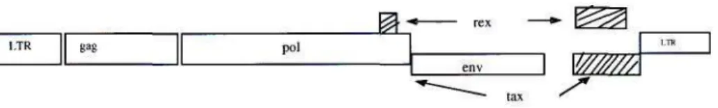
The core consists of a diploid RNA genome of high molecular weight (approximately 9056 bp long) with structural features common to all retroviruses, namely the genes for group specific antigen (gag), reverse transcriptase (pol) and envelope protein (env), and flanked by long terminal repeat (LTR) sequences on either end, (Figure
D
nThe LTR comprises three distinct domains, U3, R and U5, in a 5' to 3' direction. The length of the repeat I sequence (228bp) of the LTR region is no longer than in the other retroviruses. HTLV-I also contains unique regulatory genes encoding for transactivating proteins (tax and rex) enveloped between the 3' untranslated end of the provirus and the env gene. The tax gene stimulates transcription of all genes from the 5' LTR sequences.
The rex gene is a positive post-transcriptional regulator for the gag and env expression and is also a negative regulator capable of inhibiting expression and replication of HTLV- I in vivo.12
Transmission of HTLV-I is by cell-cell contact. The receptor(s) for entry of HTLV-I into the host's cell are unknown. More recently, studies have been reporting GLUT as the likely receptor for entry of HTLV-I into the cell.1 As a provirus within the infected cell,
DNA by reverse transcriptase. Laboratory studies have shown that T cells are then transformed and immortalized. Unlike other type C transforming viruses, HTLV-I has not been found to possess cell derived oncogenes.
[ | h « rex — r $ ^ g 1 pol
] mm
Figure 1: A schematic map of the HTLV-I genome
1.3 Epidemiology
Infection with HTLV-I is a global epidemic affecting 10-20 million people
worldwide.2 HTLV-I infection, is particularly endemic in southern Japan, but foci of infection are found in geographical clusters, the Caribbean, parts of Africa, the Middle East, South America, the Pacific Malanesian Islands, and Papua New Guinea. In USA and Europe HTLV-I infection is found in the carriers among immigrants from endemic areas.3 The virus is endemic in KZN.15
1.3.1 Seroprevalence
There are very few population based studies conducted on the epidemiology of HTLV-I infection. In areas where the studies were conducted, it was shown that HTLV-I
seroprevalence ranges from 3-6% in Trinidad, Jamaica, and other Caribbean Islands to 30% in rural Miyazaki, southern Japan.3 USA and Europe has the lowest seroprevalence rate, found to be less than 1% among low risk populations.3 Population studies have shown that HTLV-I seroprevalence increases with age and is twice as high in females.3
There is paucity of epidemiological evidence for HTLV-I in Africa. A few population studies conducted in parts of Africa have indicated an overall seroprevalence of 4%. This can be broken down into 1-15% prevalence in Zaire, 0.9% in Ghanaian refugees in
19
Belguim and 0.2% in pregnant women in South Africa. Small studies conducted in South Africa (KZN) have shown an HTLV-I seroprevalence of 0.5-3.3% in different ethnic and geographical areas.15
HAID incidence and prevalence remains unknown. The incidence rates of HAID in countries where it has been reported have not been published. However Trinidad's rates of ATLL and HAM/TSP are similar to Jamaica, whereas Japan has similar rates of ATLL but lower rates of HAM/TSP (0.4/100 000). Incidence rates for HTLV-I associated diseases in Columbia and Brazil have not been reported. However, the prevalence of HAM/TSP is known to be high (100/100 000 population) in Tumaco, Columbia.16
Reports of HTLV-I positive ATLL in Africans remain scanty, probably because laboratory facilities and pathologists are scarce.17
1.4 Geographical Subtypes
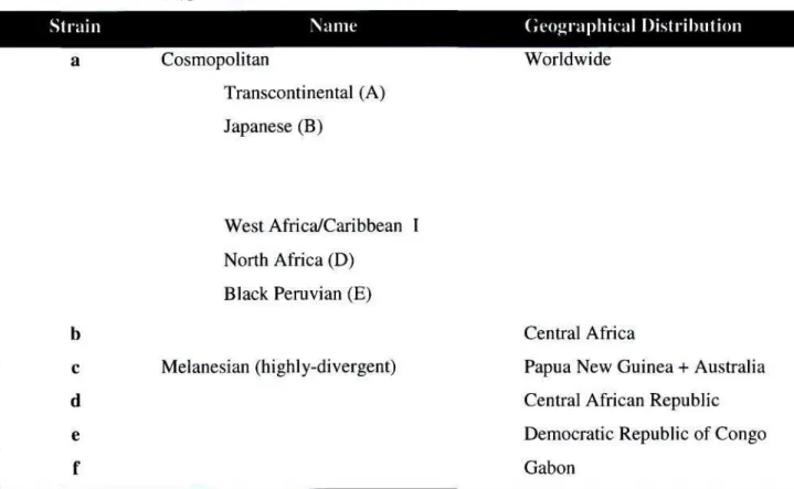
Six different genetic subtypes of HTLV-I have been proposed based on phylogenetic analyses, summarised in Table 3: a - or Cosmopolitan which is distributed worldwide;18
b - from Central Africa;19 c - a highly divergent Melanesian strain from Papua Guinea and Australia;20 d - isolated from Central African Republic (CAR) pygmies, and from
9 1 99
two patients in Cameroon and Gabon; ' e - isolated in a single sample from an Efe pygmy in the Democratic Republic of Congo (DRC); and subtype f - detected in an individual from Gabon.23 The most widespread and best studied subtype, Cosmopolitan, is further divided into five subgroups based on geographical distribution :
Transcontinental (A), Japanese (B), West African / Caribbean (C), North African (D) and Black Peruvian (E).18'24'25'26 Previous studies have reported that HTLV-I strains from KwaZulu Natal, South Africa belong to the A subgroup of the Cosmopolitan
(HTLV-Ia subtype).27
Table 3: HTLV-I Strain
a
b c d e f
subtypes
Name Cosmopolitan
Transcontinental (A) Japanese (B)
West Africa/Caribbean I North Africa (D)
Black Peruvian (E)
Melanesian (highly-divergent)
Geographical Distribution Worldwide
Central Africa
Papua New Guinea + Australia Central African Republic Democratic Republic of Congo Gabon
1.5 Clinical Features of HAID
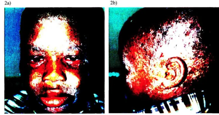
HAID is a chronic and recurrent eczema occurring during childhood and adolescence. It is distinctive, often beginning with a rhinitis labelled by the mother as a "cold".28 This is followed by an oozing, weeping eruption on the scalp, ears, neck, axillae, umbilicus, groin, perineum and natal cleft often associated with a blepharo-conjunctivitis. The full clinical picture is that of a severe exudative dermatitis with crusting of the scalp, neck, axillae, groin, external ear, and retro-auricular areas; watery nasal discharge, and/or crusting of the anterior nares; generalized fine papular rash, culture from the anterior nares or skin showing Staphylococcus aureus (S.aureus) and/or beta-haemolytic streptococcus (BHS); and prompt response to appropriate antibiotic therapy and equally
rapid relapse, if such antibiotics are withdrawn. Figures 2a and 2b show the typical clinical appearance of a patient during a relapse.
2a) 2b)
Figure 2a and 2b: Clinical picture of a relapse in a patient with HAID
1.6 Pathogenesis
The pathogenesis of HAID is poorly understood. It is the resistance to treatment, the frequent exacerbations and the infections with bacteria that are usually non-virulent in those affected which raises the possibility that infective dermatitis may be a disorder of immunosuppression.7
Data demonstrate evidence of altered immune function with hyperactivity of both humoral and cellular immune systems but the precise immunological abnormality remains to be elucidated.
The question of why some children infected with HTLV-I develop HAID while others are asymptomatic remains unanswered. This observation suggests the role of other factors in the development of HAID, the possibilities being environment or lifestyle related to socioeconomic status since patients are usually from the lower socioeconomic sectors of
29
the population, as well as the immuno-genetic background.
Postulated so far is that HTLV-I alters the immune system of affected patients, rendering them incapable of overcoming infection with Staphylococcus and BHS resulting in chronic bacterial infections.28
The fact that immune suppression played a role in the pathogenesis was suggested as early as in the cases described by Walshe. She postulated that children with this disease were immunosuppressed and suggested malnutrition as a possible cause of the
immunosuppression. However, she herself noted that only a minority were in fact obviously malnourished.
HTLV-I is tropic for cells with a CD4 phenotype and infected cells can express type MHC haplotypes (HLA-DR) as well as CD25.30
Recent data suggest HTLV-I infection might be the cause of the immune dysfunction among HAID patients. 29 by inducing an increased expression of interleukin -2 (IL-2) receptors in HTLV-I infected cells. This may result in preferential binding of soluble IL-2
o
with consequent reduction in the effective concentration of IL-2. In addition, a change in the functional phenotype induced by HTLV-I creates a deficiency in cell-cell interaction resulting in immune dysregulation. Genes of several proinflammatory cytokines such as interleukin 1, interleukin 6 and tumour necrosis factor a, are transactivated by the viral tax protein. It is postulated that the secretion of such cytokines by infected cells amplifies and/or maintains the inflammatory reaction in the skin and that this may be responsible for the recalcitrant nature of HAID.31
Genetic studies done in a single family, a mother and her two sons, indicate that there is a possible genetic predisposition contributing to the development of HAID. The results in this study showed the index case and her two sons to be the only family members to share a common haplotype namely DRB1*DQB1* (1101- 0301). This is one of the haplotypes associated with HAM/TSP among Japanese patients which correlates with high immune response and high antibody titers to HTLV-I. It is postulated that haplotype
DRB1*DQB1* (1101-0301) may determine susceptibility to HMD.29 These observations point to the similarities of genetic background between patients with HAID and those with HAM/TSP. This therefore suggests that susceptibility to these diseases observed in patients with HAID could marked by these HLA haplotypes.16
Whether HTLV-I is involved in the pathogenesis of the skin lesions or is present in the skin because inflammatory cells containing virus migrated to the lesion remains a question. The skin cells in addition to lymphocytes may be infected by the virus.32
1.7 Pathology
The histopathological features characterizing this entity have yet to be determined. There is a paucity of literature on this subject. The investigators who searched for specific features of this entity found that pathological aspects were similar to other types of
o
chronic eczema.
More recently, 19 patients with HAID were studied histologically and
immunohistologically using the following antibodies: anti-CD3, CD45RO, CD20, CD79a, CD4, CD8, CD57, TIA-1, granzyme-B and perforin A.33 Chronic dermatitis features similar to that of seborrhoeic dermatitis was observed in 15 of these patients, whereas architecturally aspects mimicking mycoses fungoides (MF) were observed in the remaining 4 patients. The specific features that characterize HAID remain to be
answered. It was with this background above that we set out to investigate histological features of HAID.
1.8 Disease Transmission
HTLV-I infection is blood borne and can be transmitted by blood-blood contact as well as by sexual contact. Transfusion is the most efficient mode of virus transmission. It has been shown that the probability of seroconversion in a recipient of contaminated blood can be as high as 40-60% with the median time to seroconversion of 51 days. Sexual intercourse is recognized as an important factor for HTLV-I transmission.34 Sexual intercourse is recognized as an important factor for HTLV-I transmission.34 HTLV-I has been detected in the semen and cervical secretion of infected persons. Most HTLV-I infections are attributable to transmission from mother to child with the mother's milk being a major risk factor of infection.35 The probability of mother to infant transmission is 18-30%.3 Maternal risk factors include higher HTLV-I antibody titre, prolonged ruptured membranes during delivery and low socioeconomic status. Breastfeeding for more than 6 months has been associated with transmission which has led to the
hypothesis that shortening the duration of breastfeeding may reduce the risk of HTLV-I transmission. However infection still occurs in about 3% of patients who are not
breastfed.3
1.9 Modes of Detection
HTLV-I infection can be easily detected by screening blood for specific antibodies, using enzyme-linked immunoassay (EIA or ELISA) techniques. Gelatin particle agglutination (GPA or PA), immunoflouresence (IF), radioimmunoprecipitation assays (RIPA) are other common serological screening used in the diagnosis. Samples that react repeatedly in anti-HTLV-I screening assays need to be retested in an immunoblot assay. Highly sensitive HTLV-1/2 immunoblot assays like the Line immunoassay (LIA) and Western blot (WB) are most commonly used.35 Advanced methods, such as the HTLV- specific polymerase chain reaction (PCR) tests in combination with T-cells culture may be of additional value since they are able to detect the viruses with increased sensitivity.
Guidelines from the US Public Health Service, WHO and other international groups such as the HERN (HTLV European Research Network)36, recommend that newly identified
seropositive individuals have additional blood collected for repeat testing to eliminate possible technical errors, also to distinguish HTLV-I from HTLV-II. HTLV-II has 65%
sequence homology to HTLV-I. HTLV-II infection is present predominantly amongst intravenous drug users (IVDU) and is usually a co-infection with HIV-I. It is difficult to distinguish the two from one another unless virus specific reagents are used. The
distinction is important because HTLV-II is less pathogenic than HTLV-I.
1.10 HTLV-I associated diseases
HTLV-I was the first human retrovirus described as causing disease. It was first isolated from cell lines from patients with ATLL.37 Since then HTLV-I has been shown to be aetiologically associated with a number of diseases, Table 4. Of these ATLL, TSP/HAM and HAID are most widely researched.
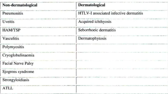
Table 4. HTLV-I associated diseases 2,3,38 Non-dermatological
Pneumonitis Uveitis HAMATSP Vasculitis Polymyositis Cryoglobulinaemia Facial Nerve Palsy Sjogrens syndrome Strongyloidiasis ATLL
Dermatological
HTLV-I associated infective dermatitis Acquired ichthyosis
Seborrhoeic dermatitis Dermatophytosis
„ _. „,....„,„.-..-.,„...„-,„.,,.-..„ ... .„,„., .,,,. ,,..., .
1.10.1 Adult T cell leukaemia / lymphoma (ATLL)
ATLL is a uniformly fatal T-cell malignancy. Among HTLV-I carriers, less than 5% of individuals develop ATLL. HTLV-I is an indolent virus. A long latency period between
Q
infection and subsequent development of disease has been documented. ATLL occurs predominantly in the age range of 40-70 years with average age of onset being about 60 years in Japan but only 40 in Jamaica, Trinidad and Brazil. This difference is
unexplained.3 It is rapidly progressive, usually resistant to chemotherapy. Most patients die of this disease within a few months. It appears to be restricted to individuals with a lifelong infection.39 Combination chemotherapy has been used to treat ATLL, but long term survival has been very limited especially in the acute and lymphoma types.
1.10.2 HTLV-I Associated Myelopathy / Tropical Spastic Paraparesis (HAM/TSP) A chronic disabling demyelinating neurological disorder characterized by slowly
progressive spastic paraparesis and bladder disturbances.1 The disease commonly follows adult acquired infection by either sexual contact or through blood transfusion but may follow a childhood infection. Symptom progression seems to be more rapid in blood transfusion associated HAM/TSP than in the cases of mother to child transmission.2 The incubation period from infection to the onset of myelopathic symptoms is believed to range from months to decades. This is a progressively disabling disorder with studies showing that one-half of patients with this disease become wheel chair bound within 10 years of acquiring it.17 There is no definite therapy for this condition.2 Secondary complications may lead to death after many years.
1.10.3 HTLV-I Associated Uveitis
This intraocular inflammatory disorder has been associated with a variety of infectious causes including tuberculosis, syphilis, cytomegalovirus, toxoplomosis or non-infections causes such as Behcet's, sarcoidosis and Vogt-Koyanagi-Harada syndrome(reviewed in
). In about 40% of cases, a firm cause is not identified. It was a high number unexplained
cases of uveitis (idiopathic uveitis) in HTLV-I endemic areas that led to speculation that HTLV-I might be the cause. An association with HTLV-I was established when 35% of patients with idiopathic uveitis were found to be HTLV-I positive compared with 10% of uveitis cases where another cause has been identified. A patient with HTLV-I uveitis presents with a variety of clinical symptoms including blurred or foggy vision and acute, sudden onset of "floaters" (reviewed in ). Iritis, vitreous opacity, retinal vasculitis, retinal exudates and haemorrhages are all signs that may be found on ophthalmologic examination. PCR is used to establish clinical diagnosis through detection of proviral DNA in mononuclear cells in peripheral blood and vitreous humour. Topical and systemic corticosteroids may improve visual acuity (reviewed in ).
1.11 Complications
Long term HTLV-I infection may be entirely asymptomatic but clinical or subclinical consequences may affect various organ systems. Complications occur in 30-35% of patients.
Asymptomatic carriers of HTLV-I have been reported to harbour various infections including strongyloidiasis, trypanosomiasis and leishmaniasis.8 Other complications include: scabies, corneal opacities, chronic bronchiectasis, regressing atypical histiocytosis, glomerulonephritis and lymphocytic interstitial pneumonia. ' Some patients with HAID where HTLV-I is endemic may go on and develop severe HTLV-I related illnesses such as TSP/HAM or ATLL."
In addition to HAID, Strongyloides stercoralis (Ss) infection has been proposed as a cofactorof ATLL.40
1.12 Natural History
The steps leading from virus infection to the development of the different HTLV-I associated conditions are partly understood.40 Some patients become carriers of the disease while others go on to develop HTLV-I related illnesses.
Although HTLV-I infection is frequently asymptomatic, the risk of disease in long term infection has been under recognized yet it could have significant health implications.
Early diagnosis of HTLV-I allows neurological and lymphoreticular symptoms to be taken into account in the clinical care of patients and makes it possible to provide
•50
preventative counselling to reduce the likelihood of infection transmission.
It is estimated that the cumulative lifetime risk of developing a life threatening or
debilitating disease as a result of HTLV-I is approximately 5% increasing to 8-10% when the patient has other illnesses.
Epidemiological data suggest that HAID is not only a marker for childhood HTLV-I infection but also a possible harbinger of more serious HTLV-I associated disorders later in life as there have been reports on occurrence of ATLL in patients 12-25 years after a diagnosis of HAID.
A suggested postulate is that an exaggerated host response to the presence of HTLV-I, coupled with the virus's ability to immortalise T cell-clones predisposes them to
malignant transformation due to accumulation of genomic mutations." Among HTLV-I carriers less than 5% of individuals develop ATLL or HAM/TSP and there is usually a long latency of decades between infection and subsequent development of disease with the exception of transfusion associated HAM/TSP which can develop several weeks to months following infection from contaminated blood components.3
1.13 Treatment
Treatment of HAID is currently aimed at controlling infection with S. aureus and BHS by using appropriate antibiotic therapy. This measure alone keeps the dermatitis fairly well controlled and may require addition of mild topical steroids for full control. Prolonged use of antibiotics is recommended in the literature until puberty at which time the severity of the bacterial infection seems to lessen. Relapses always occur following the
withdrawal of the drug treatment. A combination of artificial feeding, prophylactic immunoglobulin and perhaps antiretroviral therapy need to be investigated for possible use in the control of infection. Some investigators are also exploring the feasibility of an HTLV-I vaccine.3
1.14 HIV / HTLV-I co-infection 1.14.1 Historical Review
A year after the discovery of HTLV-I, a cluster of patients with a novel disease of acquired cellular immunodeficiency, later known as the acquired immunodefiency
syndrome (AIDS), was first described.17 In the following 2 years several groups isolated a retrovirus from patients with AIDS. This virus was called lymphadenopathy - associated virus (LAV), Human T cell lymphoma virus type III (HTLV-III) or Aids Related Virus (ARV). In 1986 these isolates were grouped under the name Human Immunodeficiency Virus (HIV). This later discovery of HIV has given rise to the massive interest in HIV and AIDS worldwide.
1.14.2 Structural Differences
Structural features are common to all retroviruses, (Figure 3). The core proteins of HIV and HTLV-I have similar molecular weights and are designated pi5, pl7 and p24 (p26 in HIV-II).17 Glycoprotein's 120 (gpl20) and 130 (gpl30) are major glycoproteins of HIV-I and HIV-II is respectively. In contrast to HIV, the size of the cleaved glycoprotein precursor of HTLV's gp68 is much smaller. The HIV virus encode additional proteins
of virion proteins (rev) and a negative factor (nef) which inhibit the replication of the HIV. In contrast, HTLV have a rex and tax which encode a p40 and p27/p21 molecule respectively which is not present within the HIV structure. The p40 augments viral RNA expression, while p27/p21 enhances the expression of viral genes.16 Despite the
similarities, HIV is classified as belonging to the family of Lentivirinae as it is associated with slowly progressive inflammatory and degenerative disorders while HTLV belongs to the family Oncovirinae as these viruses are associated with malignancies.17
•< rex
LTR gag pol
env
%t
Will/
LTR
tax / HTLV-I/n
LTR gag
pol
HIV-I
Figure 3: A schematic map of HTLV-I/II genome in comparison with HIV genome 14
Similar modes of transmission are observed with both retroviruses and are by three routes, namely, (1) intimate sexual contact, (2) perinatally (predominantly through breastfeeding and (3) parentrally (through blood transfusion or exposure to needles and syringes contaminated with blood). The risk of male infection is increased with
concomitant penile ulcers and concurrent syphilis, which holds true for both retroviruses.3
Co-infection with HTLV-I is common in some populations infected with HIV,
particularly from regions with high HIV prevalence, e.g. KZN in South Africa. Whether HTLV-I /II influences the outcome in patients with HIV remains to be known.4
HIV positive patients co-infected with HTLV-I seem to have more severe
immunosuppression than do the HTLV-I seronegatives.8 HTLV-I can increases HIV replication in vitro and several studies suggest that HTLV-I accelerates the progression of HIV and in turn progression to AIDS.43 Several other studies have suggested that HTLV- I does not appear to affect HIV viral load, currently considered to be the best marker of HIV disease progression.4
Several mechanisms have been proposed concerning HTLV-I and HIV co-infections (in vitro studies): CD4 lymphocytes infected with HTLV-I are immortalized via stimulation of IL-2 and its receptor. Translocation of the replicating factor, NFKB in the nucleus activates the T lymphocytes. The product of HTLV-I tax gene will also have a
transactivating effect on the provirus HIV-LTR replication. Finally infection with HTLV- I may facilitate HIV by inducing CD4, expression in non-expressing cells.44
1.14.3 Local Experience
The first cases of HAM/TSP in the Orange Free State were reported by van der Ryst and colleagues where 18% of HTLV-I positive patients with spastic myelopathy had
HAM/TSP.45 Most work has been carried out by Bhigjee and colleagues in the
Ngwelezana district in 1993; a 2.6% seroprevalence was found .15 A follow up study at Ubombo showed a seroprevalence rate of 3.33%.I2 This emphasized the fact HTLV-I is endemic in KZN.
Previous studies have reported that HTLV-I strains from KZN, South Africa belong to
27
the A subgroup of the Cosmopolitan (HTLV-Ia) subtype. HTLV-I associated infective dermatitis from the southern part of Africa is not as well documented as it is in the
presenting with a typical rash neither has there been any characterizing the histopathology in South Africa.
CHAPTER 2 2.1. Aims
To document the clinicopathological and virological characteristics of HTLV-I associated infective dermatitis in KZN, South Africa.
2.2. Objectives
1. To describe the clinical and histological features of HTLV-I associated infective dermatitis in KZN, South Africa.
2. To determine the virological characteristics of HTLV-I in KZN, South Africa.
3. To assess for HTLV-I / HIV co-infection.
2.3 Methods
This was a prospective study carried out in the dermatology outpatient department of King Edward VIII Hospital, a major tertiary referral hospital in KZN, South Africa. The study was carried out over a 3 year period starting from January 2003 till December 2005. This study commenced following ethics approval from the University of KZN's Biomedical Research Ethics Committee (HI81/03).
All patients who presented with clinical features in keeping HTLV-I associated infective dermatitis were informed of the study and were invited to participate. Only those who gave informed consent were recruited. Patients with features of seborrhoeic dermatitis who were HTLV-I negative, were found to be HIV infected. We did not come across patients who had clinical features of HAID who were HTLV-I and HIV negative. They had features of HIV seborrhoeic dermatitis and were therefore an unsuitable comparison group for HAID.
Those recruited were then subjected to confirmatory HTLV-I testing. Where possible,
The various components of the study were:
2.3.1 Clinical examination (see appendix) 2.3.2 Laboratory investigations
2.3.3 Histological investigation 2.3.4 PCR and sequencing
2.3.1 Clinical examination
Clinical assessments performed for all patients included a medical history, a general and a detailed dermatological assessment. The diagnosis of HTLV-I associated infective dermatitis was made according to previously established criteria. Ophthalmologic examination was performed on all patients who had visual complaints. All patients with signs of neurological abnormalities were sent to the neurology department for a detailed neurological evaluation.
2.3.2 Laboratory investigation
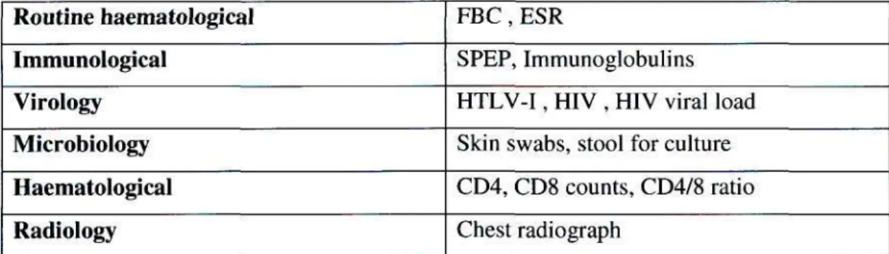
The following laboratory tests were performed for patients with suspected HAID: These have been summarised in Table 5, below.
Table 5. Laboratory tests performed on participants
Routine haematological Immunological
Virology Microbiology Haematological Radiology
FBC, ESR
SPEP, Immunoglobulins HTLV-I, HIV , HIV viral load Skin swabs, stool for culture CD4, CD8 counts, CD4/8 ratio Chest radiograph
Haematological analysis included complete blood count with differential white cell count (WCC) and measurement of erythrocyte sedimentation rate (ESR). Serum protein
electrophoresis was used to measure albumin levels and globulin fractions. Measurement of serum levels of IgA, IgE, IgG, IgM. Skin swabs were taken from nasal, perinasal and lesional skin. These were referred for bacteriological studies. Skin scrapings were carried out for those patients with clinical features suggestive of scabies in search of scabies mites/ova or faecal pellets. Stool samples were examined for parasitic organisms, ova and/or cysts.
Diagnosis for HTLV-I infection was determined according to the recommended stepwise procedure for the diagnosis of HTLV-I or II, i.e.screening for antibodies to HTLV-I was done using a particle agglutination test (Serodia ATLA, Fuji Rebio, Tokyo) as has been discussed in section 1.9. Positive sera were confirmed using the most widely used
immunoblot assay, WB (HTLV Blot 2,4 Genelabs Diagnostics, Singapore). This test was used also in order to distinguish HTLV-I from HTLV-II.
HIV-I positive sera were identified on all HTLV-I seropositive patients using Vironostika HIV-1 IMPVD Microelisa system (Biomerieux, Durham, NC). HIV-I viral loads using Nuclisens EasyQ HIV-I system (Biomerieux) were determined. CD4 and CD8
lymphocyte subsets counts were calculated and the CD4/CD8 ratio was determined. A chest radiograph was done on all patients in search of chest related HTLV-I complication such as lymphocytic interstitial pneumonia.
2.3.3 Histological Investigations
4mm punch biopsy skin specimens were taken from lesional skin on all those enrolled patients who gave consent for histological analysis. One dermatopathologist was
responsible for the histological analysis. This pathologist was blinded as to the HIV status of the patients.
Tissue specimens for histological examination were examined by light microscopy.
Biopsies were fixed in buffered formalin and processes routinely for paraffin-wax embedding. Up to 15 serial histological sections were obtained from each biopsy specimen.
2.3.4 PCR and Sequencing
DNA was extracted from the peripheral blood morphonuclear cells DNA using QiaAmp Blood kit (Quiagen). Amplification of HTLV LTR region was performed as two overlapping fragments, an LTR-gag product of 473 bp and a tax-LTR product of 458 bp.
Nested hot-start PCR and AmpliTaq Gold were used with cycling conducted on a Perkin Elmer 9700 thermal cycler under standard reaction conditions. Following separation on
1% agarose gels, the PCR products were purified using a Qiaquick Gel Extraction kit (Quiagen) and subjected to cycle sequencing on an Applied Biosystems 3100 capillary sequencer. Edited sequences were aligned and analysed using a variety of different phylogenetic software programs (Maximum Likelihood, Neighbour Joining and PAUP methods). Molecular clock evaluation was tested using the Likelihood Ratio Test (LTR) of Felsenstein.
CHAPTER 3
RESULTS
Among the patients who presented at King Edward VIII outpatients department during the period of January 2003 and December 2005, 60 patients had clinical features suspected to be due to HAID. Of these thirty three (33) met the diagnostic criteria for HTLV-I associated infective dermatitis based on the following: HTLV-I seropositivity, onset of disease in early childhood, eczema with crusting of the scalp, external ear and retro-auricular area, relapsing and remitting clinical course during withdrawal and resumption of therapy respectively. Evidence consistent with minor criteria included a generalized fine papular rash, generalized lymphadenopathy, anaemia, raised erythrocyte sedimentation rate, raised CD4 count, CD8 count and CD4/8 ratio.
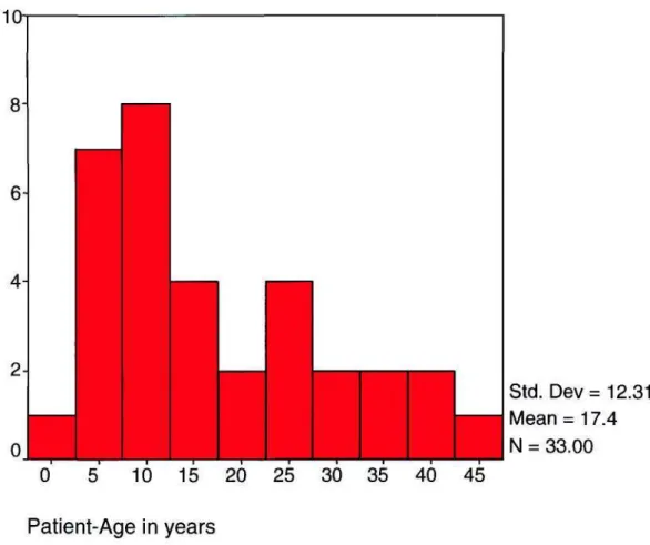
3.1 Demography 3.1.1 Age
The mean age of patients with HAID was 17 years (SD 12.3) range (8 months- 46 years).
One third (30.3%) of the patients fell between the ages 6 and 10 years, represented in (Figure 4), below.
10i
8-
0
Std. Dev= 12.31 Mean = 17.4
33.00
• Mei
| N = 10 15 20 25 30 35 40 45 Patient-Age in years
Figure 4: A histogram of the ase distribution in the cohort, age on the x axis and number of individuals on the y axis.
3.1.2 Gender
In the sample, 24 (72.7%) were female while 9 (22.27%) male, hence a female to male ratio of 2.6:1. In the age group < 12yrs, the female to male ratio was 1:1. In the over 12 year group, 93.7% were females and only 6.7% were males, (Figure 5).
Age group
childhood (<=12 years) adulthood (>12 years)
Male Female
Sex-patient
Figure 5: Gender distribution between adults and children in patients with HAID
3.1.3 Racial distribution
All 33 (100%) of patients were of African origin.
3.2 Study patients
Of the 60 patients screened, 33 were confirmed HTLV-I associated infective dermatitis cases. A total of 27 patients were excluded from this study either because their HTLV-I serology was negative (n=22) or because their HTLV-I results could not be retrieved (n=4), while one (n=l) patient was HTLV-I positive but did not have other features of HAID. Refusal rate was nil.
The 33 patients who fulfilled the clinical criteria had eczema in the following sites:
exudative eczema on the scalp, neck and groin, a generalized papular rash and nasal
discharge and/or crusting of the anterior nares. An example of some of these features is shown in (Figures 6a and 6b).
Figure 6a and 6b: A typical exudative eczema with nasal crusting.
3.3 Dermatological examination
The scalp was the commonest site of involvement, with 77.4% of all patients affected.
This site was closely followed by ear involvement, with both the external ear and
retroauricular areas involved, making up about 71%. The axillae were affected in 65% of patients (Figure 7). Paranasal involvement 58%, groin 55%, eyelid 52% and neck 39%.
These skin lesions were erythematous, scaly and exudative with adherent yellowish crusts. Retroauricular fissures were also seen. Blepharoconjuctivitis was observed in 3 (8.26%) of patients. A disseminated follicular papular eruption was found in 16 patients (23.5%), see (Figure 8). Lymphadenopathy was present in 18 (54.5%) patients. Chronic nasal discharge was observed in 16 (48.4%) patients, whereas crusting of the anterior nares was found in 13(39.3%). The remaining four patients (12.1%) exhibited neither chronic nasal discharge nor crusting of the anterior nares. Figure 9 graphically illustrates the frequent sites of skin involvement in our cohort with HAID.
Figure 7: Picture of patient
demonstrating flexural involvement
Figure 8: A seneralised papular eruption in HAID
T r
Scalp Axil Groin Ear Eyelid l r
Paran neck
Figure 9: Anatomical sites involved in the HAID patients
3.4 Complications
A total of 11(33.3%) individuals in our cohort had complications associated with HAID.
These were corneal opacities in 3 (8.26 %) patients {Figures 10a and 10b). Scabies was confirmed in 6 (18.1%) patients, while 2 (6%) patients had HAM / TSP. None of the patients had evidence of lymphocytic interstitial pneumonitis on chest radiograph.
10a) 10b)
Figures 10a and 10b: Pictures demonstrating corneal opacities affectinz some patients with HMD
3.5 Microscopy results 3.5.1 Skin swabs
As skins swabs were taken from more than 1 site in some patients, a total of 41 specimens were collected. A single swab was taken from the perinasal skin, 18 swabs from anterior nares while 22 were taken from lesional skin. The single swab taken from perinasal skin revealed colonisation with Streptococcus pneumoniae. From the nasal skin swabs, results of the 18 swabs taken revealed that 14 (77.7%) of those were colonised by S. aureus. Streptococcal species were isolated in only 2 (11.1%) specimens taken from this site. Multiplicity of organisms was also found in 2 (11.1%) specimens. Findings from the swabs taken from lesional skin showed that S. aureus was present in 20 (90.9%) specimens while 15 (68.1%) of which also had Streptococcus species. Multiplicity of organisms was found in 1(4.5%) swab. These findings are summarised in Figure 11. The streptococcal species, isolated from the different regions were a combination of:J3- haemolytic streptococcus (BHS), groups A, B, C and G, together with streptococcus pyogenes. The most common of these was BHS group G, which was found in 5/15
(33.3%) swabs taken from the lesional skin.
• Total
• S aureus
n Streptococci
>=> Multiple organisms
Nasal Peri nasal Lesional
Figure 11: A histogram showing the distribution of bacteriology results
3.5.2 Stool sample results
Stool samples were collected in 12 of the 33 patients and neither parasites nor ova were isolated from any of these specimens.
3.6 Bloods Results 3.6.1 Haematology
A complete blood count could be retrieved in 25 of the 33 patients. Haemoglobin levels ranged from 8.0 to 14 g/dl. They had a mean Hb of 11.5g/dl. This was indicative of a mild anaemia which was defined as haemoglobin < 12 g/dl. The mean white cell count, differential counts and the platelet counts were within the normal limits, Table 6.
Table 6: Summary of the blood count results
Normal
Mean Levels ( n = Minimum
Maximum
= 33)
Haemoglobin 11.5-13.5 g/dl
11.5 g/dl 8 g/dl 14g/dl
White cell count 4.0-11.0 x 10A9/1
10.1 x 10A9/1 5.3 x 10A9/1 17.4 x 10A9/1
Platelet count 150-450x 10A9/1
405 x 10A9/1 116 x 10A9/1 607 x 10A9/1
3.6.2 Erythrocyte Sedimentation Rate (ESR)
ESR results were available in 14 of the 33 patients. Of these, the mean was 53 mm/hr (range 10-130 mm/hr) and 12 (85.7%) patients had elevated (ESR > 15mm/hr) levels.
3.6.3 HIV/HTLV-I co-infection
All 33 (100%) patients with features of HAID were seropositive for HTLV-I serology.
HIV results were retrieved in 30 of the 33 patients tested for this. Of these 9 (30%) were HIV positive, 21 (70 %) were HIV negative. Of the 9 HIV positive patients 8 were adults (age range 15- 41 years) and 1 was a child aged 8 months. Eighty-eight percent (88%) of the HIV positive patients were female. The mean viral load in this group was 52 000 copies/ml.
3.6.4 Immunology
IgG levels were high in 16 (48.4%) of the patients evaluated. IgA was high in 8 (24.2%) of patients while IgM was found to be elevated in 2 (6%) of patients. The mean levels are depicted in Table 7. Levels for the IgD and IgE could not be determined due to the non- availability of reagents in our laboratory,
Table 7: Immunologic parameters among patients with HAID Variable (normal range)
IgA (0.68 - 3.78 g/1) IgG (6.94- 16.18 g/1) IgM (0.60 - 2.63 g/1)
Mean Levels 3.94 g/1 21.41 g/1
1.62 g/1
Table 8: Results of serum electrophoresis inpatients with HAW Variable (normal range)
Albumin (32-50 g/1)
Alpha 1 globulin (1.72 - 3.30g/l) Alpha 2 globulin (4.2 - 8.7 g/1) Beta globulin (5.2-10.5 g/1) Gamma globulin (7.1 - 14.5 g/1)
Mean levels 31.00 g/1 4.0059 g/1 11.1412 g/1
11.494 g/1 26.781 g/1
Tafo/e S include the results of the serum electrophoresis in all the enrolled patients, this is further illustrated in {Figure 12) below.
[31]
1 0 1
0 0
0
Albumin Alpha 1 globulin Alpha 2 globulin Beta globulin Gamma globulin
Figure 12: Schematic diagram of Protein Electrophoresis Result
3.6.5 CD4/ CD8 (entire group)
CD4 counts were tested in 18 of the 33 patients. Their mean CD4 count levels was 1730 cells/u.1 (range 114 - 3134 cells/ul) with a mean CD8 count of 1299 cells/u.1 (range 436 - 3433 cells /u.1). The CD4:CD8 ratio of the group was therefore 1.33. CD4 and CD8 counts were checked for in 6 of the 9 patients co-infected with HIV. In this group CD4 count levels were 1505 cells/ul, a CD8 level of 1704 cells/ul and therefore a ratio of 1:15.
These results are shown in Table 9.
Table 9: CD4 and CD8 count analysis
CD4 cells/ul CD8cells/uI CD4/CD8 ratio
Normal 550-1955 250-1200 0SA HTLV-1 1730 1299 1.33 (n=18)
HTLV-1+HIV 1505 1704 1.15 (n=6)
3.7 Virology
All 33 patients were HTLV-I positive (particle agglutination test, confirmed on Western Blot). Only 12 of the 33 were subtyped, the predominant strain was found to be the Cosmopolitan, Subtype la.
3.8 Histopathology results
Histopathological examination was performed on punch biopsies of 31 patients. These biopsies were of various lesions in different stages of evolution and included papules, patches, eczematous areas and macules. The average number of biopsies per patient was two. A wide spectrum of histopathological features ware present and the salient features that were assessed were as follows:
• superficial perivascular dermatitis (SD)
• deep perivascular dermatitis (PVD)
• superficial and deep perivascular dermatitis (S/D-PVD)
• lichenoid inflammatory dermatitis (LID)
• interface dermatitis (ID)
• seborrrhoeic dermatitis features (seb derm)
• leucocytoclastic vasculitis (LCV)
• chronic folliculitis (CF)
Strict definitions of the above terms were used when assessing the biopsies e.g.
Superficial perivascular dermatitis when viewed at scanning magnification is one of the most common reaction patterns in inflammatory skin pathology , characterized by the presence of inflammatory cells around venules in the upper reticular dermis. The
inflammation may be confined to a zone around the venules (perivascular only) and may also occupy the interstitium (perivacular and interstitial) where the inflammatory cells are scattered within the collagen bundles. If the inflammation involved the venules deep within the dermis then this was called deep perivascular dermatitis.
When epidermis was involved by the inflammatory infiltrate in a manner that obscured the dermoepidermal junction then this pattern was called interface dermatitis and if the inflammation was continuous along the interface with associated vacuolar alteration then this was called a lichenoid inflammatory dermatitis.
Seborrhoeic dermatitis was diagnosed when mounds of scale crust in the epidermis were demonstrated with occasional neutrophils. Additional features were intercellular oedema causing widening between keratinocytes beneath the epidermis (spongiotic dermatitis). A dermal superficial perivascular and interstitial dermatitis was also present in some of the specimens.
Exudation of neutrophils with fibrin around dermal venules with nuclear dust and extravasation of red cells was designated leucocytoclastic vasculitis.
When lymphocytes were identified within the hair follicules especially infundibular epithelium, this was designated chronic folliculitis.
Superficial and deep perivascular dermatitis (S/D-PVD) was a major histological feature in our cohort with HAID. It was found present a single feature in 12 (38%) of the 31 cases (Figure 13). S/D-PVD was also noted associated with a lichenoid inflammatory dermatitis (LID) in 28% of cases (Figure 14). Chronic dermatitis characterised by SVD- PVD with interface dermatitis (ID) was found in 14 (12.9%) of the specimens. ID is shown in Figure 15. Two (6.4%) of the skin specimens had features in keeping with seborrhoeic dermatitis (seb derm), (Figure 16a and 16b). Seb derm is one clinical condition that can be mistaken for HAID.
S/D-PVD was found associated with chronic folliculitis (CF) (Figure 17), in one (3.2%) case. There single cases (3.2%) of LID, CF and CF together with LID, respectively. One patient (3.2%) showed features of leucocytoclastic vasculitis (LCV). The patients who were co-infected with HIV showed similar features dominated by S/D-PVD but in addition their histology sections were studded with eosinophils.
Figure 13: Superficial and deep perivascular dermatitis (S/D-PVD)
Figure 74: SVD-PVD + Lichenoid inflammatory dermatitis (LID)
| Figure 15: Interface Dermatitis (ID)
Fieure 16a: Seborrhoeic dermatitis: Skin demonstrating mounds of scale crust with
occasional neutrophils. Beneath this there is pallor of keratinocytes. A dermal superficial perivascular and interstitial dermatitis is also seen.
Fieure 16b: Skin demonstrating sponsiotic dermatitis in seborrhoeic dermatitis
Figure 17a: Chronic folliculitis (CF ): Skin demonstrating lymphocytes within follicular especially infundibular epithelium.
Figure 17 b: A deey section demonstrating chronic folliculitis
The spectrum of the histological features that were found in this group of patients is summarized in a pie diagram below (Figurel8).
Figure 18: Histological characteristics of HAID
M S/D-PVD
• S/D-PVD +LID
• S/D-PVD +ID D S/D-PVD +CF 1