Page | 1
TO IDENTIFY THE CHANGES IN THE HAEMODYNAMICS IN PATIENTS WITH PRE-ECLAMPSIA
USING BRAIN NATRIURETIC PEPTIDE AND DOPPLER STUDIES
A dissertation submitted to the Faculty of Health Sciences University of Kwa Zulu-Natal
In partial fulfilment of the requirements for the Master of Medical Science degree
By: Dr SB Fayers (MBChB)
Supervisor: Professor DP Naidoo
Page | 2
This study is dedicated to my
Lord and Saviour Jesus Christ and to my family.
Thank you for believing in me.
Your love and support has carried me through.
Page | 3
STATEMENT OF DECLARATION
I declare that this dissertation is my own unaided work. It is submitted in partial fulfilment for the Master of Medical Science Degree at the University of Kwa Zulu-Natal. It has not been submitted before for any degree or examination at any other educational institution.
__________________ October 2011
Samantha Fayers
Page | 4
ACKNOWLEDGEMENTS
I am sincerely grateful to the many individuals who contributed to the completion of this study.
I would like to thank my supervisor, Prof DP Naidoo, whose passion for the subject and warm fatherly nature encouraged me along this journey.
My sincere gratitude to Prof J Moodley. Thank you for your ‘open door’; always being there to offer advice, valuable recommendations and stability.
Mr Shaun Khedun, you have proved to be reliable, faithful and diligent in promptly providing assistance when necessary.
Thank you Mr Govender for your words of encouragement and for making this thesis look the way it does; well done.
Mr Mondi Mia, you came into my life at the right time. Thank you for your timeous efforts and faithfulness – you’re simply the best.
Thank you, Mrs S Naidoo for persevering and competently carrying out the echocardiographs amidst your busy schedule.
To our ultrasonographer, Ms N Bayat, your pleasantness in completing the task is much appreciated.
Dr P Naidoo and the lab team at IALCH, thank you so much for your willingness to assist and the many hours spent analysing specimens and efficiently keeping good records.
To the Obstetrics and Gynaecology Department at Prince Mshiyeni Memorial Hospital – without your assistance, this study would not have been possible.
Thank you, Mrs T Esterhuizen, Mr Tlou and Mr Stephan, for assisting with the statistical analysis.
Page | 5
Contents
Contents ... 5
ABBREVIATIONS ... 7
CHAPTER 1... 11
INTRODUCTION ... 11
Epidemiology ... 11
Haemodynamics and cardiac changes in PE ... 15
Investigative evaluation of maternal haemodynamics ... 16
The evaluation of ventricular function ... 18
Timing of testing ... 19
Ultrasound and doppler studies ... 20
Types of doppler studies ... 23
Correlating central haemodynamics with uteroplacental circulation ... 26
Effect of treatment ... 27
Rationale for this study... 29
CHAPTER 2... 31
STUDY DESIGN AND METHODS ... 31
2.1. Aim and objectives ... 31
2.2 Study Design ... 31
2.3. Inclusion and Exclusion Criteria ... 32
2.4. Methodology ... 32
CHAPTER 3... 39
RESULTS ... 39
3.1 Demographic Data ... 39
HIV Status ... 39
Body Mass Index (BMI) ... 39
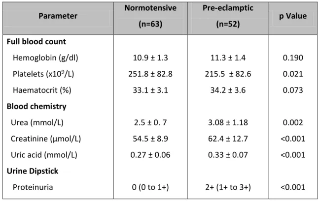
3.2. Laboratory Investigations ... 40
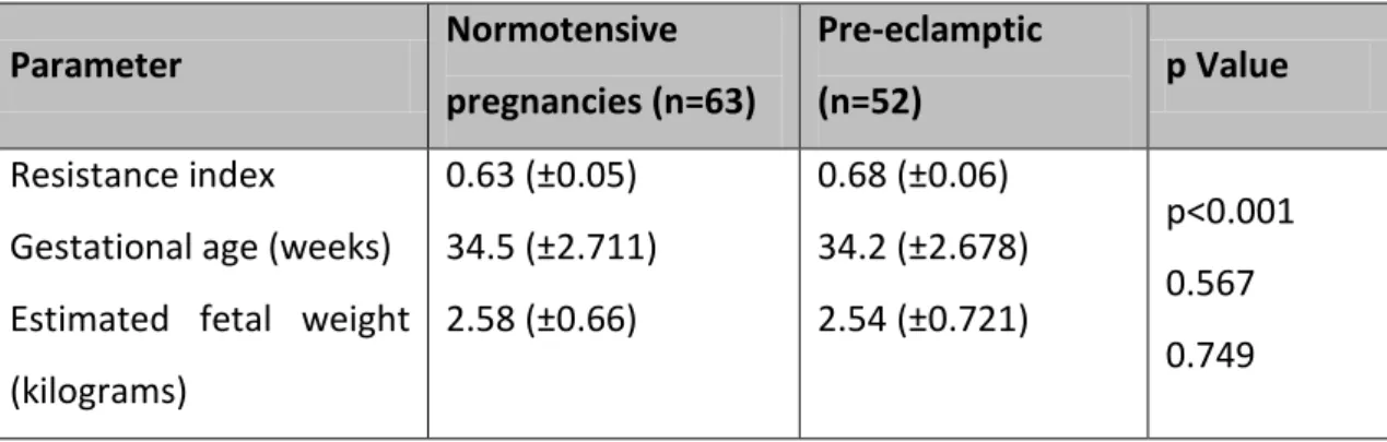
3.3. Fetal ultrasound at initial assessment ... 41
3.4. Echocardiography findings ... 42
Page | 6
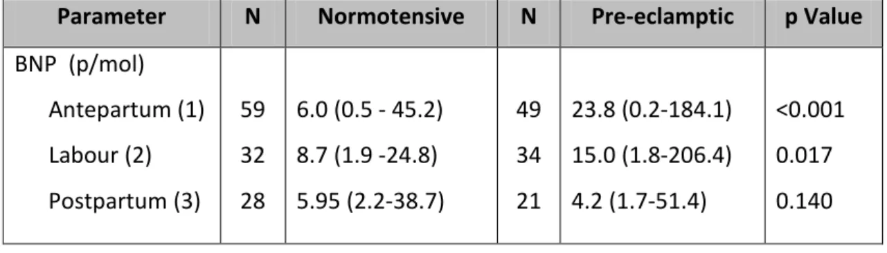
3.5. B-TYPE Natriuretic Peptide Levels ... 43
3.6. Correlation Studies (Table 7) ... 44
3.6. Pregnancy Outcomes ... 45
CHAPTER 4... 50
DISCUSSION ... 50
CHAPTER 5... 54
CONCLUSIONS, RECOMMENDATIONS AND LIMITATIONS ... 54
5.1. Conclusion ... 54
5.2. Recommendations ... 54
5.3. Limitations ... 54
5.4. Disclosure of Interest ... 55
REFERENCES ... 56
APPENDIX ... 69
a) INFORMED CONSENT ENGLISH ... 69
b) INFORMED CONSENT (ISIZULU) ISIVUMELWANO SOKUBA INGXENYE YOCWANINGO ... 70
Page | 7
ABBREVIATIONS
AEDF Absent End Diastolic Flow
AIDS Acquired Immunodefieciency Syndrome ANP Atrial Natriuretic Peptide
BMI Body Mass Index
BNP Brain Natriuretic Peptide
BP Blood Pressure
CPD Cephalopelvic Disproportion
DIC Disseminated Intravascular Coagulation
DOH Department of Health
ECHO Echocardiography
EDTA Ethylenediaminetetraacetic acid
E/ Ea ratio Mitral valve Doppler inflow velocity (E) / Mitral annulus n Tissue Doppler (Ea)
EFW Estimated Fetal weight
ENND Early Neonatal Death
FBC Full Blood Count
FIG Figure
GA Gestational Age
HELLP Syndrome: Haemolysis/Elevated Liver enzymes/ Low Platelets
HIV Human Immunodeficiency Virus
IE Imminent Eclampsia
IOL Induction of Labour
Page | 8
IUGR Intra Uterine Growth Restriction
LA Left Atrium
LV Left ventricle
MSL Meconium Stained Liquor
NICU Neonatal Intensive Care Unit
PE Pre-eclampsia
PI Pulsatility Index
PROM Prolonged Rupture of Membranes REDF Reversed End-Diastolic Flow
ROM Rupture of Membranes
SA South Africa
SB Stillbirth
SD Standard Deviation
TDI Tissue Doppler Index
U/S Ultrasound
UNK Unknown
Page | 9
TO IDENTIFY THE CHANGES IN THE HAEMODYNAMICS IN PATIENTS WITH PRE-ECLAMPSIA USING BRAIN
NATRIURETIC PEPTIDE AND DOPPLER STUDIES
ABSTRACT AIM:
To determine the haemodynamic changes in pre-eclampsia by using echocardiography and cardiovascular markers i.e. Brain Natriuretic Peptide (BNP);
and relating these changes to perinatal outcomes.
MATERIAL AND METHODS:
A prospective study was conducted at a large regional hospital in Durban, KwaZulu Natal. One hundred and fifteen primiparous patients were studied; 63 were normotensive pregnancies and 52 were pre-eclamptics. Patients were matched for age and gestational age. Study participants were examined during pregnancy, labour and within the puerperium. Transthoracic echocardiography, tissue Doppler imaging, umbilical artery Doppler, and laboratory investigations were performed. The ratio of the mitral inflow (E) to the early diastolic tissue Doppler velocity (Ea) was measured as a marker of the left ventricular filling pressure.
RESULTS:
BNP levels were significantly increased in the antepartum (23.8 (2 - 184.1) vs 6.0 (0.5 - 45.2) pmol/L; p<0.001) and during labour (15 (1.8-206.4) vs 8.7 (1.9 -24.8) pmol/L;
p=0.01) in the pre-eclamptic group when compared to the normotensive pregnancies. In the postpartum period, median BNP levels were 4.2 (1.7-51.4) and 5.95 (2.2-38.7) pmol/L in the pre-eclamptic and normotensive groups, respectively.
There was no statistically significant difference in the tissue Doppler E/Ea between the pre-eclamptic when compared to the normotensive group of patients (11.02 ± 5.6 vs 9.16 ± 2.6; p=0.058). The caesarean section rate was 47% in the pre-eclamptic group and 32% in the normotensive group. Eight subjects were lost to follow-up, therefore no inference could be made from this data. There were 2 stillbirths in the pre-eclamptic group, none in the normotensive group.
Page | 10 CONCLUSION:
In pregnancies complicated by pre-eclampsia, changes in the haemodynamic state are accompanied by raised blood levels of BNP in comparison to normotensive pregnancies. These revert to baseline values in the puerperium.
Page | 11
CHAPTER 1
INTRODUCTION
EPIDEMIOLOGY
HYPERTENSIVE DISORDERS OF PREGNANCY
Hypertensive disorders of pregnancy complicates approximately 10-16% of pregnancies and is one of the leading cause of maternal, fetal and neonatal morbidity and mortality worldwide (1, 2). A recent population-based study estimates that hypertensive disorders of pregnancy affect 12% of pregnant women in Durban, South Africa (SA) (3). Hypertensive disorders of pregnancy accounts for about 9% of maternal deaths in Africa and Asia, and about 25% of maternal deaths in Latin America and the Caribbean. The maternal deaths are mainly due to pre-eclampsia and eclampsia (4). According to the recent National Confidential Enquiry into Maternal Deaths (NCCEMD) report in SA, which assessed avoidable factors and missed opportunities in deaths that occurred at health facilities from 1 January 2005 until 31 December 2007, approximately 15% of all deaths were due to hypertension and its complications. Hypertensive disorders of pregnancy were the commonest direct cause of maternal deaths in SA. It was found that over one half (52.7%) of all hypertensive-related deaths were avoidable. The highest number of deaths (21.2%) occurred in the province of Kwa-Zulu-Natal. Furthermore cardiac disease accounted for 2.2% of all deaths (5).
Hypertensive disorders of pregnancy have been classified by The National High Blood Pressure Education Program of the National Health Lung and blood institution (NHLBI) into the following categories: gestational hypertension, chronic hypertension, PE and superimposed PE (6). Gestational hypertension is a working definition used when an elevated blood pressure (BP) (systolic BP >/= 140mmHg and diastolic >/= 90mmHg) is first detected after the twentieth week of pregnancy (in the absence of proteinuria) and returns to normal in the peuperium. Women diagnosed with gestational hypertension may eventually fulfill diagnostic criteria for PE if proteinuria subsequently develops. In the absence of proteinuria, chronic hypertension is diagnosed when elevated blood pressures are present before 20 weeks gestation and remain elevated postpartum. Pre-eclampsia is defined as the presence of hypertension, and proteinuria exceeding 0.3 g/day after the twentieth week of pregnancy in a previously normotensive woman. PE occurs in 2-5% of
Page | 12
pregnancies worldwide but it complicates up to 10% of pregnancies in developing countries where emergency care is often inadequate or lacking (6).
PRE-ECLAMPSIA
The cause of PE remains unclear. Pre-eclampsia is characterised by the new onset of hypertension (systolic and diastolic blood pressure of ≥ 140 and 90 mmHg respectively on two occasions at least 6 hours apart) with proteinuria (protein excretion of ≥ 300mg in a 24 hour urine collection, or a dipstick of ≥ 2+) that develops after 20 weeks gestation in a previously normotensive woman (7, 8). The cure is delivery of placenta. Known maternal characteristics such as obesity, ethnicity (black race), diabetes, collagen vascular disease, multiple pregnancies, previous episodes of PE, thrombophilias, molar pregnancy and extremes of age (<20 or >40 years) increase the risk for PE. Women with chronic hypertension have a 15–25%
increased risk of developing superimposed PE (9). The prognosis for mum and baby depends on the severity of the pre-eclampsia. In mild disease, there is a good prognosis. In severe disease, complications include abruption placenta, eclampsia, renal and liver disease, disturbances of haemostasis and the HELLP syndrome. A higher incidence of pre-eclampsia has been found among women who conceive with assisted reproduction techniques, nulliparous women, and in women with autoimmune conditions demonstrating the probable influence of an inexperienced maternal immune system (10, 11). Women with pre-existing metabolic, vascular or renal disease are at increased risk for superimposed PE (12, 13). This is possibly due to their increased sensitivity to the physiological changes of pregnancy.
Identifying “at-risk” women is important because modern obstetric care places emphasis upon the primary care setting for expectant women. A marker which identifies high-risk women would allow for closer supervision in secondary care. Such a marker would also facilitate recruitment for trials of potential therapeutic agents, for accurate diagnosis, and for timely intervention whenever problems develop.
PATHOPHYSIOLOGY OF PRE-ECLAMPSIA
The placenta is considered central in the pathogenesis of PE (14). This is known to be the case because PE occurs only in pregnancy and resolves after delivery of the placenta. Furthermore, it can occur in the absence of a viable fetus for example, in molar pregnancies (15).
Page | 13
The pathophysiology of PE it is believed to be multifactorial. The teaching has been that PE develops due to an immune maladaptation between the mother and the fetus. Poor placentation is considered the first step in the two-stage theory (16).
Trophoblastic invasion of the maternal spiral arterioles occurs early in pregnancy and results in the formation of wide-bore vessels carrying maternal blood to the developing fetus. Defective trophoblastic invasion, possibly due to genetic or immune mechanisms, results in the spiral arterioles remaining as narrow conduits.
The result is tissue anoxia due to hypoperfusion (17). The consequence of the tissue anoxia is the release of apoptotic cells, trophoblastic debris and angiogenic factors that causes widespread vascular endothelial damage (18, 19). This leads to the second step of the disorder which is the development of maternal signs and symptoms affecting multiple systems of the body (20).
BIOCHEMICAL MARKERS OF PRE-ECLAMPSIA
The ideal marker would be non-invasive, cheap, with a high sensitivity and specificity and suitable for resource limited settings. Based on the etiology of this life- threatening pregnancy disorder, a major focus of research has recently been maternally expressed proteins and the identification of placental factors showing abnormal expression in pre-eclamptics placentas (21, 22). Their potential use for non-invasive early prediction or early detection would be invaluable. The availability of such markers could potentially improve surveillance of high risk patients and result in earlier referrals and better outcomes. The impact and cost of treating the complications of PE (e.g prolonged ICU admissions, etc) for both the family and the heath system could potentially be reduced.
Misdiagnosis and failure to recognise warning symptoms is still an issue in our health care system owing partly to the multiple clinical symptoms associated with the syndrome (23). The availability a reliable biochemical indicator might thus help in making the clinical diagnosis. Biochemical markers would allow the classification of pre-eclamptic patients into categories based on their severity and this would direct the clinical management and improve the pregnancy outcomes (24).
IDENTIFICATION OF NOVEL BIOMARKERS
Although a number of promising biomarkers already exists, a lot of effort is being made to find novel candidates that bear a greater potential to identify women at risk for PE, in order to provide the best possible care for these mothers and children (25, 26). Several approaches have been made (27, 28, 29). Microarrays screen the placental transcriptome for up- and down-regulated transcripts in pre-eclamptic samples and compare these to healthy controls. Comparative transcription analyses
Page | 14
have been performed by numerous groups (30, 31, 32). These groups were however, studying the molecular mechanisms of PE rather than potential biomarkers. The microarray based screening for RNA molecules in maternal circulation that are transcribed in placenta has been reported as a potential source for pregnancy related biomarkers (33, 34), including PE (35, 36). Gene experiments are being performed frequently due to the rapid evolution of the microarray technology.
Another direction of research in the field of complex diseases is metabolomics. It involves the analysis of endogenous and secreted metabolites in a biological system.
The reports investigate the placental metabolome using varying oxygen tensions in the plasma of pre-eclamptic patients. It is suggested that this technology may bring new insights into placental function (37, 38).
THE NATRIURETIC PEPTIDE SYSTEM
Most of the molecular markers that have been described above relate to the pathogenesis of the PE process. The early sequelae in PE involve well documented haemodynamic changes which have been investigated and corrobated by invasive and non-invasive techniques. The naturetic peptide system, first discovered in the early 1980s to regulate salt and body fluid balance, has been shown to closely reflect fluid dynamics in the circulation (39).
Human cardiocytes manufacture a family of structurally related peptide hormones that include atrial natriuretic peptide (ANP), brain (or B-type) natriuretic peptide (BNP), and their metabolically associated peptides (40). Release of the natriuretic peptides is stimulated by haemodynamic stress, such as wall stretch, ventricular dilation, and increased pressures resulting from fluid overload. The natriuretic peptides have powerful natriuretic, diuretic, antimitotic, and vascular smooth muscle relaxing actions. The natriuretic peptides are antagonists for the sympathetic nervous system and the renin-angiotensin-aldosterone axis (41).
BNP has emerged as a superior biomarker to ANP for clinical applications involving heart failure and left-ventricular dysfunction (42). In a study by Cowie and colleagues (43), BNP showed a greater predictive power as an indicator of heart failure when compared with either ANP or its metabolites. Contributing factors are BNP's longer half-life, activation at the gene level and greater quantity in left ventricular tissue.
Also, BNP has a 2- to 3-fold more powerful natriuretic and blood pressure lowering effect compared to ANP (44). Thus in vitro diagnostics for BNP and associated metabolites have been the focus for clinical applications (45).
Page | 15
BIOSYNTHESIS AND SECRETION OF BNP AND NT-proBNP
Human BNP is derived from 134 amino-acid precursor termed pre-proBNP1-134.
Haemodynamic stress activates cleavage of a 26-aa signal peptide sequence from the N-terminus of pre-proBNP1-134. The remaining proBNP1-108 prohormone is then cleaved by corin; the N-terminal pro-BNP1-76 (NT-proBNP) fragment and active 32-peptide, C-terminal BNP77-108 (BNP) hormone are then released into the circulation. BNP and the metabolically inert NT-proBNP are released in a 1:1 molar ratio.
The release and metabolism of BNP and NT-proBNP is a far more complicated process. The intact prohormone proBNP1-108 may be released into circulation and may cross-react with immunoassays for BNP and NT-proBNP. Also, N-terminal proBNP1-76 in blood may be comprised of numerous fragments and not a single 77 amino-acid molecular entity. Furthermore, BNP appears be undergo metabolism following release into the circulation. Truncation of the 32-amino acid peptide at either the C-terminal or N-terminal arms to an inactive form of BNP has been reported (46).
MECHANISM OF ACTION OF NATRIURETIC PEPTIDES
The primary function of ANP and BNP is to regulate blood volume and pressure.
Under high blood volume or pressure, ANP and BNP are released into the circulation.
In target organs such as the kidneys and peripheral blood vessels, the peptides activate their receptor, natriuretic peptide receptor –A (NPR-A), and increase intracellular cGMP production. This leads to natriuresis, diuresis, and vasodilation resulting in the lowering of blood volume and pressure. ANP and BNP also suppress the renin angiotensin release, which is an additional mechanism to regulate vascular tone (47). The blood levels of BNP and NT-proBNP, reflect haemodynamic stress, and as such, may serve as a marker of early haemodynamic changes that accompany PE.
HAEMODYNAMICS AND CARDIAC CHANGES IN PE
During normal pregnancy, plasma volume increases steadily throughout the first two trimesters and reach a plateau at approximately 32 weeks gestation. In a singleton pregnancy at term, plasma volume is nearly 50% more than that seen in a non- pregnant woman. Total peripheral vascular resistance decreases in normal
Page | 16
pregnancy, cardiac output increases, reaching a peak of 30-50% of the pre- pregnancy level by the end of the 2nd trimester. There is usually no further increase (48). In patients with PE, there is a further rise in cardiac output as well as an increase in total peripheral resistance (49).
There is evidence that cardiac function is altered in PE. Significant changes in the volume status and systemic resistance have been documented in patients with pre- eclampsia. Left ventricular mass (and index) in PE patients is higher than normotensive patients and is associated with increased left ventricular wall thickness. There appears to be no difference in left ventricular diastolic filling rates between PE and normotensive patients (49). A further significant increase in left ventricular end-systolic and end-diastolic volumes and reduction in contractile function has been found in PE women. Under these conditions, changes in volume loading may affect ventricular function. There are limited data available on the changes in cardiac structure and function during PE. Most studies focus on changes in normotensive patients (50). A study found that there was reduced cardiac function in pregnancies complicated by pre-eclamptic IUGR when compared to normotensive IUGR pregnancies (51). This was due to reduced intrinsic contractility and reduced diastolic filling. Some markers of the changes in diastolic filling would help to understand the haemodynamic changes of PE. Left ventricular diastolic filling may precede changes in systolic function. Therefore, assessment of diastolic function may provide an early marker of PE.
INVESTIGATIVE EVALUATION OF MATERNAL HAEMODYNAMICS
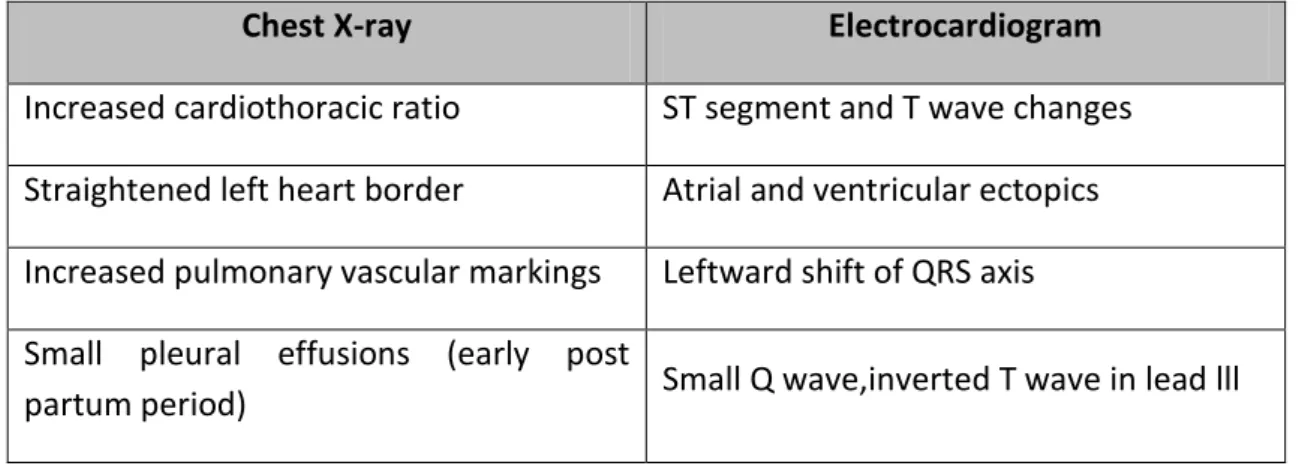
Several diagnostic investigations have been used to evaluate the cardiac changes that occur in PE, but because of the physiological changes seen even in normal pregnancies (Table 1), interpretation of these findings is often difficult.
Page | 17
Table 1: Chest X Ray and Electrographic Changes in normal Pregnancy
Chest X-ray Electrocardiogram
Increased cardiothoracic ratio ST segment and T wave changes Straightened left heart border Atrial and ventricular ectopics Increased pulmonary vascular markings Leftward shift of QRS axis Small pleural effusions (early post
partum period) Small Q wave,inverted T wave in lead lll
TISSUE DOPPLER ECHOCARDIOGRAPHY
What is required is a sensitive measure of the cardiac changes that occur during PE, which may easily be measured non-invasively. Until recently most of the non- invasive studies of the hemodynamic changes of PE have employed echocardiography with the focus being on cardiac dimensions and systolic function.
Tissue Doppler imaging of the heart is a technique that has enabled the evaluation of changes in ventricular filling at a much earlier stage than can be detected by conventional echocardiography. This technique measures the frequency of ultrasound returning from the moving myocardium to estimate the velocity of the myocardial wall. Isaaz et al (1989) were the first to describe the clinical applications of tissue Doppler in cardiac disease, demonstrating that low myocardial velocities at the posterior mitral annulus correlated with abnormal posterior wall motion on left ventricular angiography (52). Gulati et al (1996) demonstrated that tissue Doppler systolic mitral annular velocities correlated with global left ventricular ejection fraction as assessed by radionuclide ventriculography (53). Diastolic tissue Doppler velocities reflect myocardial relaxation, and in combination with conventional Doppler measurements, ratios (transmitral early diastolic velocity / mitral annular early diastolic velocity (E/Ea) have developed to non-invasively estimate left ventricular filling pressure. It has been shown that Tissue Doppler derived Ea correlated with the invasively measured left ventricular time constant of relation (Tau), establishing Ea as a relatively load independent measure of myocardial relaxation in patients with cardiac disease (54).
Echocardiography is a good diagnostic tool as it allows for the structural as well as functional assessment of the heart. Furthermore the cardiovascular changes that have been documented appear to coincide with increases in plasma BNP and have
Page | 18
been observed to be linearly related to the left ventricular structural and functional changes observed in patients with PE (49).
THE EVALUATION OF VENTRICULAR FUNCTION
Our study was designed to evaluate BNP changes in PE as a measure of ventricular function. As previously mentioned, BNP and ANP are polypeptide neurohormones synthesized by cardiac atrial and ventricular myocyes in response to stretch (55).
They have a regulatory and modulatory role in the cardiovascular system. They are secreted as prohormones, proANP and proBNP. They are then split into equimolar amounts of active ANP and BNP, and inactive amino-terminal fragments NT-proANP and NT-proBNP (56). BNP levels reflect long-term intravascular volume status. It shows less affinity for clearance receptors and therefore has a longer half life than ANP. ANP is more dependent on atrial filling volume than filling pressure and is rarely measured in clinical practise. BNP and NT-proBNP are more commonly measured, often interchangeable, based on clinician’s preference and laboratory availability (57). There is evidence however, that NT-proBNP measurement is superior in detecting mild systolic or diastolic heart failure or asymptomatic left ventricular dysfunction (LVD) (58). There is fast synthesis and release into the blood by myocardial stretch which is increased in LVD. Left ventricular stretch activates neuronal and hormonal pathways that results in BNP being secreted directly into the blood during ventricular dilatation. Increased plasma levels of BNP have been found in patients with LVD from various causes. BNP has a high sensitivity, specificity and predictive value for LVD and is an approved marker for the diagnosis of congestive heart failure in patients with dyspnoea in an acute care setting (59). During normal pregnancies, haemodynamic changes do not place significant stress on the heart.
However, increased ANP and BNP levels of statistical significance were found in patients with PE as compared to normotensive pregnant patients. A study which evaluated maternal, umbilical plasma and amniotic fluid BNP and ANP levels in nineteen normotensive pregnant women and thirty five pre-eclamptic patients suggested that increased ANP and BNP levels were a sequel of PE pathophysiology and did not play an etiological role in PE (60). Another study looked at the trends in BNP levels during pregnancy. BNP blood levels were taken during the 1st, 2nd and 3rd trimester in normotensive as well as in patients with PE. In normotensive pregnancies, median BNP levels are < 20pg/ml and stable throughout gestation. An increase in BNP levels in pregnancies complicated by mild PE and an even higher increase in severe pre-eclamptics was found. This may reflect ventricular strain and/
or subclinical cardiac dysfunction associated with pre-eclampsia (61). In a study
Page | 19
investigating the role of BNP in the circulation of patients with pregnancy induced hypertension, they found that BNP and ANP participate in maintaining homeostasis of the maternal circulation. This cross-sectional study looked at thirty six normal pregnancies and compared levels of BNP and ANP to seventeen women with pregnancy induced hypertension. They found statistically significant increased levels of BNP and ANP in patients with pregnancy induced hypertension. In patients with severe pregnancy induced hypertension, an eight-fold increase in BNP levels were found. There was a positive correlation between the plasma BNP levels and the mean arterial blood pressure (r=0.62, P<0.001) (62). Numerous studies have confirmed the definite increase in BNP levels in patients with PE (63, 64). In a study looking at the natriuretic peptides and PE, it was found that the high BNP concentrations found in pre-eclampsia reflect cardiac strain caused by high afterload (64). In a review of the charts of fifteen obstetric patients who presented with acute dyspnoea, seven of them had PE with elevated BNP levels. This correlated with acute ventricular overload and they responded well to volume management and diuresis.
In two patients, markedly elevated serum BNP levels and significant left ventricular dysfunction was found that was not apparent by standard clinical evaluation. It was thus concluded that serum BNP levels provided valuable information that could be used in the management of obstetric patients with acute dyspnoea and may assist in identifying patients with cardiac dysfunction that might have been otherwise missed on clinical evaluation (65).
TIMING OF TESTING
So, if serum BNP is activated during pregnancies complicated by PE, the crucial question is “when is the optimal time to test these levels”; and “when do they return to normal?” In a study comparing proBNP levels in pre-eclamptic versus normotensive obstetric patients antenatally, and then later in the puerperium, the proBNP level in the pre-eclamptic group showed a seven-fold increase to that of the control group (p > 0.05). Of note is that in the puerperium, these levels normalised (66). In the non-pregnant situation, a study by Bayes-Genis et al., found that there was a good association between the New York Heart Association functional class and NT pro-BNP concentrations; NT pro-BNP concentrations in the emergency rooms did not significantly differ from those after 24 hours; NT pro-BNP concentrations on day 7 did not differ from that at 6 or 12 months and could be considered as the basal stable level for that particular patient (67).
Page | 20
We know that there are dramatic changes in circulating volume in the perinatal period which would influence BNP levels in relation to labour and uterine contractions. Not much is known about changes in BNP on an hour to hour basis.
Vasoactive hormones are said to play an important role in the pathogenesis of pre- ecalmpsia thus linking placental hypoperfusion with hypertension, systemic disease and proteinuria. In a study evaluating the diurnal patterns of vasoactive hormones in PE and comparing these to a normotensive comparable control group, it was found that in the control group there was diurnal variations (blood samples were taken every 2 hours over a 25 hr period) in serum BNP levels amongst others. The BNP concentrations in the PE group tended to be higher throughout the 25 hours but had blunting of the normal diurnal variation (68). Although this study did show certain trends, in clinical practise it is difficult for serum sampling to be done at exactly the same time in our patients.
ULTRASOUND AND DOPPLER STUDIES
Ultrasound allows for a detailed, painless and safe analysis of the placenta and fetus.
Numerous investigative modalities have been used by researcher’s e.g X-rays by Panigel demonstrating placental maternal blood jet lags, Magnetic Resonance Imaging, 2-dimensional grey-scale ultrasound, colour imaging and now 3- dimensional ultrasound imaging (69). Several medical disorders of the pregnant woman or her fetus begin or end in the placenta. Examples of these include PE, IUGR, triploidy, etc. Ultrasound is the optimal investigation method because of the benefits mentioned above as well as the fact that it allows for placental structural and functional assessment. Three dimensional ultrasound permits evaluation of the placenta in several planes, more precise depiction of internal vasculature and a more accurate assessment of volume.
Pre-eclamptic vascular changes are seen relatively early in pregnancy. At the level of the uterine circulation, significant haemodynamic changes have been documented.
Harrington et al confirmed that the development of PE was associated with failure to modify the uterine circulation in early pregnancy as shown by abnormal Doppler values done between 12-16 weeks gestation (70). In another study investigating the vascular mechanical properties and endothelial function in PE with special reference to bilateral uterine artery notch (bilateral uterine artery notch is associated with ischaemic pathology), it was found that a significant number of placentas in the group with PE did not show noteable ischaemic or other morphological changes that could explain the role of the placenta in the development of PE. Women with PE
Page | 21
showed signs of endothelial dysfunction significantly more pronounced in women with the bilateral uterine artery notch (71).
Inadequate placental perfusion has lead to the use of Doppler ultrasonography to assess the velocity of the blood flow in the uterine arteries. A persistence of an early diastolic notch after 24 weeks of gestation or abnormal flow velocity ratios has been associated with inadequate trophoblast invasion. Pregnancies associated with an abnormal uterine Doppler after 24 weeks of gestation (high pulsatility index and/or presence of an early diastolic notch) are associated with a more than six fold increase in the rate of pre-eclampsia (72).
Among high-risk patients with a previous PE, Doppler ultrasound of the uterine arteries has an excellent negative predictive value, thus it is an important tool in patient management and care which is of paramount benefit for patients with PE in a previous pregnancy. A systematic review evaluated the use of Doppler ultrasonography in PE. A total of 74 studies were included. Of these studies, 69 were cohorts, 3 were randomized controlled trials and 2 were case-control studies. These studies involved a total of 79547 patients, 2498 of whom developed PE. The authors showed that Doppler ultrasonography of the uterine arteries were more accurate in the second trimester than in the first trimester. The most predictive measurement of the Doppler ultrasonography to predict PE was shown to be the pulsatility index (alone or in combination with a persistent notching after 24 weeks of pregnancy) (73).
A study showed that the combined measurement of uterine perfusion in the second trimester and analysis of angiogenic markers have a high detection rate for pre- eclampsia (74). However, data does not support the use of Doppler ultrasonography for routine screening of low risk patients for PE. A multicentric study involving nine hundred and sixteen low risk pregnancies were followed up with end points being PIH, low birth weight for gestational age, small for gestational age with abnormal outcome and PIH requiring delivery. The study was blinded. Umbilical and uterine artery Dopplers were performed nineteen to twenty four weeks gestation and at twenty six to thirty one weeks gestation. This study found the positive predictive value to be low. It did, however, identify the severe forms of PIH and small for gestational age fetus’ in mid pregnancy but could not be used as a screening tool in a low risk population where the prevalence of the disease is low (75). Doppler velocimetry of the umbilical cord has been found to be a more specific and sensitive method of fetal surveillance than cardiotography. It is a fast and convenient method for monitoring fetal well being in high risk pregnancies (76).
Page | 22
Ultrasound and Umbilical Artery Dopplers have also been well studied and their importance in predicting fetal outcomes is well established (77, 78). The use of Doppler studies even in mild PE has been shown to be essential and prompt response to abnormalities validated as shown in the following case report. A pregnant woman with mild PE and severe growth restriction, with reverse end- diastolic flow velocity detected on uterine arteries by colour Doppler was managed expectantly. Follow up Doppler studies of the internal iliac and umbilical arteries also showed reverse flow. This fetus died two weeks after initial abnormality detected (79).
Another study estimated systolic/diastolic ratios in a low risk population to see if this could be used as a screening tool. It was found not to be useful as the positive predictive value was calculated to be between zero and five percent. It was also found to be of no clinical use in predicting IUGR in this population group. One thousand six hundred and sixty five Doppler examinations were performed on five hundred and sixty five women in this study. Dopplers were performed at 27-31, 32- 36 and 37-42 weeks gestation. Low dose aspirin (60mg) was also used in this double blinded prevention trial from twenty four weeks gestation. Aspirin did not affect the relation between the Doppler indices and outcomes using the logistic regression model (80). In our study, we looked at the Resistance Index (RI) in patients in the third trimester.
“Reverse Flow” as a sonographic finding, indicates the appearance of retrograde blood flow in the diastolic part of the cardiac cycle and is an indicator of deterioration of the fetal condition (81). Reversed end-diastolic flow velocity is well known to be an ominous finding. A study conducted on thirty patients during the third trimester with reversed end diastolic flow found a perinatal mortality rate of fifty percent. All newborns were admitted to the NICU. Statistically significant complications included caesarean sections, fetal acidosis and small for gestational age babies (82). It is thus imperitive that intensive and frequent surveillance be carried out and prompt aggressive management plans be implemented in an attempt to alleviate these devastating outcomes when reverse end-diastolic flow is identified.
Abnormal Doppler umbilical artery waveform is a strong predictor of perinatal mortality and is associated with a poor perinatal outome. The study, conducted over 2 year period, included 43 patients with severe PE. Of these, twenty-two (52%) had abnormal Doppler umbilical pulsatility index. The abnormalities included: positive end diastolic velocities with elevated pulsatility index values (13 patients/ 59%), absent end diastolic velocometries (7 patients/ 32%) and reversed flow (2 patients/
Page | 23
9%). There were 6 perinatal deaths in this group with abnormal umbilical artery Dopplers, two in the reversed flow (100%), two in the absent flow (28%) and 2 in the positive end diastolic flow (15%). There were no perinatal deaths in the group with normal umbilical Doppler waveform. Of note is that neonates with abnormal pulsatility index had lower birth weights, lower APGAR score at 5 minutes, higher admission rates to the neonatal intensive care units and significant neonatal morbidity as compared to those with normal velocimetry (p<0.05) (83).
Interestingly, in a study looking at the independent contribution of absent or reversed end-diastolic umbilical artery Doppler flow in the prediction of adverse neonatal outcomes, only an increased frequency of hypoglycaemia (OR 1.7) and polycythemia (OR 1.7) were found whereas other negative outcomes could be explained by prematurity and IUGR. Two hundred and seventy pregnancies complicated by PE with either absent or reversed flow where followed up. Other outcomes included low APGAR scores, thrombocytopaenia, jaundice, neonatal mortalities, etc. Gestational age, oligohydramnios and IUGR were controlled for (84).
An abnormal Doppler result usually preceeds the appearance of an abnormal biophysical profile. Fetal well being by weekly assessing umbilical artery Dopplers and biophysical profiles were investigated in patients with pre-eclampsia in their third trimester. It was found that thirty eight percent of patients had an abnormal Doppler on the last evaluation before delivery. In this group, significantly higher blood pressures and serum uric acid were recorded, lower platelet counts, higher incidence of IUGR, lower APGAR score at five minutes, a higher incidence of perinatal deaths and higher operative delivery rates. Abnormal Doppler results clearly help to identify fetus at risk requiring intensive surveillance (85).
TYPES OF DOPPLER STUDIES
So which Doppler examination is the best in evaluating underperfusion and should be used in the management of patients with PE? Numerous sites have been evaluated with often contradicting results. Intraplacental villous artery Doppler measurements have been found to be of no value in the clinical management of patients with severe pre-eclampsia (86). A study looking at forty three patients;
twenty normotensive and twenty three with severe PE found that intraplacental villous flow were within normal limits even in patients with abnormally high umbilical artery resistance indices. There was no difference in the intraplacental villous artery resistance indices between the two groups. Failure to detect villous artery Doppler flow signals was associated with fetal compromise (86).
Page | 24
Whether uterine artery Doppler, umbilical artery Doppler or both should be examined in high risk pregnancies has been a topic of much debate with conflicting results. A study examined two hundred and eighty women with singleton pregnancies in the third trimester. While blood flow in the cerebral and umbilical arteries remained unchanged, Doppler indices from the main uterine branches showed increased resistance. This study concluded that increased uterine artery resistance is an early indicator of increased risk to the fetus when compared with umbilical and fetal cerebral artery Dopplers (87). Another study involving one hundred and forty two patients with intra-uterine growth restriction (birth weight
<10th percentile) and/ or PE/ HELLP Syndrome, was conducted where they examined the umbilical artery Doppler and bilateral uterine artery Dopplers. Patients were divided into six groups based on Doppler findings i.e. uterine artery unilaterally/
bilaterally normal/ pathological and umbilical artery normal/ pathologic. The median birth weight, gestational age at delivery and frequency of occurrence of complications were calculated. The birth weight and gestational age at delivery were significantly lower where there was a pathologic result in all three vessels. PE/ HELLP Syndrome were found in only six percent of patients who had pathological findings in the umbilical artery Doppler. However, ninety percent of patients who had pathologic findings on uterine artery Doppler developed PE/ HELLP syndrome. The umbilical artery Doppler was normal in twenty eight percent of this high risk group.
Without performing the uterine artery Dopplers, the impaired placental haemodynamics would have been missed. The authors advised examination of both uterine and umbilical artery Doppler as an important tool in assessing placental performance and high risk groups (88). In another study confirming the importance of both uterine and umbilical artery Doppler examinations in predicting adverse outcomes in pregnancies complicated by PE, a correlation between maternal blood pressure, proteinuria and placental vascular resistence was sought. One hundred and eighty pre-eclamptic patients were included in the study. They found that the uterine artery velocimetry was not related to the maternal blood pressure or the degree of proteinuria. However, abnormal arterial blood velocity waveforms on both sides of the placenta were significantly related to increased caesarean section rates, younger gestation at delivery, lower birth weight and increased frequency of admissions to the neonatal intensive care unit (NICU). Bilateral rather than unilateral uterine artery notches were predictive of poor perinatal outcome. This study concluded that the best predictor of adverse outcomes was a combination of umbilical and uterine artery Doppler waveform results. They could also be used to predict the need for operative interventions during labour and delivery (89).
Page | 25
To support the value of performing both uterine and umbilical artery Dopplers, a retrospective study looked at the frequency of increased uterine artery vascular impedence in the third trimester, its relationship to abnormal umbilical artery Doppler and adverse pregnancy outcomes. Five hundred and seventy pregnancies complicated by PE were analysed. Increased umbilical artery vascular impedence was seen in 59 cases (10.4%) whereas increased uterine artery pulsatility index or notching, or both were seen in 207 patients (36.3%). Signs of increased uteroplacental vascular impedence were more common in severe than in mild PE (57.4% vs. 31.4%). This study showed that only one-third of pre-eclamptic cases analysed showed signs of increased uterine artery vascular impedence in the third trimester. Increased vascular resistance was more frequent in the uterine than in the umbilical artery and was strongly related to adverse outcomes of pregnancy (90).
Uterine artery Dopplers are technically difficult to perform in the third trimester and are discomforting for the patient especially when the presenting head has engaged into the pelvis.
The use of uterine artery Doppler waveform in the third trimester in growth restricted infants delivered at ≥ 34 weeks gestation was looked at. This study was conducted over four years and all consecutive euploid singleton fetuses with accurate dating diagnosed with IUGR were included into the study. After PE was controlled for, it was found that in growth restricted infants delivered at ≥ thirty four weeks gestation, the presence of abnormal Doppler waveforms at the uterine arteries at diagnosis is associated with a four-fold increased risk of adverse neonatal outcomes (these events included increased rates of admission to NICU and low birth weights) (91).
However, another study found that an abnormal umbilical artery waveform was a strong and independent predictor of adverse perinatal outcomes in patients with PE (92). This study looked at seventy two consecutive patients admitted with severe PE.
Doppler velocimetry was performed within seven days of delivery and perinatal outcomes noted. Adverse perinatal included necrotising enterocolitis, APGAR score <
seven at five minutes, perinatal death, acute renal failure and significant neonatal morbidity. After confounding variables were adjusted for, it was found that an abnormal umbilical artery waveform was a significant independant predictor of adverse perinatal outcomes (OR 14.2; p < 0.005) (92). Another study supporting the use of umbilical artery flow velocimetry alone included one hundred and seventy one women with hypertensive pregnancies at thirty six to forty two weeks gestation.
The Doppler ultrasonography profile included umbilical artery Dopper flow velocimetry waveforms, amniotic fluid volume, fetal movements, placental grading
Page | 26
and fetal growth pattern. Doppler ultrasonography profiles were correlated with the neonatal outcome (one minute APGAR score or admission to the NICU as the main outcomes). Only four patients had PE. The majority of patients (one hundred and forty five) had gestational hypertension, while twenty two had chronic hypertension.
Doppler ultrasonography was found to have a high predictive value in assessing fetal wellbeing in hypertensive pregnancies. They also concluded that the umbilical artery Doppler flow velocimetry alone is a significant variable in the multiple logistic regression analysis (93).
CORRELATING CENTRAL HAEMODYNAMICS WITH UTEROPLACENTAL CIRCULATION A study using Doppler velocimetry measurements was designed to correlate the relationship between the uteroplacental circulation, central haemodynamics and perinatal outcomes in pregnancies complicated by severe PE. Patients enrolled into the study had no other medical conditions. Doppler investigations were performed on the maternal central haemodynamics as well as umbilical and uterine arteries on admission. Patients were divided into three groups based on their systemic vascular resistance levels. They were then followed up and perinatal outcomes analysed. An inverse relationship was noted between the left ventricular function and cardiac index, and vascular resistance. As the vascular resistance increased, the left ventricular and cardiac index decreased. Small for gestational age was found to be statistically significant in the group with high resistance and was postulated to be due to reduced blood flow to the uteroplacental circulation. The authors concluded that in order to maintain adequate uteroplacental perfusion in pregnancies with high uterine artery resistance, a high cardiac output in low systemic-vascular-resistance might compensate (94).
Two studies have shown a correlation between intra-uterine weight and maternal ventricular function. The first study was performed to compare the maternal cardiac function in patients with pregnancies complicated by intra-uterine growth restriction and those with small for gestational age babies. This study showed that in the intra- uterine growth restriction group, maternal cardiac output was lower while the total vascular resistance was higher when compared to the mums in the small for gestational age group. A model using total vascular resistance, left atrial diameter, fetal middle cerebral artery pulsatility index and gestational age yielded a sensitivity of 96.2% and a specificity of 84.6% for the detection of intra-uterine growth restriction.The authors concluded that maternal echocardiography provides a
Page | 27
sensitive tool for identifying pregnancies complicated by intra-uterine growth restriction (95).
A second study showed that certain haemodynamic changes in pregnancies complicated by intra-uterine growth restriction preceded the clinical manifestation of the intra-uterine growth restriction. While physiologicial changes in blood pressure, heart rate and stroke output were seen in the small for gestational age group, in the IUGR group, the total vascular resistance (TVR) was increased. A significant inverse linear correlation was observed between TVR and weight (r=0.83;
p<0.001) (96).
EFFECT OF TREATMENT
Methyldopa is the most common antihypertensive drug used in pregnancy in our setting. A study looking at its effects on umbilical artery blood flow found that it decreases umbilical artery vascular resistance in mild PE and chronic hypertensive pregnant women. The study involved ninety patients who were subclassified as either treated or untreated chronic hypertensives, pre-eclamptics or normotensive patients. They were commenced onto treatment with methyldopa twenty four hours after admission. The pulsatility index was measured on two occasions i.e. day zero and day seven. This study showed a statistically significant decrease in the BP recordings in the chronic hypertensive patients only (p <0.05). The PI in the umbilical artery decreased in the treated pre-eclamptic and chronic hypertensive group, while there was no difference in the normotensive and untreated pre-eclamptics and chronic hypertensives. After treatment, there was no difference in the umbilical artery PI in all five groups and birth weights were similar (97).
There has been contradicting results from studies looking at the effects of plasma volume expansion and drugs on Doppler velocimetry (98, 99, 100). For example, plasma volume expansion was not shown to influence the pulsatility indices of the umbilical and middle cerebral arteries in a study involving two hundred and sixteen patients with severe pre-eclampsia and other hypertension related complications.
Patients were infused with 250mL hydroxyethyl starch 6% twice daily in 4 hours and NaCl 0.9% between these doses and with intravenous medication while the control group did not receive any plasma expanders. Measurements of the umbilical and middle cerebral pulsatility indices were done on 4 occasions and not found to be any different between the groups. The only difference was a significant decrease in the haemoglobin concentration as would be expected in the treatment group (98). A randomised trial of plasma volume expansion in hypertensive disorders of pregnancy
Page | 28
looked at the changes on the pulsatility indices of the fetal umbilical artery and middle cerebral artery after treatment was initiated. It was found that there was a significant decrease in the umbilical artery pulsatility index after both plasma volume expansion and dihydrallazine administration (99). The response to acute versus chronic antihypertensive drugs has also been evaluated. In patients with pregnancy- induced hypertension, acute and chronic blood pressure reduction did not change the systolic / diastolic ratios in the umbilical or maternal brachial artery. This study did however show a transient rise in the uteroplacental systolic/diastolic ratio (100).
Another study looked at the effect of a single oral dose of nifedipine compared to that of an infusion of hydralazine on central haemodynamics in pregnancies complicated by severe pre-eclampsia. There was a similar reduction in blood pressure and systemic vascular resistance and a similar rise in heart rate and cardiac output. There was a greater decrease in pulmonary capillary wedge pressures with dihydralazine (101). Magnesium sulphate and qingxintong administration have been found to increase the time average velocity and volume of blood flow (p<0.05) through the uterine and umbilical arteries in patients with pregnancy induced hypertension. This study compared thirty one patients with PIH and seventy one normotensive patients. Initially the TAV and Q values were low (p<0.05) but subsequently improved after treatment. Interestingly, this study also showed elevated levels of serum estriol and human placental lactogen levels (102). As discussed earlier, colour flow Doppler imaging is clearly a valuable predictive index of fetal and placental function (88). Another study mentioned above looked at the role of low dose aspirin (60mg) in the prevention of pre-eclampsia in a low risk population. Healthy nulliparous patients with a singleton pregnancy were enrolled into the study at twenty four weeks gestation. They were followed up throughout pregnancy with regular Doppler examinations of the umbilical arteries and adverse pregnancy outcomes noted. Logistic Regression model was used and treatment with aspirin was not found to affect the relation between Doppler indices and adverse pregnancy outcomes (80).
ADVERSE MATERNAL AND FETAL OUTCOMES IN PRE-ECLAMPSIA
In the mother, PE can manifest either as a mild syndrome with proteinuria and hypertension late in pregnancy or a severe disease with widespread endothelial dysfunction and end organ damage. It is a multisystem condition. Endothelial dysfunction is believed to be due to an imbalance in circulating vasoactive substances. This results in ‘leaky’ capillary beds which produces clinical edema. This may be generalised and patients are prone to pulmonary oedema. When cerebral
Page | 29
oedema is present, patients present with headaches, visual changes (e.g. blurrying of vision) and seizures. The hallmark renal lesion of PE is termed glomerular endotheliosis. It is associated with reductions in glomerular filtration rate (GFR) and proteinuria. Thrombotic microangiopathy, also known as The HELLP syndrome is characterised by Haemolysis, Elevated Liver enzymes and Low Platelets and develops in 10–20% of pregnancies complicated by severe PE (103).
Women who previously experienced PE remain at increased long-term risk for cardiovascular and cerebrovascular events, relative to women with normotensive pregnancies (104). In a retrospective study of more than 1 million Canadian women, those who previously suffered PE, gestational hypertension, placental abruption, or placental infarction were twice as likely to develop premature cardiovascular disease at a median follow-up of 8.7 years, compared with those without a maternal placental syndrome (105).
Furthermore infants of affected mothers are at risk for the complications of premature delivery, intrauterine growth restriction, oligohydramnios and still births (106).
RATIONALE FOR THIS STUDY
Non-invasive methods for cardiac monitoring have limitations; therefore there is a need for reliable biomarkers of cardiac function and risk assessment. However the available biomarkers to date have not consistently been able to to discriminate among the different clinical phenotypes in hypertensive disorders of pregnancy.
Hence there is a need for exploration of other biomarkers of PE. Given that the vasoconstriction and volume retention that characterises hypertensive disorders of pregnancy, it is possible that the biomarkers of cardiac stress may be useful in diagnosing women with these disorders. BNP is a protein released by the ventricles in the presence of myocytic stretch, and has been correlated to left ventricular filling pressures and, independently, to other cardiac morphological abnormalities. In addition, BNP is significantly affected by age, sex, renal function and obesity. Given its correlation with multiple cardiac variables, BNP has high sensitivity, but low specificity, for the detection of elevated left ventricular filling pressures. The clinical use of BNP to diagnose patients with heart failure is now well established but little is known about the clinical use of this marker in hypertensive disorders of pregnancy.
There is evidence that cardiac function is altered in PE. Significant changes in the volume status and systemic resistance have been documented in patients with PE.
Given that BNP is a protein released from the cardiac ventricles in response to
Page | 30
myocytic stretch, elevated left ventricular diastolic pressures cause elevation of plasma BNP. We performed Doppler studies of the heart and measured plasma BNP levels in pre-eclamptic and normotensive patients and correlate these findings with perinatal outcomes.
Page | 31
CHAPTER 2
STUDY DESIGN AND METHODS
2.1. AIM AND OBJECTIVES AIM:
The aim of this study is to identify haemodynamic changes in patients with pre- eclampsia by evaluating serum BNP levels and performing Doppler studies of the heart and umbilical cord.
OBJECTIVES:
1) To evaluate the changes in volume status in PE using BNP and echocardiographic changes
2) To evaluate the role of BNP in the management of PE
3) To relate changes in BNP to fetal outcome as assessed by the APGAR score and fetal condition.
2.2 STUDY DESIGN
Study population:
Consenting primigravidae in the third trimester of pregnancy
Sampling strategy:
Convenient sampling
Statistical planning (variables/ confounders):
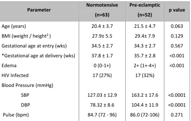
52 pre-eclamptic and 63 normotensive pregnant patients
Sample size:
115 patients
Page | 32
2.3. INCLUSION AND EXCLUSION CRITERIA
Inclusion criteria:
1) Women in their first pregnancy who had a blood pressure of at least 140mmHg systolic and 90 mmHg diastolic pressure on 2 occasions for the first time after the twentieth week of pregnancy. All women satisfying this criteria, had to have at least one plus of proteinuria on urinary dipstick.
2) Patients in whom informed consent can be obtainable
Exclusion criteria:
1) Multiple pregnancy 2) Multigravidae
3) History or clinical evidence of pre-existing hypertension/ cardiac or renal disease
2.4. METHODOLOGY
2.4.1 Data Collection Methods And Tools
Selection of suitable participants was undertaken after discussion and obtaining informed consent (see appendix 1) from antenatal patients attending Prince Mshiyeni Memorial Hospital antenatal clinic in Durban. A full history and clinical examination was performed. Baseline blood investigations included a full blood count, urea and creatinine, urates and BNP levels. Proteinuria was tested for by the dipstick method (Ames). Echocardiography (including a Tissue Doppler Imaging) was performed by an experienced sonographer at recruitment. An obstetric ultrasound was performed on all patients. Fetal wellbeing was assessed at ultrasound in terms of appropriate growth for gestation and placental sufficiency with the aid of placental Doppler flow measurements. Pregnancies were followed and timing and mode of delivery noted. Plasma BNP levels were repeated during labour (or within 24 hours post delivery). The baby at birth was assessed in respect of APGAR scores, admission to neonatal intensive care and condition on Day 7 following delivery.
Maternal serum BNP was collected within the puerperium.
Page | 33
2.4.2. Data Analysis Techniques
a) Echocardiography and Tissue Doppler
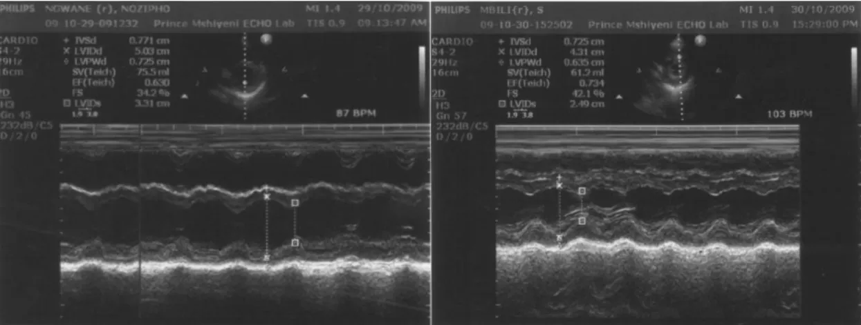
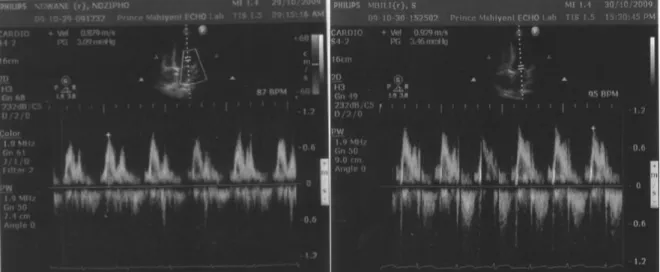
All patients had standard M-mode 2-dimensional Doppler echocardiograms performed by the same highly skilled examiner who was unaware of the patient’s blood pressure values. Doppler echocardiography was performed using a HD imaging system (Philips) with phased array transducer and an emission frequency of 3.0 megaHertz with the patient in the left decubitus position. Parameters were measured e.g. the LV end-systolic and end-diastolic dimensions, LV wall thickness, and left atrial (LA) dimensions were measured according to American Society of Echocardiography guidelines using the leading edge method (107). The left atrial volume was estimated using the biplane ellipsoid formula. The LV end-systolic and end-diastolic volumes and the ejection fraction were measured from the apical four- chamber view using the Modified Simpson’s method (108). Tissue Doppler Imaging (TDI) was performed with transducer frequencies of 1.8–3.6 MHz with minimum optimal gain as possible to obtain the best signal to noise ratio. The mitral inflow pattern was used to measure the peak early (E) and late (A) filling velocities End expiratory tissue Doppler views were obtained of the inferoseptal side of the mitral valve in pulsed mode. The angle between the Doppler beam and the longitudinal motion of the annulus was kept o a minimum in order to obtain the highest tissue velocities with the spectral pulse wave Doppler velocity adjusted to an approporiate range. The diastolic tissue velocity pattern at the annulus as used to measure the peak early (Ea) and late (Aa) tissue velocities. The ratio of the peak early mitral inflow velocity to the peak early tissue velocity (E/Ea) was calculated as an estimate of the left ventricular filling pressure. The following figures are shown of 2 randomly selected study patients.
Page | 34
Figure 1a: M-Mode recording in a normotensive patient (A) and in a pre-eclamptic patient (B).
Patient A (Normotensive) Patient B (Pre-eclamptic)
Figure 1b: Doppler recordings in a normotensive patient (A) and in a pre-eclamptic patient (B) across the mitral valve.
Page | 35
Figure 1c: Tissue Doppler recordings in a normotensive patient (A) and in a pre- eclamptic patient (B) across the lateral wall of the mitral valve annulus.
Patient A (Normotensive) Patient B (Pre-eclamptic)
b) Brain Natriuretic Peptide
Three venous blood samples were taken in plastic specimen tubes containing ethylenediaminetetraacetic acid (EDTA). Three specimens were collected from participants i.e. at the time of recruitment, intra-partum and the last specimen was collected from day 7 post delivery until the end of the peuperium. The samples were transported on ice to the laboratory where they were centrifugued. Standard handling of specimens was practised (109). The plasma was stored at −20°C and NT- proBNP was assayed in batches by standard electrochemiluminescence immunoassay (“ECLIA”) using the Modular Analytics E170 (ELECYS module) and Elecsys 1010/2010 analyzer (Roche diagnostics,). Samples from cases and controls were stored for the same duration and were handled together, identically. According to the National Committee for Clinical Laboratory Standards (NCCLS) (109), the resting BNP values considered normal for this methodology lie below 100 pg/mL. The within-assay and total precision coefficients of variation for NT- proBNP mean 208 pmol/l is 0.8% and 4.5% respectively. The reading sensitivity is <2.0-5000pg/ml (0.58- 1445pmol/L)
Page | 36
c) Fetal Ultrasound and Umbilical Artery Doppler
Routine biometrical ultrasound was performed on patients using a Toshiba (Nemio) scanner in B-Mode. In B-Mode, the image shows all the tissue that is transversed by the ultrasound probe in a 2-dimensisonal view. It is also known as B-Mode sections.
If multiple B-Mode images are watched in rapid sequence, they become ‘real time’
images (110). A low frequency (3.75 MegaHertz) curvilinear probe was utilised with patients lying supine. The sonographer was blinded to the study. Standard principles of measurements were used (111). The estimated fetal weight (EFW) was automatically calculated after measurement of the abdominal circumference (AC), femur length (FL) and biparietal diameter (BPD). BPD was measured as the distance between the parietal eminences on either side of the skull. The projection of the femur in a transverse section was identified. It was scanned at 90 degrees to contain a longitudinal section. Measurement was made from one end to the other end. The abdominal circumference was measured in a cross sectional view (as round as possible), at the level of the fetal liver. Antero-posterior and transverse diameter from the outer edge of the fetal abdomen on one side to the outer edge on the other side were used. The abdominal circumference was measured by multiplying the sum of the two dimensions by 1.57 i.e. AC= (AP diameter + T diameter)/1.57.
Umbilical artery Doppler studies were then performed on all patients at the time of the fetal ultrasound. Pulsed wave Doppler was used to measure the speed of flow in the umbilical artery. The Resistance Index (RI) was computed by measuring the peak of systole and then dividing it by the sum of measurements at peak systole and diastole. RI= systole/ (systole+diastole)l as an average over three cardiac cycles (111).