Investigations into the morphometrics, uterine tissue adaptation, maternal fluid biochemistry, heavy metal offloading and early embryonic teeth development impacting the reproductive strategy
of the female Ragged-tooth shark (Carcharias taurus)
Kristina Naidoo
B.Sc., B.Med.Sc. (Hons), M. Med.Sci
Submitted in fulfilment of the requirements for the degree of Doctor of Philosophy (Medical Biochemistry), School of Laboratory Medicine and
Medical Sciences, College of Health Sciences, University of KwaZulu-Natal
2018
This project was a collaborative undertaking with the following institutes:
New South Wales Department of Primary Industries, Port Stephens Fisheries Institute, Taylors Beach, New South Wales, Australia
KwaZulu-Natal Sharks Board, Umhlanga, South Africa
Discipline of Medical Biochemistry, School of Laboratory Medicine and Medical Sciences, College of Health Sciences, University of KwaZulu-Natal, South Africa
DECLARATION I, Kristina Naidoo, declare that:
i) The research reported in this thesis (unless otherwise indicated) is my original work, has not been submitted for any degree or examination at any other university, does not contain other persons’ data, diagrams, graphs, or other information, unless specifically being acknowledged as sourced from other persons, does not contain other persons’ writings unless being acknowledged as being sourced from other researchers. Where other written sources have been quoted then:
a) Their work has been re-written, but the information sourced has been referenced to the authors. Where their exact words have been used, their writing has been placed within quotation marks and referenced.
ii) Where I have reproduced a publication of which I am author, co-author, or editor, I have indicated in detail which part of the publication was written by myself alone and have properly referenced such publication.
iii) This thesis does not contain text, graphics or tables copied and pasted from the internet, unless specifically acknowledged and the source being detailed in the thesis and reference section
Signed
27-AUGUST-2018
Kristina Naidoo Date
ACKNOWLEDGEMENTS
Prof Chuturgoon: My deepest gratitude to you prof, for your years of mentorship and encouragement and support. Your guidance has been invaluable and strengthening.
Prof Gregory: Your guidance and support during this research endeavour was invaluable.
Thank you for all your effort and time.
Mr Cliff: Your enthusiasm for the marine field has never dwindled. Thank you for introducing me to the marine world; you created an environment devoid of boundaries. I thank you for your trust, countless conversations and guidance.
Dr Singh, Dr Ellis and Dr. Otway: Thank you for the opportunity to take on such a project.
Thank you for mentoring me.
To my parents and family: Thank you for years of unconditional support, love and unwavering understanding and patience during my studies. Your framework of hard work and commitment engrained in me ensures I go further than I think is humanly possible.
My friends (old and new): A humble thank you to each one of you for your encouragement, laughs and keeping me sane. Special thanks to Dr. M Singh, Dr. S Gounden, Dr. A Phulkdharee, Mrs B Reddy and Ms. P Dudhrajh for the motivation and laughs. To Dr. K Dhuness for your patience and unwavering encouragement to keep pushing forward. To Mr K Singh and Mrs R Sobaren Singh for all your assistance. You all kept me going. Thank you so much.
KZNSB Research Division: Mr P Zungu, Mr E Makhathi, Mr B Zungu, Mrs A Ganesan, Mr T Mlambo, Ms. Naidu, Mrs S Wintner and Dr M Dicken and Mr P von Blerk. It’s been an honour working with you all. Thank you for the assistance and advice over the years.
Department of Medical Biochemistry, MMU and Physiology at UKZN: Thank you for the support and encouragement. A special thank you to Mr. S Naidu and Mr. Bharuth, Mr P Christopher and Mrs S Singh for the in between laughs during the work; it made for a wonderful environment to work in.
LIST OF PUBLICATIONS JOURNAL PUBLICATIONS
(1) Possible maternal offloading of metals in the plasma, uterine and capsule fluid of pregnant Ragged-tooth sharks (Carcharias taurus) on the east coast of South Africa K Naidoo, AA Chuturgoon, G Cliff, SD Singh, M Ellis, N Otway, AVosloo, MA Gregory (2017). Environmental Science and Pollution Research 24 (20): 16798-16805
(2) Dentition facilitates release of encapsulated R agged-tooth (Carcharias taurus) embryos
K Naidoo, AA Chuturgoon, G Cliff, MT Ellis, NM Otway, MA Gregory, SD Singh, SL Naidu (2017). Environmental Biology of Fishes 100:1343-1354
(3) Histology of non-gravid and gravid Ragged-tooth female sharks (Carcharias taurus) on the east coast of South Africa
K Naidoo, AA Chuturgoon, G Cliff, MT Ellis, NM Otway, MA Gregory, SD Singh (2018). Prepared for Environmental Biology of Fishes
(4) Biochemistry of non-gravid and gravid Ragged-tooth female sharks (Carcharias taurus) on the east coast of South Africa
K Naidoo, AA Chuturgoon, G Cliff, MT Ellis, NM Otway, MA Gregory, SD Singh (2018). Prepared for Environmental Biology of Fishes
LIST OF PRESENTATIONS
(1) Observations of early capsule escape of the Ragged-tooth shark embryos (Carcharias taurus)
Naidoo K1, Chuturgoon AA1, Cliff G2, Ellis MT3, Otway NM4, Singh SD5, Gregory MA6.
1Discipline of Medical Biochemistry, 5Biomedical Resource Unit, 6School Life Sciences, University of KwaZulu-Natal (UKZN), 2KwaZulu-Natal Sharks Board (KZNSB), Umhlanga, Durban, South Africa; 3Gladstone Ports Corporation, Gladstone, Queensland, Australia, 4New South Wales Department of Primary Industries, Port Stephens Fisheries Institute, Taylors Beach NSW, Australia. 2nd Sharks International Conference 2014 [Poster presentation].
(2) Uterine changes in the different sexual stages of the Ragged-tooth (Carcharias taurus)
Naidoo K1, Chuturgoon AA1, Cliff G2, Ellis MT3, Otway NM4, Singh SD5, Gregory MA6.
1Discipline of Medical Biochemistry, 5Biomedical Resource Unit, 6School Life Sciences, University of KwaZulu-Natal (UKZN), 2KwaZulu-Natal Sharks Board (KZNSB), Umhlanga, Durban, South Africa; 3Gladstone Ports Corporation, Gladstone, Queensland, Australia, 4New South Wales Department of Primary Industries, Port Stephens Fisheries Institute, Taylors Beach NSW, Australia. 2nd Sharks International Conference 2014 [Poster presentation].
(3) Possible early dentition and capsule release Ragged-tooth (Carcharias taurus) embryos
Naidoo K1, Chuturgoon AA1, Cliff G2, Ellis MT3, Otway NM4, Singh SD5, Gregory MA6.
1Discipline of Medical Biochemistry, 5Biomedical Resource Unit, 6School Life Sciences, University of KwaZulu-Natal (UKZN), 2KwaZulu-Natal Sharks Board (KZNSB), Umhlanga, Durban, South Africa; 3Gladstone Ports Corporation, Gladstone, Queensland, Australia, 4New South Wales Department of Primary Industries, Port Stephens Fisheries Institute, Taylors Beach NSW, Australia. 3rd South African Shark and Ray Symposium 2015 [Oral presentation].
(4) Possible heavy metal transfer between Ragged-tooth female and embryo
Naidoo K1, Chuturgoon AA1, Cliff G2, Ellis MT3, Otway NM4, Singh SD5, Gregory MA6.
1Discipline of Medical Biochemistry, 5Biomedical Resource Unit, 6School Life Sciences, University of KwaZulu-Natal (UKZN), 2KwaZulu-Natal Sharks Board (KZNSB), Umhlanga, Durban, South Africa; 3Gladstone Ports Corporation, Gladstone, Queensland, Australia, 4New South Wales Department of Primary Industries, Port Stephens Fisheries Institute, Taylors Beach NSW, Australia. Western Indian Organisation for Marine South Africans (WIOMSA) (2015) [Oral presentation].
(5) Observation of early-staged Ragged-tooth (Carcharias taurus) embryos
Naidoo K1, Chuturgoon AA1, Cliff G2, Ellis MT3, Otway NM4, Singh SD5, Gregory MA6.
1Discipline of Medical Biochemistry, 5Biomedical Resource Unit, 6School Life Sciences, University of KwaZulu-Natal (UKZN), 2KwaZulu-Natal Sharks Board (KZNSB), Umhlanga, Durban, South Africa; 3Gladstone Ports Corporation, Gladstone, Queensland, Australia, 4New South Wales Department of Primary Industries, Port Stephens Fisheries Institute, Taylors Beach NSW, Australia. National Association of African-American Studies and Affiliates International Research Congress 2017 [Oral Presentation].
(6) Possible maternal offloading of metals in the plasma, uterine and capsule fluid of pregnant Ragged-tooth sharks (Carcharias taurus) on the east coast of South Africa.
Naidoo K1, Chuturgoon AA1, Cliff G2, Ellis MT3, Otway NM4, Singh SD5, Gregory MA6.
1Discipline of Medical Biochemistry, 5Biomedical Resource Unit, 6School Life Sciences, University of KwaZulu-Natal (UKZN), 2KwaZulu-Natal Sharks Board (KZNSB), Umhlanga, Durban, South Africa; 3Gladstone Ports Corporation, Gladstone, Queensland, Australia, 4New South Wales Department of Primary Industries, Port Stephens Fisheries Institute, Taylors Beach NSW, Australia. 4th South African Shark and Ray Symposium 2017 [Oral Presentation].
TABLE OF CONTENTS
DECLARATION...iii
ACKNOWLEDGEMENTS ... iv
LIST OF PUBLICATIONS JOURNAL PUBLICATIONS ... v
LIST OF PRESENTATIONS ... vi
TABLE OF CONTENTS ...viii
LIST OF FIGURES ... xii
LIST OF TABLES ... xvi
LIST OF ABBREVIATIONS ... xx
ABSTRACT ... xxii
CHAPTER 1 Introduction ... 1
1.1 Rationale/Aim ... 3
1.2 Hypothesis ... 3
1.3 Aims….. ... 3
1.4 Summarised study design... 4
1.5 Novelty of the study ... 5
1.6 Setup of the thesis ... 6
CHAPTER 2 Literature Review ... 7
2.1 Taxonomy and background of Carcharias taurus ... 7
2.1.1 Assessment of C. taurus ... 7
2.1.2 Geographic distribution and diet ... 8
2.1.3 Physical description and movement... 8
2.1.4 Biology ... 9
2.1.5 The importance of C. taurus and protection strategies ... 9
2.2 Reproduction ... 10
2.2.1 Migration... 10
2.2.2 Reproduction and ovulatory cycles ... 10
2.2.3 Reproductive tract ... 11
2.2.3.1 Uterus ... 13
2.2.3.2 Gestation ... 13
2.2.3.3 Uterine structure ... 14
2.2.3.4 Reproductive mode: viviparity, oophagy and adelphophagy ... 17
2.2.3.5 Uterine modifications ... 18
2.2.3.6 Uterine Fluid (UF) ... 19
2.2.3.7 Intracapsular Fluid (ICF) ... 20
2.2.3.8 Plasma ... 20
2.2.4 Reproductive endocrinology ... 21
2.2.5 Maternal offloading ... 21
2.3 Reasons for the study ... 22
2.4 References ... 24
BRIDGE CHAPTER1/2 to CHAPTER 3 ... 37
CHAPTER 3 Morphometrics of non-gravid and gravid female Ragged-tooth sharks (Cacharias taurus) on the east coast of South Africa ... 38
3.1 Abstract ... 38
3.2 Introduction ... 39
3.2 Materials and Methods ... 40
3.2.1 Sampling and criteria ... 40
3.2.2. External measurements ... 40
3.2.3 Internal measurements ... 40
3.2.4 Reproductive staging ... 41
3.2.5 Migration... 42
3.2.6 Data Analysis ... 42
3.3 Results ... 44
3.3.1 Captured Sharks ... 44
3.3.2 Morphometric relationships ... 44
3.3.3 Seasonal movements ... 46
3.3.4 Morphometrics of C. taurus females ... 47
3.3.4.1 Comparison and the correlation of length and weight ... 47
3.3.4.2 Comparison of the uterus width (UW) and its correlation to other morphometric indices ... 48
3.3.4.3 Comparison and correlation of hepatosomatic and gonadosomatic indices ... 48
3.3.5 Capsules and embryos... 50
3.4 Discussion ... 53
3.5 Conclusion ... 56
3.6 Acknowledgements ... 56
3.7 References ... 58
BRIDGE ... 61
CHAPTER 4 Histology of non-gravid and gravid female Ragged-tooth sharks (Carcharias taurus) on the east coast of South Africa ... 62
4.1 Abstract ... 63
4.2 Introduction ... 64
4.3 Materials and Methods ... 65
4.3.1 Sampling and classification ... 65
4.3.2 Uterine processing ... 66
4.4 Results ... 67
4.4.1 Shark sampled ... 67
4.4.2 Uterine transformation ... 67
4.4.2.1 Uterine wall ... 67
4.4.2.1.1 Description of the uterine wall in NGF………68
4.4.2.1.2 Description of the wall in GF……… 68
4.4.2.2 Uterine epithelial projections ... 68
4.4.2.2.1 NGF: description of the immature females (RS1) ... 68
4.4.2.2.2 NGF: description of the mature non-active females (RS2B) ... 71
4.4.2.2.3 NGF: description of the mature sexually active females (RS3)... 72
4.4.2.2.4 Measurements of the epithelium of NGF ... 74
4.4.2.2.5 Epithelium description of GF that only contain capsules (RS4) ... 74
4.4.2.2.6 GF: Description of epithelium in females with pre-hatched/encapsulated ... 75
embryos (RS5A) ... 75
4.4.2.2.7 GF: description of epithelium of females with hatched embryos during the ... 77
intrauterine cannibalistic phase (RS5C) ... 77
4.4.2.2.8 GF: description of epithelium in females with single oophagous embryo in ... 78
each uterus (RS5D ... 78
4.4.2.2.9 Measurements of the epithelium of GF ... 79
4.4.2.2.10 NGF: scanning electron microscopy of the uterine epithelium ... 80
4.4.2.2.11 GF: scanning electron microscopy of the uterine epithelium ... 81
4.5 Discussion ... 85
4.6 Conclusion ... 87
4.7 Acknowledgements ... 88
4.8 References ... 89
BRIDGE ... 92
CHAPTER 5 Biochemistry of the maternal fluid (plasma, intracapsular and uterine fluid) of non-gravid and gravid female Ragged-tooth female sharks (Carcharias taurus) on the east coast of South Africa ... 93
5.1 Abstract ... 94
5.2 Introduction ... 95
5.3 Material and Methods ... 96
5.3.1 Shark sampling and staging ... 96
5.3.2 ICF, UF and blood sampling and processing ... 96
5.3.3 Analysis... 97
5.4 Results. ... 98
5.4.1 Sampling ... 98
5.4.2 Fluid composition ... 99
5.4.2.1 Biochemical analytes (minor and major stress analytes) in the plasma of NGF and GF ... 101
5.4.2.2 Biochemical analytes (major stress analytes) in plasma, UF and ICF of GF ... 102
5.4.2.3 Biochemical analytes (minor stress analytes) in ICF and UF of GF ... 102
5.4.3 Reproductive hormones in the plasma of NGF and GF ... 105
5.4.4 Rank in plasma and UF analytes ... 106
5.5 Discussion ... 107
5.5.1 Stress analytes ... 108
5.5.2 Biochemistry analytes ... 108
5.5.3 Endocrinology ... 112
5.5.3 Post-mortem references ... 115
5.6 Conclusion ... 115
5.7 Acknowledgement ... 116
5.8 Reference ... 117
BRIDGE ... 124
CHAPTER 6 Possible maternal offloading of metals in the plasma, uterine and capsule fluid of pregnant Ragged-tooth sharks (Carcharias taurus) on the east coast of South Africa ... 125
6.1 Abstract ... 126
6.2 Introduction ... 127
6.3 Materials and methods ... 129
6.4 Results. ... 130
6.5 Discussion ... 134
6.6 Conclusion ... 137
6.7 Acknowledgements ... 138
6.8 References ... 139
BRIDGE ... 144
CHAPTER 7 Dentition facilitates the release of encapsulated Ragged-tooth shark (Carcharias taurus) embryos ... 145
7.1 Abstract ... 146
7.2 Introduction ... 147
7.3 Materials and Methods ... 148
7.4 Results…. ... 150
7.5 Discussion ... 160
7.6 Conclusion ... 163
7.7 Acknowledgement ... 163
7.8 References ... 164
BRIDGE CHAPTER 7 TO CHAPTER 8-10 ... 166
CHAPTER 8 DISCUSSION ... 167
8.1 C. taurus NGF ... 167
8.2 C. taurus GF: ... 168
8.3 References ... 172
CHAPTER 9 CONCLUSION AND PROSPECTIVE FUTURE WORK ... 175
CHAPTER 10 APPENDIX ... 177
LIST OF FIGURES
CHAPTER LEGEND PGS
CHAPTER 2
Figure 2.1 Physical Images of C. taurus. Image reference:
(http://www.oceansafrica.com/ragged-tooth-shark/)... 8 Figure 2.2 Illustration B shows the reproductive movements of C. taurus
shark. Insert A (box) illustrates the KZNSB bather protection region along the coastline. Image adapted from
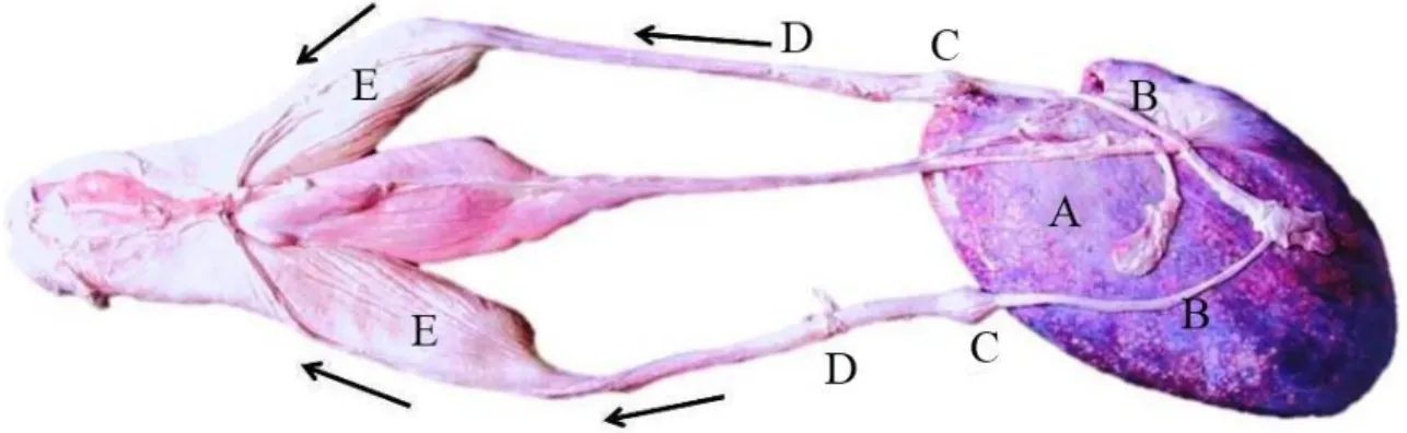
(http://www.booktravel.travel/index.php)... 12 Figure 2.3 Reproductive tract of female C. taurus. The bi-tract consists of
the A) ovary, B) anterior tube/oviduct, C) O.G, D) isthmus and E) uterus on the left and right sides of the female. Arrows (left
and right sides) represent movement of ova………. 13 Figure 2.4 C. taurus embryology. Stage I-II: A) ova encapsulated with
blastodisc, B) embryo encapsulated (EE) filled with ICF, C) a free-floating embryo (FFE) out of its capsule (15 mm TL) and Stage III: D) a longer FFE (65 mm TL) with yolk sac attached.
Stage IV: E) embryo and capsules in uterus showing evidence of intrauterine cannibalism and F) embryo/pup (166 mm TL) with yolk in its mouth. Stage V: G) oophagous embryos from the left and right uterus (535 mm and 550 mm TL). Stage VI:
H) embryo (900 mm TL) with slender features and protruding
belly filled with yolk………... 16
CHAPTER 3
Figure 3.1 The median A) HSI and B) GSI values for each reproductive stage of C. taurus females. Medians with the same superscripted letters (K-L, N-R, T-U and W) indicated a
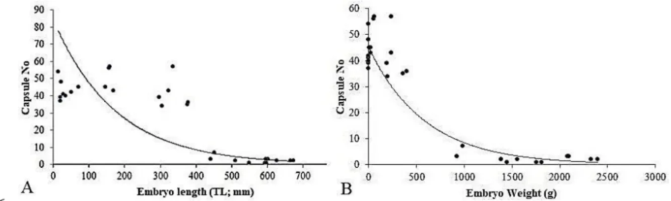
significant difference between stages (p ≤ 0.05)……… 49 Figure 3.2 Capsule and embryo length (A) and weight (B) relationships in
gravid C. taurus female sharks (RS4-RS5D)……… 50 CHAPTER 4
Figure 4.1 Figure 4.1: PAS-AB images demonstrating the difference in wall thickness between C. taurus NGF: Images A-E) are immature females (RS1), Images F-L) are mature but virgin females (RS2B) and Images M-R) are mature and mating females (RS3). Images A, F and M represents the mucosa section (i.e. epithelial and LP layer); Images B, C, G-I and M-P represents the middle wall section (i.e. MM layer); as well as Images C-D, J-K and P-Q that represents the areolar tissue layer). Images E, L and R-S represent the serosa section (Scale
Bar = 100 µm for all images)……... 69 Figure 4.2 Figure 4.2: PAS-AB stained images of uterine tissue
demonstrated the difference in the thickness of the wall between early C. taurus GF (RS4; Images A-F) and late G (RS5D; Images G-K). Images A and G represents the mucosa
section (i.e. epithelial and LP layer); Images B-C and H-I represents the middle wall section (i.e. MM layer). Images D and J also represents the middle wall section (i.e. areolar tissue layer). Images F and K represents the Serosa section (Scale Bar = 100 µm for all images)………
70 Figure 4.3 Images A and C (H&E staining) and Images B and D (PAS-
AB staining) of the immature C. taurus NGF (RS1) epithelium and wall of the uterus. Images A and B indicate the basal lamina (BL), basal cells (BC), and Lamina propria (LP) in the Mucosa (M). The SubM (submucosa) which is part of the middle wall area, is shown below the M region, as shown in Images C and D. Scale Bar = 100 µm for all
images………. 71
Figure 4.4 Images A and C (H&E staining) (Scale Bar = 100 µm) shows the epithelium and the wall of the uterus while Images B and D (PAS-AB staining) (Scale Bar = 50 µm) shows the wall of the uterus in the mature, inactive C. taurus NGF (RS2B). Image A and B shows the uterine lamellae (UL) lined by basal cells (BC) on the basal lamina (BL) and lamina propria (LP) pushing into the lumen of the uterus that forms the Mucosa (M). This formation creates troughs (T) between the UL. The submucosa (SubM) and muscularis mucosa (MM), layers that form the middle wall area also shown below the M region with small BV. Images C and D show a closer view of the UL, BC
and BL areas……….. 72
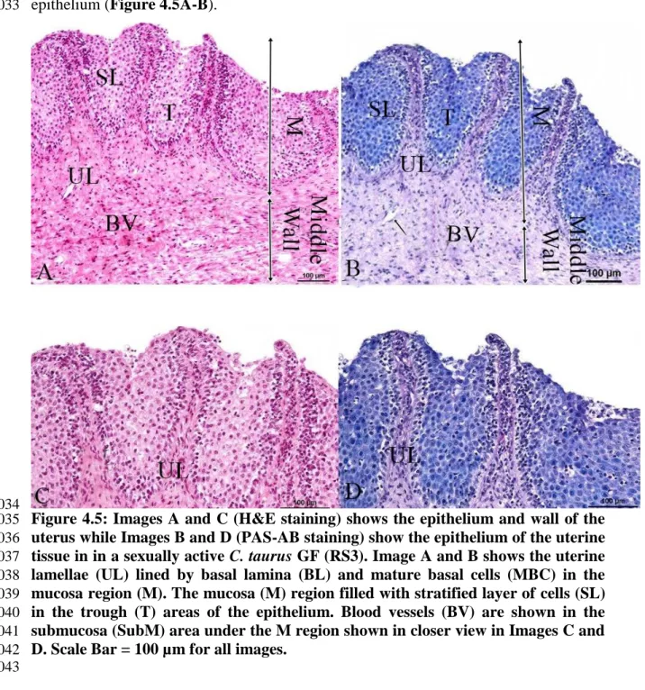
Figure 4.5 Images A and C (H&E staining) shows the epithelium and wall of the uterus while Images B and D (PAS-AB staining) show the epithelium of the uterine tissue in in a sexually active C.
taurus GF (RS3). Image A and B shows the uterine lamellae (UL) lined by basal lamina (BL) and mature basal cells (MBC) in the mucosa region (M). The mucosa (M) region filled with stratified layer of cells (SL) in the trough (T) areas of the epithelium. Blood vessels (BV) are shown in the submucosa (SubM) area under the M region shown in closer view in
Images C and D. Scale Bar = 100 µm for all images…………. 73 Figure 4.6 Image A (H&E staining) and Image B (PAS-AB staining) of
the epithelium of the uterus with the presence of blood vessels (BV) within the uterine lamellae (UL) in a sexually active C.
taurus GF (RS3) (Scale Bar = 100 µm for all
images)……… 74
Figure 4.7 Images A, C and E (H&E stains), Images B, D and F (PAS- AB) stains of the wall and uterine lamella (UL) the epithelium of the uterus in C. taurus GF (RS4). Image A and Image B (Scale Bar = 500 µm) shows the UL in the mucosa (M) and submucosa (SubM) and muscularis (MM) layers of the middle wall. Image C and Image D (Scale Bar = 100 µm) depicts the UL lined with basal cells (BC) and shows signs of arterial loops (AL) (indicated by arrows). Images E and F (Scale Bar =
50 µm) shows a closer view of the UL shows the BC stained, especially at the top end of the cell (indicated by
asterisk)…... 76 Figure 4.8 Uterine epithelium and wall of C. taurus GF (RS5A). Image A
and C (H&E stain, Scale = 500 µm) shows the epithelium and the wall of the uterus while Image B and D (PAS-AB stain, Scale = 100µm) shows the epithelium. Images A and B shows the thin, long UL extending into the lumen. Images C and D shows a closer view of the increased bud like/arterial loops
(AL), along the periphery of the
UL……… 77
Figure 4.9 Image A and Image C (H&E stain, Scale Bar = 500 µm) shows the uterine epithelium and wall and Image B and D (PAS-AB stain, Scale Bar = 50 µm) shows the epithelium of the uterus in the G C. taurus female (RS5C). Images C and D shows the increased bud like blood projections along the periphery of UL
filled with red blood cells (RBC)……… 78 Figure 4.10 Uterine epithelium of C. taurus GF (RS5D) is represented in
Images A and C (H&E stain, Scale Bar = 100 µm) and Image B and D (PAS-AB, Scale Bar = 50 µm). All images show increase in length of bud like blood projections of blood vessels (BV) filled with red blood cells (RBC) along the
periphery of each uterine lamella (UL)……… 79 Figure 4.11 SEM images of C. taurus NGF (RS1-RS3). Shows the
presence of “plate-like” structure of the uterine lamellae, mature basal cells (MBC), stratified layer of cells (SL) and secretory substance (SC) on some of the epithelium of the immature (RS1: Image A, Scale Bar = 20 µm) and Image B, Scale Bar = 10 µm), mature and inactive (RS2B: Images C and D, Scale Bar = 100 µm, Images E-F, Scale Bar = 20 µm) and Image G, Scale Bar = 1 µm) and the mature, sexually active (RS3:Images I, (Scale Bar = 100 µm and Images J-K, Scale
Bar = 20 µm) ………. 82
Figure 4.12 SEM images of uterus of C. taurus GF (RS4-RS5). Shows the uterine lamellae (UL) surrounded by micro-ridge (MR) structures that become prominent as pregnancy progresses in RS4: Images A and B, RS5A: Images C and D, RS5C: Images E and F and RS5: Images G and H. Scale Bar = 100 µm for all
images except Image G (Scale Bar = 200 µm)……… 84 CHAPTER 5
Figure 5.1 Plasma hormone levels of A) LH, B) FSH, C) progesterone and D) oestradiol trend within the plasma of all females.
Letters A-T (in the graph) represents significant p
value……… 106
CHAPTER 6
Figure 6.1 Encapsulated C. taurus embryos (EEs) found within ICF- filled, semi- translucent egg capsule. Both embryos showed the presence of eye structures as well as an attached yolk sac. The embryos measured A) 28 mm and B) 35 mm TL
respectively………. 131
Figure 6.2 The ratio of each metal in the different compartments: A) ICF:
Plasma, B) UF: Plasma and C) ICF: UF. Abbreviation: Al:
Aluminium; As: Arsenic; Cd: cadmium; ICF: intracapsular
fluid; Pb: Lead; Se: Selenium; UF: uterine fluid……….. 133 CHAPTER 7
Figure 7.1 Stereo (A-B) and SEM images (C-D) of the tooth locations, measured on the labial side, of the upper jaw (pq) (A, C) and lower jaw (Mc) (B, D) of E3 (52 mm FFE) from shark 4.
Image B) shows the naming the teeth from the centre of the
jaw moving distally……… 150
Figure 7.2 Digital image of a 34 mm FFE in shark 2. Asterisks indicate the rudimentary tooth-like structures. Arrow indicates
remnants of forming gill filaments……….. 152 Figure 7.3 Stereo image of teeth location on the left and right sides of the
A) upper jaw (pq) and B) lower jaw (Mc) of E6 (aborted 35
mm EE) from shark 4……… 154
Figure 7.4 Stereo image A) shows the dental like structures (indicated by asterisks) in E4 (36 mm FFE) from shark 4. SEM images B) show clearer dentition (indicated by the asterisks). Teeth location shown on the left and right sides of the upper jaw (pq)
SEM image……… 155
Figure 7.5 SEM image A) of E2 (46 mm FFE) from shark 4. The asterisks indicate the small peg-like structures along the gum line. SEM image B) shows teeth location the left and right sides of the upper jaw (pq). The lower jaws (Mc) did show a few teeth but
tissue appeared damaged therefore not shown……… 156 Figure 7.6 Stereo image A) and SEM images B) and C) are based on the
E1 (50 mm EE) from shark 4. The asterisks in stereo image A) shows the embryo peg like tooth structures. The arrow in A) also indicates the presence of gill filaments. SEM images (B-
C) shows teeth……… 157
Figure 7.7 SEM images of E3 (52 mm FFE) from shark 4. Image (B) is an enlargement of the boxed area 1 in Image A. Image C is an enlargement of the boxed area 2 in Image A. Asterisks indicates tooth- like structures. Teeth location on the upper
(pq) and lower (Mc),……….. 158
CHAPTER 8
Figure 8.1 Illustration of hormone trends for progesterone (dashed blue curve) and oestradiol (solid curve) with three possible ovulatory periods where rate of ovulation increases (indicated by arrows) through the non-gravid (NGF) and gravid (GF) stages. Image adapted from reproductive cycle illustration in
Koob and Callard (1999)……… 171 APPENDIX A Redness of the gill structures indicates freshness of the
shark……….. 177
APPENDIX B Depth of the recession of both eyes indicates freshness of the
shark………. 177
APPENDIX C Dissection Form………. 178
APPENDIX D Capsule Form……… 180
APPENDIX E Key Form and Uterus histology drawing……… 181, 182
LIST OF TABLES
CHAPTER LEGEND PGS
CHAPTER 2
Table 2.1 Chondrichthyan reproductive modes………... 17 CHAPTER 3
Table 3.1 A summary of the reproductive stage definitions used to divide C. taurus females into their respective maturity stages. A more detailed description can be found in APPENDIX . Description of RS5A-RS5D is an extension of the embryological stages in Gilmore et al. (1983) ………...
43 Table 3.2 Morphometric relationships of length, weight, hepatosomatic
index (HSI) and gonadosomatic index (GSI) in C. taurus NGF (RS1-RS3) and GF (RS4-RS5) based on the larger KZNSB
database (APPENDIX )……….. 45
Table 3.3 Morphometric relationships of length, weight, HSI and GSI for C. taurus embryos <100 mm TL and > 100mm TL.
Measurements based on this study and KZNSB database……. 46 Table 3.4. Seasonality and sample size of catches of C. taurus NGF and
GF……….. 47
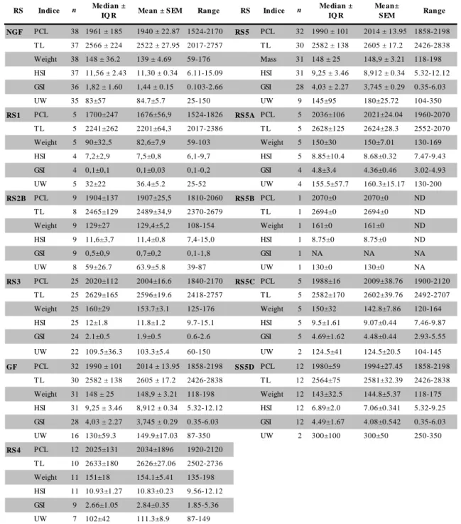
Table 3.5 Median ± IQR; Mean ± SEM and Ranges for the PCL, TL (mm), Weight (kg), HSI (%), GSI (%), UW (mm) of all C.
taurus females captured in this study………. 51 Table 3.6 The capsule and embryo data from gravid C. taurus females
(RS4-RS5D)……… 52
CHAPTER 4
Table 4.1 Median measurements (µm) of the uterine epithelium and wall
of C. taurus NGF (RS1-RS3) and GF (RS4-RS5D) stages……. 80 CHAPTER 5
Table 5.1 Methods used for measuring analytes in the plasma of all
females C. taurus females……….. 98
Table 5.2 Descriptive statistics (Median ± IQR; Mean ± SEM and
ranges) for the NGF and GF C. taurus maternal plasma……… 100 Table 5.3 Descriptive statistics (Median ± IQR, Mean ± SEM and
ranges) for the stress analytes in the plasma, UF and ICF in GF
(C. taurus)………. 102
Table 5.4 Descriptive statistics for the UF analytes in all C. taurus
GF………... 103 Table 5.5 Descriptive statistics for the analytes found in the ICF of
RS5A females (C. taurus) ………. 104
Table 5.6 Rank of analytes between the plasma and UF in C. taurus
GF………... 107
Table 5.7 Comparison of immature and mature NGF C. taurus [i.e., Ragged-tooth female sharks (dead in this study) vs. Sand tiger
females (live from Otway (2015) ……….……. 111
Supporting Tables for CHAPTER 5
APPENDIX F Description of sexual staging of C. taurus NGF (RS1-RS3) and GF (RS4-RS5D). Data originated from the KNZSB
database……… 183
APPENDIX G Descriptive statistics (Median ± IQR, Mean ± SEM and range) of plasma analytes in C. taurus NGF sub-groups
(RS1 and RS2B)……… 190
APPENDIX H Descriptive statistics (Median ± IQR, Mean ± SEM and ranges) of plasma in C. taurus NGF sub-group
(RS3)………. 191
APPENDIX I Descriptive statistics (Median ± IQR, Mean ± SEM and range) of the plasma analytes in C. taurus GF sub-groups
(RS4 and RS5)………. 192
APPENDIX J Descriptive statistics (Median ± IQR, Mean ± SEM and range) for the plasma analytes in GF C. taurus sub-groups
(RS4 and RS5)……….. 193
APPENDIX K Descriptive statistics (Median ± IQR, Mean ± SEM and range) for the UF analytes in C. taurus GF sub-groups (RS4
and RS5)……….. 194
APPENDIX L Descriptive statistics (Median ± IQR, Mean ± SEM and range) for the UF analytes in C. taurus GF sub-groups
(RS5A and RS5B)………. 195
APPENDIX M Descriptive statistics (Median ± IQR, Mean ± SEM and range) for the UF analytes in GF C. taurus sub-groups
(RS5C and RS5D)………. 196 CHAPTER 6
Table 6.1 Details of the three captured C. taurus sharks (shark 1- shark 3) examined and their respective encapsulated
embryos (EEs) and floating embryos (FFEs)……….. 130 Table 6.2 The median and mean concentrations [mg/L] of metals in
the three fluid compartments i.e., maternal plasma, ICF and
UF of three C. taurus……… 132
Table 6.3 Tabulated Spearman rs and p values for metal correlations in the ICF of embryo length and weight against metal
concentration……… 133
CHAPTER 7
Table 7.1 The total length (TL), sex (S), status (EE vs. FFE) and location (right or left uterus or aborted) of each embryo found in four pregnant C. taurus (shark 1- shark 4).The total length (TL), sex (S), status (EE vs. FFE) and location (right or left uterus or aborted) of each embryo found in four
pregnant C. taurus (shark 1-shark 4)………. 152 Table 7.2 Median measurements (µm) of the uterine tissue (the
epithelium and wall) of C. taurus NGF (RS1-RS3) and GF
(RS4-RS5D) stages………... 153
Table 7.3 The Mean, Standard Deviation and Range of the relative percentages of elements calcium (Ca), phosphorus (P), fluorine (F) and oxygen (O) that were analysed on four separate areas of the enameloid layer (SLE layer) of each
tooth on the upper and lower jaws of E1-E6…………... 159
LIST OF ABBREVIATIONS Species
A. vulpinus Alopias vulpinus
(common name: Thresher shark)
M.
mustelus
Mustelus mustelus (Smooth-hound shark) C. carcharias Carcharodon Carcharias
(Great white shark)
M.
antarcticus
Mustelus antarcticus (Gummy shark) C. obscurus Carcharhinus obscurus
(Dusky shark)
O. ornatus Orectolobus ornatus (Wobbegong ark) C. taurus Carcharias taurus
(Ragged-tooth shark)
R. longurio Rhizoprionodon longurio (Pacific sharpnose shark) G. galeus Galeorhinus galeus
(School shark)
S. canicula Scyliorhinus canicula (Small-spotted catfish) I. oxyrinchus Isurus Oxyrinchus
(Shortfin mako shark)
S.
acanthias
Squalus acanthias (Spiny dogfish shark) L. nasus Lamna Nasus
(Porbeagle shark) Abbreviations
AB+ Alcian Blue positive NSW DPI New South Wales,
Department Primary Industries (in Australia)
Al Aluminium NW North-Western
Al arteriole loops O. G oviducal gland
As Arsenic O Oxygen
AU artificial uterus OD oesteodentine layer
BC basal cells p Phosphate
BD Becton Dickson (Company) p significant value set at
< 0.05
BL Basal Lamina PAS Periodic Acid Schiff
BV blood vessels PAS+ Periodic Acid Schiff
positive
Ca Calcium PAS&AB Periodic Acid Schiff and
Alcian Blue
Capn capsule sample size Pb Lead
Cd cadmium PCL precaudal length
DAAD Deutscher Akademischer Austauschdienst
ppm parts per million
ECF extra capsular fluid pq paratoquadrate
EDX Energy dispersive X-ray r2 trendline value
EI enameloid inner r correlation value
(Spearman/Pearson)
EE encapsulated embryos RBC red blood cell
E1-E6 embryo 1- embryo 6 RT room temperature
e.g., example S serosa
ESEM Environmental SEM S serosa
Fn female sample size SA South Africa
FE field emission SC secretory substance
FFE free-floating embryos Se Selenium
g/G Gram/Gauge SEM scanning electron microscopy
GF Gravid females SD standard deviation
GSI gonadosomatic index SDD Silicon drift detector
g/L grams/litre SL stratified layer of cells
H&E Haematoxylin and Eosin SLE shiny layer enameloid
Hg Mercury SubM Submucosa
HSI Hepatosomatic index T trough/s
ICF intracapsular fluid TH tooth height
ICP-MS Inductively-Coupled Plasma Mass Spectrophotometry
TL total length ID identification number for animal at
KZNSB TEM Transmission Electron
Microscopy
IQR interquartile range TW tooth width
IUCN International Union for Conservation of Nature
µM micrometre
in-utero in the uterus U unidentified
in vitro in a glass UC upper caudal
kg kilogram UF uterine fluid
km kilometre UKZN University of KwaZulu-
Natal
KZN KwaZulu-Natal UL uterine lamella/e
KZNSB KwaZulu-Natal Sharks Board USA United States of America
LM light microscopy U/L units/litre
LP Lamina Propria UW uterus width
M male/Mucosa/molar umol/L micromole/litre
MBC mature basal cells Vtg Vitellogenin
Mc Meckels cartilage VP variable pressure
mg/L milligram/litre v/v volume/volume
ml millilitre x mean
MMU Microscopy and Microanalysis Unit Vtg Vitellogenin
MM Muscularis Mucosa VP variable pressure
mmol/L Millimole/litre v/v volume/volume
mIU/ml milli-international units/ millilitre x mean
n sample size xd median
NA not applicable
NGF non-gravid females I-VI Embryology stage one-six
of C. taurus NISD Nikon instrument documentation 0C celsius degree
nmol/L nanomole/litre % percentage
No number Xg centrifuge rotations per
minute RS Reproductive Stage/s Ca5(PO4)3 Fluoroapatite
ABSTRACT 1
2
“Vulnerable” status of ragged-tooth sharks (Carcharias taurus) in South Africa caused 3
by overexploitation, late maturity and low fecundity suggests an intervention to increase 4
the size of this population is needed. Achieving this will require an understanding of all 5
aspects linked to this species maternal-embryonic relationship.
6 7
Morphometric relationships, uterine histology and maternal fluid biochemistry were 8
assessed in C. taurus through all the respective reproductive stages (RS) from non- 9
gravid (immature to mature-sexually active; RS1-3) to gravid (i.e. only capsules found;
10
RS4 or capsules and pups found; RS5A-5E) females. Examination of metals in the 11
maternal fluids and embryonic dentition were only examined in early-staged gravid 12
females (i.e. RS5A).
13 14
Haematoxylin/Eosin and Periodic Acid Schiff-Alcian Blue stains in conjunction with 15
light microscopy was used to assess the uterine epithelium and wall while scanning 16
electron microscopy further evaluated the epithelium. These techniques revealed an 17
increase of the uterine lamellae (folds) protruding into the lumen lined with micro- 18
ridges containing blood vessels. The close proximity of blood vessels to the lumen filled 19
with uterine fluid and the decrease in wall thickness as pregnancy progressed suggests 20
an adaption for the exchange of respiration and osmoregulation in the developing 21
aplacental embryos. Although there is no evidence for uterine secretion through 22
structural adaptations, the female supports the embryos nutritional requirement through 23
embryonic tissue (intrauterine cannibalism) and yolk (oophagy) provisions. The pivotal 24
interplay of the liver and ovary, during vitellogenesis, that impact on yolk formation 25
was evident during the morphometric evaluation of hepatosomatic and gonadosomatic 26
indices. Length, weight, uterine width, capsule production and migration trends of the 27
females as well as length and weight relationship of the embryos were tabulated.
28 29
Reproductive hormones, assessed in maternal fluids (i.e. plasma (in RS1-5D females), 30
uterine fluid (in RS4-5D females) and intracapsular fluid (in RS5A females), showed 31
that follicle-stimulating-, progesterone- and oestradiol hormones were responsible for 32
promoting vitellogenesis and encapsulation which led to three main stages where the 33
rate of ovulation increases in the female. Clinical biochemistry analysers confirmed the 34
composition and concentration of biochemical analytes in same maternal fluids, which 35
were found to be higher in the plasma.
36 37
Finally, heavy metals were found to be present in all three fluids, but found highest in 38
the plasma, using inductively coupled mass spectrophotometry. In addition, variable 39
pressure (VP) SEM confirmed the dental composition of the embryonic teeth found in 40
the jaws of some embryos that appeared to escape encapsulation earlier than previously 41
documented. It would appear that the embryos are creating adaptive ways to survive the 42
intracannibalistic stage (RS5C) by escaping encapsulation early. However, the presence 43
of heavy metals in the maternal fluids that surround the embryos could compromise 44
their development over time; creating concern for a species that is Vulnerable.
45 46
This study, which serves as the first detailed analysis of the maternal-embryonic 47
relationship, may serve as areas to model in forthcoming programmes aimed at 48
increasing the numbers of this species.
49
CHAPTER 1 50
51
Introduction 52
53
Carcharias taurus (C. taurus) is a lamnoid shark species that is currently classified as 54
“Vulnerable” globally including South Africa (SA) [1-3]. Sub-populations have been 55
found to be Critically Endangered in Australia (Queensland) [2] and Southwest Atlantic 56
[41] and Near Threatened in Western Australia [2]. The trend of the Critically 57
Endangered C. taurus subpopulation is currently in decline [2,4] while other 58
populations remain unknown [3]. This species may be better known by their common- 59
names: Ragged-tooth (i.e., “raggie”) (SA), Grey nurse (Australia) and Sand tiger 60
(America). Local [5,6] and international [2,3,7] conservation and management 61
programmes have had minimal success in population recovery. Additional strategies 62
such as the application of breeding programmes [8-10] is required to increase that rate 63
of recovery; which may otherwise take decades to recover [11-15]. Understanding these 64
species biology, migration and reproductive behaviour would be crucial to the latter.
65
This C. taurus, reaches sexual maturity at six years [16], and is known to have one of 66
the lowest fertility rates among chondrichthyans [17,18], due to its unique consistent 67
practice of in utero cannibalism (/embryophagy: i.e., “eating siblings in uterus”) that 68
occurs biennially, during the early phases of a 9-12 month gestation period [11,19-22]. It 69
has, however, been recently reported that adelphophagy occasionally occurs in another 70
lamnoid Isurus oxyrinchus (I. oxyrinchus) [23]. Low fecundity, easy accessibility, 71
overfishing (industrial or sport fishing) and anthropogenic activities [12] remain a 72
detriment to their declining population regardless of the great steps to implement 73
protection acts [3], recovery plans [2] and marine protected areas [7]. Their declining 74
number is further impacted by high-levels of heavy metal environmental exposure from 75
anthropogenic activity that creates additional concern to the development of an already 76
limited progeny [24,25]. A naturally low breeding rate with a slow rebound potential to 77
grow indicates that this population may require several decades to recover [11-15,20].
78 79
Studies to date have directly or indirectly contributed to the knowledge of C. taurus 80
reproductive biology. Some of these studies looked at their reproductive behaviour, 81
migratory distribution [5,12,20], general reproductive endocrinology [26], aplacental 82
viviparous mode of reproduction that includes oophagy and intrauterine cannibalism 83
[17,21,27-30], extensive intrauterine embryology [17,21,27,31] as well as matrotrophic 84
uterine modifications in elasmobranchs [27,29-33]. Understanding the maternal- 85
embryonic relationship in C. taurus requires understanding how the female 86
physiologically supports her progeny until birth. This relationship still requires 87
clarification and assessment.
88 89
The current literature shows some ambiguities in Gilmore’s (1993) description of 90
epithelium of C. taurus as well as an extension of this species tissue presented by 91
Hamlett and Hysell (1999). Gilmore (1993) inferred images from a similar reproductive 92
mode in the lamnoid shark I. Oxyrinchus. Hamlett and Hysell (1998) did not examine 93
the uterus from the different reproductive stages of the species. In addition, biochemical 94
analysis has only been done on the plasma of C. taurus non gravid females (NGF). No 95
biochemical investigations has been reported on the blood, intracapsular (ICF) and 96
uterine fluid (UF) of C. taurus gravid females (GF)[34]. The profile of plasma 97
hormones of both NGF and gravid females (GF) as well the UF and ICF of GF has 98
never been documented in C. taurus. This study was undertaken to clarify the existing 99
ambiguity and extending the current information on the uterine tissue [32] and fluid 100
biochemistry [34] of C. taurus females through the different NGF and GF reproductive 101
stages. Reproductive stages were determined by maturity indicators (i.e. set of 102
measurements based on the female’s biology) that was divided into two main groups i.e.
103
NGF and GF. The NGF reproductive stages included the immature, immature-inactive, 104
and mature-active females. The GF reproductive stages consisted of sharks pregnant with 105
capsules alone or females pregnant with pre-hatched, post-hatched, intrauterine 106
cannibalistic or a single embryo per uterus in addition to the capsules.
107 108
The knowledge gained from the histology and fluid biochemistry of this shark, together 109
with additional findings in relation to their embryo survival, may be invested into a 110
possible breeding intervention program postulated by the New South Wales (NSW) 111
Department of Primary Industries (NSW DPI) [9]. This information will also serve as 112
the first documentation for C. taurus to better understand the reproductive strategy and 113
better management for both wild and captive species.
114 115
1.1 Rationale/Aim 116
117
To document the changes in the epithelium and wall of the uterine tissue as well as the 118
biochemical composition (including associated reproductive hormones) of the plasma 119
(and where possible the UF and ICF) through all the reproductive stages (i.e. from 120
immature to pregnant) of the developmentally classified C. taurus females in SA.
121
This information would assist in understanding the maternal-embryonic relationship in 122
C. taurus thereby advancing the elasmobranch reproductive and clinical understanding of 123
this species. This information is necessary to institute a breeding intervention program 124
aimed to increase and promote successful breeding by growing C. taurus embryos 125
within an artificial uterus (AU) that was previously postulated by the NSW DPI to 126
increase their C. taurus sharks [9]. Scientists at NSW DPI have already succeeded in 127
creating AU technology for shark development with the Orectolobus ornatus (O.
128
ornatus) embryos [8]. The hope is that the knowledge from studies investigating the 129
physiology of C. taurus (including this study) can be incorporated into existing AU 130
technology for shark development to assist with C. taurus population recovery in 131
Australia. Such an undertaking will require a fundamental knowledge of the maternal 132
environment to create proper in vitro AU for the propagation of C. taurus embryos. This 133
study provides an insight into the creation of such an AU.
134
1.2 Hypothesis 135
136
This C. taurus females support the growth of their young through transformation of the 137
uterine structure and associated fluids (i.e., plasma, UF and ICF).
138
1.3 Aims 139
140
This study aims to document the uterus and fluid (blood, ICF, UF) in the different 141
reproductive stages of C. taurus females. To clarify the extent of maternal support to the 142
embryos, the following objectives were investigated:
143
a) Record and confirm, using KwaZulu-Natal sharks board (KZNSB) records the 144
morphometric measurements (i.e., body, reproductive organs and reproductive- 145
associated structures) to determine any changes between the NGF and GF stage.
146
b) Use Light and Scanning electron microscope to examine changes in the uterine 147
epithelium and wall in the NGF and GF stages 148
c) Document the composition of plasma, with any significant trends in the analytes of 149
the NGF and GF stages 150
d) Document the composition of the ICF and UF, with any significant trends in the 151
analytes of the GF stages 152
e) Review any changes in the maternal uterus and fluid biochemistry (blood, ICF, UF) 153
that could be necessary for embryo survival.
154
f) Document additional findings during the project that may have an impact on the 155
development or survival of C. taurus embryos such as presence of heavy metals 156
and the dentition of these embryos 157
1.4 Summarised study design 158
159
This study examined NG and GF C. taurus sharks. The studied migration pattern of 160
these females and unfortunate situation of many of them being caught yearly in the 161
KZNSB bather nets (under KZN 2008 Act) [11,35,36], created a unique sampling 162
opportunity at KZNSB (Ethic no 076/10/Animal). This unfortunate opportunity allowed 163
SA to contribute to the physiological information of these sharks [21,37,38]. To the best 164
of our knowledge, the two subpopulations of C. taurus are stable along the KwaZulu- 165
Natal provincial coastline of SA [11,39,40].
166 167
The study design of investigating the nine female reproductive stages, recruited into the 168
study, was constructed on the reproductive staging (RS) that was assigned to each C.
169
taurus female. The staging was based on maturity indicators (measurements associated 170
with the female); a system used at KZNSB for all captures. The reproductive stages 171
(RS) from NGF (immature to mature-sexually active; RS1-3) to GF (i.e. only capsules 172
found; RS4 or capsules and pups found; RS5A-5D) sharks were assessed in this study.
173
Detail of the criteria related to the reproductive staging, freshness evaluation, overall 174
dissection process and sampling requirements are addressed in CHAPTER 3 for all 175
females. This chapter compared the morphometrics data of all sampled females (i.e., 176
length, weight, uterine width (UW), capsule production, HSI and GSI). The histology of 177
uterus was addressed in CHAPTER 4 for all reproductive stages of the females. The 178
wall and epithelium of the uterus was stained with Haematoxylin/Eosin and Periodic 179
Acid Schiff-Alcian Blue and analysed using light microscopy. The epithelium was 180
further analysed using scanning electron microscopy. CHAPTER 5 addressed the 181
analysis of the female’s plasma, using a clinical analyser that determined the 182
concentration and composition of biochemical analytes in all NGF and GF. Analytes 183
were also determined for the UF and ICF in respective GF sharks. The ICF, found in 184
only three early-staged GF (i.e., RS5A), together with the plasma and UF from these 185
females, were also analysed, using inductively coupled plasma mass spectrophotometry 186
to determine the presence of heavy metals in CHAPTER 6. Finally, in CHAPTER 7, C.
187
taurus shark embryos from these three females had their dental composition assessed 188
using Variable Pressure SEM.
189
190
1.5 Novelty of the study 191
The morphological, histological and fluid biochemical analysis of C. taurus female is 192
well adapted to provide the nurturing environment to develop her embryos to full term 193
predators; with the possibility that the embryos are creating adaptive ways to survive. A 194
concerning factor, is the presence of heavy metals, which the females appear to be 195
offloading onto her progeny that could lead to devastating consequences over time for 196
an already vulnerable species. It also reveals all aspects of the female’s reproduction that 197
needs to be considered when attempting to apply a physiological intervention to increase these 198
species numbers.
199
The unique contribution of this study to the current literature of C. taurus females is as 200
follows:
201
a) description of all NGF and GF reproductive stages 202
b) description and confirmation of the changes in the uterine epithelium in all 203
reproductive stages 204
c) description of the changes in the uterine wall in all reproductive stages 205
d) composition and trends of the plasma and UF in GF stages 206
e) composition of the ICF in GF presenting with pre-hatched embryos 207
f) comparison of reproductive hormone changes through all stages 208
g) metal offloading/transference into the maternal fluid 209
h) early development of embryonic teeth in C. taurus embryos 210
211
1.6 Setup of the thesis 212
213
Due to the length of information for each section, it was decided that it would be best to 214
compare the NG and GF in individual chapters dealing with morphometrics, histology 215
and biochemistry themes. References are made to different chapters within the text 216
where necessary. A detailed explanation of the possible maternal supportive role of C.
217
taurus female extends throughout this thesis as follows:
218
CHAPTER 1: a summarised breakdown of literature and flow of the thesis 219
CHAPTER 2: Literature review 220
CHAPTER 3: focuses on the morphometrics of the NGF and GF 221
CHAPTER 4: describes the uterine wall and epithelium changes in the NGF and 222
GF stages 223
CHAPTER 5: investigates the composition and concentration of the plasma 224
analytes and trends in the biochemistry of the NGF and GF stages. The 225
compositions and concentration of analytes in the ICF and UF from GF with pre- 226
hatched embryos are also reported here.
227
CHAPTER 6: reports on the presence of heavy metals in the plasma, ICF and UF of 228
GF with pre-hatched embryos.
229
CHAPTER 7: describes some embryos, found in CHAPTER 6, that were found 230
released from their capsules at a far smaller size than previously recorded. Dental 231
structures were observed and reported in these C. taurus embryos 232
CHAPTER 8: Discussion 233
CHAPTER 9: Conclusion and proposed future work 234
CHAPTER 10: Appendix 235
Bridges appear between the chapters to guide the reader between the chapters 236
237
CHAPTER 2 238
239
Literature Review 240
241
2.1 Taxonomy and background of Carcharias taurus 242
243
Carcharias taurus (C. taurus) (formerly known as Eugomphodus taurus, Odontaspis 244
taurus) is one of fifteen shark species in the family Odontaspididae, of the order 245
Lamniformes. This shark species is known as Ragged-tooth (SA), Grey-nurse 246
(Australia) and Sand tiger (United States of America). This elasmobranch species is one 247
of the most widely investigated [11,12,22,26]. C. taurus held in captivity lives for 13-16 248
years, with wild species predicted to live longer. Evidence for longer longevity was provided by 249
tagging data from a mature C. taurus female captured in Zinkwazi, (Durban, SA) 20 years 250
after being tagged in St. Lucia (Durban, SA) [41]. It is the most widely kept large shark 251
in aquariums around the world due to its adaptability and tolerance for captive habitats 252
and conditions[42].
253 254
2.1.1 Assessment of C. taurus 255
256
There has been a huge decline, in some areas, in shark numbers over the years [43,44].
257
Cacharias taurus (in Australia) was the first shark species to be protected by law.
258
Globally, this species has been assessed as Vulnerable by the International Union for 259
Conservation of Nature’s (IUCN) Red List of threatened species [3]. The risk status of 260
C. taurus population does show variation at different parts of the world. The population 261
along the east coast of Australia and southwest Atlantic are “Critically Endangered”
262
[2,4], the western Australian subpopulation are “Near Threatened” and South African 263
population is listed as “Vulnerable”[1,2]. Critically Endangered C. taurus subpopulation 264
trend is currently in decline [2,45], the Western Australian subpopulation remains stable 265
with other populations /subpopulation requiring further investigation [3].
266
2.1.2 Geographic distribution and diet 267
268
This demersal species is known to have an inshore, reef associated distribution mainly 269
in subtropical to warm temperate waters, except for the eastern Pacific, around main 270
continental landmasses [20,38]. The known cosmopolitan ocean distribution includes 271
Western and Eastern Atlantic, Western Indian Ocean (except for Madagascar), Red Sea 272
and the Western Pacific (except for New Zealand) [11,38]. They are often sighted in 273
shallow waters such as shallow bays, surf zones, coral or rocky reefs. Stomach content 274
analyses have shown that these sharks feed largely on teleosts, crabs, lobsters and small 275
elasmobranchs, although cephalopods are also eaten [37,38,46].
276 277
2.1.3 Physical description and movement 278
279
This species, C. taurus shark, is a slow-moving shark that has a stout body, cream to brown in 280
colour with large, irregular darker spots which fade with age (Figure 2.1). It has a 281
pointed snout with a mouth that bears razor-like teeth that gives this animal a menacing 282
look and false reputation of being dangerous. Females are generally longer than males.
283
A distinguishing feature of this shark is that the anal and both dorsal fins are the same 284
size. The tail has a long upper lobe and a shorter lower lobe.
285
286
Figure 2.1: Physical Images of C. taurus. Image reference:
287
(http://www.oceansafrica.com/ragged-tooth-shark/) 288
289
Upper tail Second Dorsal fin First Dorsal fin
Lower tail lobe Anal Fin
Pelvic fins
Pectoral fins