The microbiological assessment of a biofiltration system in KwaZulu-Natal (South Africa) treating
borehole water containing Mn (II) and Fe (II)
Lorika Selomi Beukes
Submitted in fulfilment of the academic requirements for the degree of
Master of Science in Microbiology
College of Agriculture, Engineering and Science, School of Life Sciences, Discipline of Microbiology, University of KwaZulu-Natal,
Pietermaritzburg Campus
28 May 2013
ii
Declaration 1 – Plagiarism
1. I hereby certify that the contents of this thesis, unless specifically indicated in the text, are the result of my own findings.
2. These findings have not been submitted previously in its entirety or in part for obtaining any other qualification.
3. This thesis does not contain any other individual’s data, images, graphs or written work, unless specifically acknowledged in the text. Where written work has been obtained from other sources, the text has been re-written and the general information obtained from these sources has been referenced. Where text has been used directly from other sources, the text has been placed within quotation marks and referenced.
4. This thesis does not contain graphics or tables copied from books, journals or the internet, unless specifically acknowledged and the source being detailed in the thesis and in the reference sections of each chapter.
_____________________ _____________________
Lorika Selomi Beukes Date
iii
Declaration 2 – Publications
Details of publications that form part and/or include research presented in this thesis:
Publication 1 (Chapter 3):
Beukes, L. S., and Schmidt, S., (2012). Isolation and characterization of a manganese oxidizing bacterium from a biofiltration system for the treatment of borehole water in KwaZulu-Natal (South Africa). Eng. Life Sci. 12: 544-552.
Additional data to this chapter is provided in the form of an appendix to chapter 3.
_____________________ _____________________
Lorika Selomi Beukes Date
Ms Lorika Beukes performed the experimental work and drafted the manuscript of the above publication.
_____________________ _____________________
Prof Stefan Schmidt Date
iv
Table of Contents
Section
Page
List of Tables v
List of Figures vi
Abstract xii
Acknowledgements xv
Chapter 1 General Introduction 1
Chapter 2 Literature Review 6
Chapter 3 Isolation and characterization of a manganese-oxidizing bacterium from a 54 biofiltration system for the treatment of borehole water in KwaZulu-Natal (South Africa)
Chapter 3 (Appendix) MALDI-TOF MS analysis of Acinetobacter sp. LB1 66 Chapter 4 Do microorganisms enhance Fe (II) oxidation in the Nottingham road 76
biofiltration systems at circumneutral pH?
Chapter 5 The detection of active biofilms and the assessment of the bacterial 107 diversity in a biofiltration system treating borehole water in KwaZulu-Natal (South Africa)
Chapter 6 Concluding remarks 137
This thesis represents a compilation of manuscripts where each chapter is an individual entity and some repetition between chapters has therefore been unavoidable.
v
List of Tables
Section Page
Chapter 3 – Appendix
Table 1: Score values generated on the Bruker Microflex MALDI-TOF MS bench 68 top system for Acinetobacter sp. LB1 and E. coli (control) using whole cells in comparison to the best matched organism in the database.
Table 2: Description of the score ranges generated on the Bruker Microflex 68 MALDI-TOF MS bench top system.
Table 3: Main spectra projections for cell extracts of Acinetobacter sp. LB1 in 69 relation to other species within the same genus from the literature.
Chapter 5
Table 1: Partial 16S rRNA gene sequences representing OTU clones established 120 from the biofilm material on the filter matrix and the closest related taxa from RDP for each phylotype.
vi
List of Figures
Section Page
Chapter 2
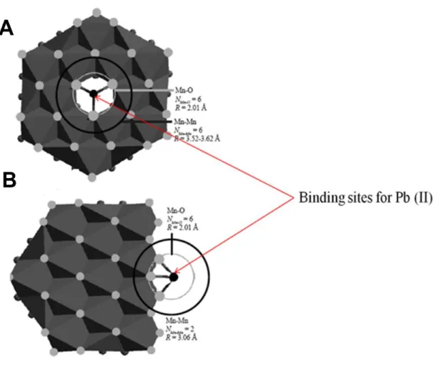
Figure 1: (Adapted from Spiro et al., 2008) Binding of Pb (II) ions onto two 12 crystal structures of bacterial manganese oxides. (A) triple-corner-sharing and (B) double-edge-sharing inner sphere surface complexes formed respectively above cation vacancy sites and at sheet edges.
Figure 2: (Adapted from Spiro et al., 2008) Crystal structure of the bacteriogenic 14 Mn oxide - Buserite. (A) Sheet structure of Buserite, with hexagonal symmetry, highlighting a monoclinic unit cell (a-b). (B) Sheet structure (a-b), illustrating di-μ-oxo bridging between neighboring Mn atoms. Manganese atoms illustrated as large black circles and oxygen atoms as the grey smaller circles.
Figure 3: (Adapted from Haferburg and Kothe, 2007) Overview of the four 32 microbial metal resistance mechanisms. (X) – Cell constituents interacting with metal cations, (M) - Metal cation.
Chapter 3
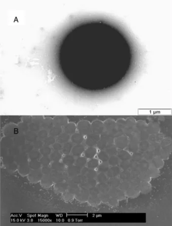
Figure 1: TEM (A) and ESEM (B) image of the manganese oxidizing isolate 58 Acinetobacter sp. strain LB1. (A) Negatively stained single cell. (B) Sputter coated cell aggregate.
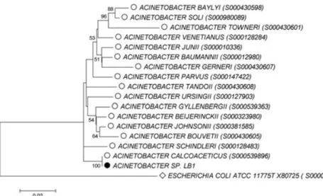
Figure 2: Phylogenetic affiliation of the strain LB1 (black circle) based on the 59 comparison of its 16S rRNA gene sequence with 16 selected 16S rRNA gene sequences for type strains of the genus Acinteobacter (open circles). The alignment of selected sequences and the construction of the tree are specified
vii in section 2. The scale bar represents two estimated changes per 100 nucleotides. Numbers shown at nodes indicate calculated bootstrap values (only values > 50% are shown). Escherichia coli was used as an out-group (open diamond).
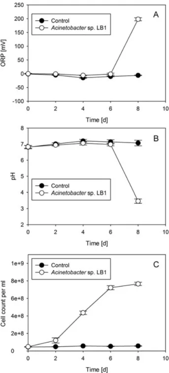
Figure 3: Impact of Mn (II) oxidation on ORP (A) and pH (B) and correlation with 60 growth (C) over time. Data shown are the means of experiments performed in triplicate in the presence of actively growing cells of the manganese-oxidizing strain Acinetobacter sp. strain LB1 (open symbols) in comparison to controls (heat-inactivated cells, filled symbols). Error bars indicate the standard deviation.
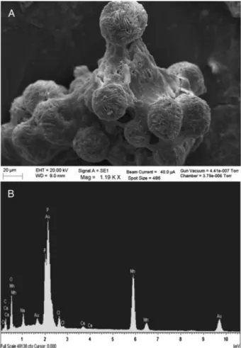
Figure 4: ESEM image (A) of a representative crystal formed in MSVP medium 60 with 1 mM manganese sulfate in the presence of Acinetobacter sp. strain LB1 after 8 days incubation. Corresponding EDX spectrum of the crystal analyzed (B).
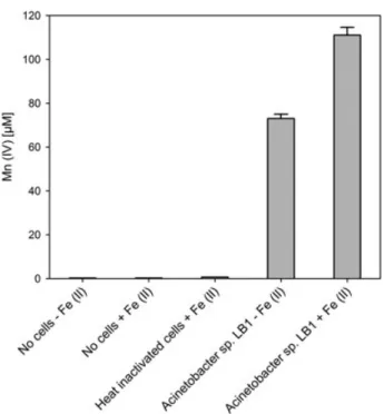
Figure 5: Oxidation of 1mM Mn (II) after 5 days incubation in the presence and 61 absence of 1 mg/L Fe (II) by Acinetobacter sp. strain LB1 in LBB medium in comparison to controls without cells in the presence and absence of 1 mg/L Fe (II) and heat-inactivated cells in the presence of 1 mg/L Fe (II). Error bars indicate the standard deviation.
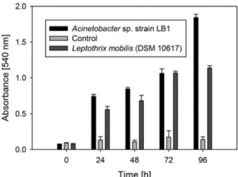
Figure 6: Biofilm-formation capacity of Acinetobacter sp. strain LB1 in comparison 61 to the manganese (II) oxidizing strain L. mobilis (DSM 10617) and non- inoculated controls. The data shown are the means obtained from measurements done in triplicate. Error bars indicate the standard deviation.
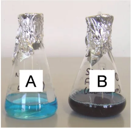
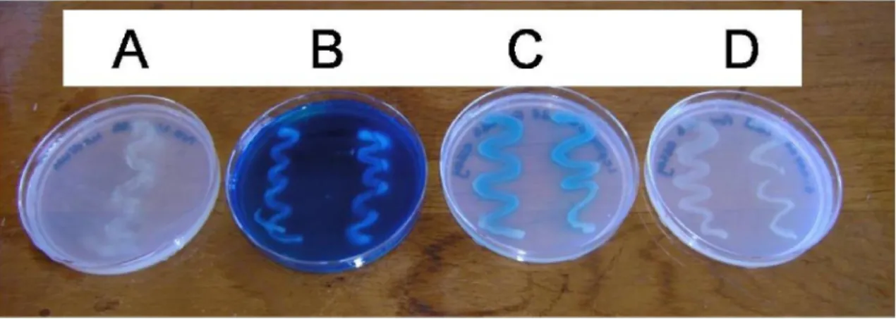
Figure S1: Qualitative detection of Mn (II) oxidation by the manganese (II) 64 oxidizing isolate Acinetobacter sp. strain LB1 using leucoberbelin blue.
[A] Non-inoculated LBB medium with 1 mM manganese sulfate after 2 weeks incubation.
viii [B] LBB medium with 1 mM manganese sulfate inoculated with Acinetobacter sp. strain LB1 after 2 weeks incubation.
Figure S2: Qualitative detection of Mn (II) oxidation by the manganese (II) oxidizing 65 isolate Acinetobacter sp. strain LB1 using leucoberbelin blue.
[A] LBB agar without 1 mM manganese sulfate inoculated with Acinetobacter sp. strain LB1 after 2 weeks incubation.
[B] LBB agar with 1 mM manganese sulfate inoculated with Acinetobacter sp.
strain LB1 after 2 weeks incubation.
[C] LBB agar with 1 mM manganese sulfate inoculated with Leptothrix mobilis (DSM10617) after 2 weeks incubation.
[D] LBB agar with 1 mM manganese sulfate inoculated with E. coli (ATCC 8739) after 2 weeks incubation.
Chapter 3 – Appendix
Figure 1: MALDI-TOF MS profile for cell extracts of Acinetobacter sp. LB1 with 70 selected m/z peak values.
Chapter 4
Figure 1: Growth of E. coli (ATCC 8739) [A] and Acinetobacter sp. LB1 [B] in 84 LBB medium in the presence of 0-10 mg/L nominal Fe (II) concentrations over an 18hr period. All data shown are the average of duplicate flasks.
Figure 2: Fe (II) oxidation at neutral pH [A] in the presence of resting cells 87 (Acinetobacter sp. LB1, 1×10⁸ cells/mL), heat inactivated bacterial cells (Acinetobacter sp. LB1, 1×10⁸ cells/mL), poisoned bacterial cells (Acinetobacter sp. LB1, 1×10⁸ cells/mL) and in the absence of bacteria. The subsequent formation of Fe (III) is shown in [B]. Error bars indicate the standard deviation. n.d. = not determined.
ix Figure 3: Fe (II) oxidation at low pH (2.42) [A] in the presence of resting cells 90 (Acinetobacter sp. LB1, 1×10⁸ cells/mL), heat inactivated bacterial cells (Acinetobacter sp. LB1, 1×10⁸ cells/mL) and in the absence of bacteria. The subsequent formation of Fe (III) is shown in [B]. Error bars indicate the standard deviation.
Figure 4: Fe (II) oxidation at alkaline pH (8.76) [A] in the presence of resting cells 91 (Acinetobacter sp. LB1, 1×10⁸ cells/mL), heat inactivated bacterial cells (Acinetobacter sp. LB1, 1×10⁸ cells/mL) and in the absence of bacteria. The subsequent formation of Fe (III) is shown in [B]. Error bars indicate the standard deviation.
Figure 5: Fe (II) oxidation in batch culture tests before and after the 7 day 94 incubation [A] and the contribution of the filter sand to the turbidity formed in the flask poisoned with 3.5% formaldehyde [B+C].
Constituents of flasks in image [A]:
[A] Borehole water + filter sand spiked with Acinetobacter sp. LB1 (1×10³ cells/mL)
[B] Borehole water + filter sand spiked with Burkholderia sp. strain LB2 (1×10³ cells/mL)
[C] Borehole water + filter sand spiked with Leptothrix mobilis (1×10³ cells/mL)
[D] Borehole water + filter sand spiked with Sphaerotilus natans (1×10³ cells/mL)
[E] Borehole water with native bacteria poisoned with 3.5% formaldehyde [F] Normal borehole water with native bacteria (7.04×10⁵ cfu/mL - iron oxidizers)
Figure 6: ESEM image [A] of crystals formed in the flask with the black precipitate 96 in borehole water containing 5.27 mg/L Fe (II) in the presence of Acinetobacter sp. LB1 after 7 days incubation and the corresponding EDX spectrum of the crystals [B].
x Figure 7: Initial and final Fe (II) concentrations (after a 7 day incubation period) 98
in borehole water in batch culture flasks. All data shown are the average of duplicate flasks.
[A] Borehole water spiked with Acinetobacter sp. LB1 (1×10³ cells/mL) [B] Borehole water spiked with Burkholderia sp. strain LB2 (1×10³ cells/mL) [C] Borehole water spiked with Leptothrix mobilis (DSM 10617) (1×10³ cells/mL)
[D] Borehole water spiked with Sphaerotilus natans (DSM 565) (1×10³ cells/mL)
[E] Normal borehole water with native bacteria (7.04×10⁵ cfu/mL - iron oxidizers)
[F] Borehole water containing native bacteria poisoned with 3.5%
formaldehyde Chapter 5
Figure 1: Confocal laser scanning micrographs of a filter matrix particle before use 115 in the biofiltration system stained with acridine orange (A) and CTC (B).
Figure 2: Confocal laser scanning split micrograph of the filter matrix after 3 116 weeks biofiltration showing the presence of apparently inactive cells in the biofilm (A), active cells (B), and a combination of the two (C), after staining with acridine orange.
Figure 3: Confocal laser scanning micrograph of a CTC stained filter matrix 117 particle showing actively metabolizing cells in red.
Figure 4: Phylogenetic affiliation of 16S rRNA genes of 15 OTU representatives 123 based on sequence comparisons to environmental strains in RDP. Numbers shown at nodes indicate calculated bootstrap values (only values > 50% are shown). The alignment of selected sequences and the construction of the tree
xi are specified in section 2. The scale bar indicates five estimated changes per 100 nucleotides.
Figure 5: Quantitative assignment of the 15 OTU’s from the 16S rRNA gene 124 clone library to their closest phylogenetic groups.
Figure 6: Rarefaction curve depicting the relationship between the number of 125 clones collected and the number of OTU’s (phylotypes) detected, computed using Estimate S.
Figure S1: [A] Representative 2% agarose gel depicting unique banding patterns 136 after ARDRA of 19 out of 100 randomly selected clones. Lane M - Molecular weight marker, lane 1-19 - samples 59-87. [B] Representative 2% agarose gel as for image A, highlighting band regions analysed using GeneSnap program version 7.09.06 (SynGene, Cambridge, United Kingdom).
Thesis supplementary figures
Figure S1: The biofiltration system employed to remove manganese and iron 142 from borehole water in Nottingham road outside the Nottingham combined school, KwaZulu Natal, South Africa.
[A] Borehole water tank [B] Aeration cascade [C] Iron biofilter [D] Manganese biofilter [E] Final receiving tank
Figure S2: Location of the biofiltration system (sampling site) in Nottingham road 143 outside the Nottingham combined school, KwaZulu Natal, South Africa. Maps adapted from http://www.maps-africa.blogspot.com.
xii
Abstract
In the following study, the potential role that microorganisms play in the removal of Mn (II) and Fe (II) was assessed using biofilter sand and water samples collected from a biofiltration system (operated by Umgeni Water in KwaZulu-Natal, Nottingham Road, at the Nottingham combined school, South Africa) treating borehole water containing manganese and iron.
Initially the presence of Mn (II) and Fe (II) oxidizing bacteria was demonstrated in the biofiltration system. Thereafter, the contribution of individual microorganisms to the overall removal of manganese and iron was assessed in the laboratory by determining the difference in metal oxidation in the presence and absence of active bacteria at neutral pH, simulating conditions in the biofilter. Controls were run to verify the elimination via physiochemical reactions occurring within the biofiltration system. Finally a diversity snapshot of the bacteria present within the biofilter matrix was established via analysis of a clone library. Viable bacterial counts for the biofiltration system were established using MSVP (minimal salts vitamins pyruvate) medium - plus added manganese sulfate or iron sulfate targeting Mn (II) and Fe (II) oxidizing bacteria - and R2A for heterotrophic bacteria.
In the first experimental chapter, batch tests using MSVP were employed to determine manganese oxidation, by measuring the pH and ORP (oxidation reduction potential) in experimental flasks and controls over time. There was a clear drop in pH and a concomitant increase in ORP when an isolated manganese oxidizing strain (designated LB1) was grown in MSVP plus added manganese sulfate, indicating manganese oxidation. Based on physiological characteristics established by the VITEK-2 system as well as by 16S rRNA gene sequence analysis and MALDI-TOF (Matrix assisted laser desorption ionization-time of flight mass spectrometry) mass spectrometry of cell extracts, the isolate was identified as a member of the genus Acinetobacter. EDX (energy dispersive X-ray analysis) analysis of crystals formed in batch culture tests, containing MSVP plus either added manganese or iron sulfate, confirmed the ability of the isolate to oxidize both Mn (II) and Fe (II). The leucoberbelin blue colorimetric assay and batch tests using MSVP both demonstrated that in the presence of the isolated strain, Acinetobacter sp. LB1, the rate of Mn (II) oxidation at neutral pH was enhanced as compared to abiotic controls.
xiii In the second experimental chapter the difference in Fe (II) oxidation between biological and abiological systems at neutral pH was determined using batch tests run with Acinetobacter sp.
LB1 and Fe (II) in saline. In addition, the rate of Fe (II) oxidation was also determined at acidic pH and at alkaline pH in experimental and control flasks. To determine Fe (II) removal under conditions simulating those in the biofiltration system, batch tests were set up using borehole water freshly collected from the biofiltration system. In order to verify the contribution of native microorganisms in the borehole water to Fe (II) oxidation, these flasks were spiked with bacterial strains isolated from the biofiltration system - Acinetobacter sp.
LB1 and Burkholderia sp. strain LB2 - and two known iron oxidizing strains Leptothrix mobilis (DSM 10617) and Sphaerotilus natans (DSM 565) were used to determine the contribution of reference iron oxidizers to Fe (II) oxidation. A separate set of the same flasks with the addition of filter sand was used to qualitatively demonstrate iron oxidation as it would occur within the biofiltration system. The ferrozine assay was employed to quantify the amount of Fe (II) in batch tests employing saline medium and in batch tests employing borehole water. EDX analysis was employed to confirm the presence of Fe (II) in oxidation products in the batch test flask with filter sand spiked with Acinetobacter sp. LB1.
In the presence of Acinetobacter sp. LB1 at neutral pH in saline medium, the rate of Fe (II) oxidation was very similar to that in the abiological controls thus demonstrating that the presence of metabolically active microorganisms does not per se enhance the oxidation of Fe (II) like in the case of Mn (II) at neutral pH. Surprisingly, in the heat inactivated control, apparently the highest amount of Fe (II) was oxidized. As expected, at acidic pH very little oxidation of Fe (II) took place and at alkaline pH almost all Fe (II) in the flasks was removed and small amounts oxidized as determined by the amount of Fe (III) produced. Batch tests using borehole water proved that native microorganisms within the biofiltration system were more efficient in the oxidative removal of Fe (II) from the system, in comparison to the reference iron oxidizing strains. In the final experimental chapter, the presence of biofilms with actively metabolizing cells was examined on a pooled sample of biofilter matrix from the manganese and iron filter using CLSM (confocal laser scanning microscopy) image analysis. DNA was extracted from the biofilm material associated with biofilter matrix to establish a diversity snapshot of the bacteria present within the biofilter matrix.
xiv ARDRA (amplified “rDNA” restriction analysis) analysis of the clone library revealed the presence of 15 unique OTU’s (operational taxonomic unit) based upon restriction patterns of amplified 16S rRNA genes of a total of 100 randomly selected clones. The majority of the clones were closely related to the genera Nitrospira and Lactococcus. Overall, 42% of the clones were assigned to the phylum Proteobacteria, 13% to the phylum Actinobacteria, 24%
to the phylum Firmicutes and 21% to the phylum Nitrospirae. Overall, the results demonstrate that bacteria present within an established biofiltration system at neutral pH can contribute to the oxidative removal of Mn (II) and, apparently only to a smaller degree, to that of Fe (II) present in borehole water and that species within the proteobacterial genus Acinetobacter are potentially involved in the geochemical cycling of these two metals.
Keywords: Biofiltration, iron and manganese oxidation, Acinetobacter sp. LB1, batch tests, 16S rRNA, MALDI-TOF MS analysis, Mn (II) and Fe (II) colorimetric assays, EDX analysis, biofilm formation, CLSM image analysis, 16S rRNA clone library
Abbreviations: MSVP (minimal salts vitamins pyruvate), ORP (oxidation reduction potential), EDX (energy dispersive X-ray analysis), MALDI-TOF MS (Matrix assisted laser desorption ionization-time of flight mass spectrometry), rRNA (ribosomal RNA), ARDRA (amplified “rDNA” restriction analysis), CLSM (confocal laser scanning microscopy), OTU (operational taxonomic unit)
xv
Acknowledgements
This thesis would not have been completed without the grace and mercy of God. I would also like to thank the following people:
My family and friends for their support and encouragement My supervisor, Prof. Stefan Schmidt for his guidance and support The technical staff at the University of KwaZulu-Natal Pietermaritzburg
The staff at Umgeni Water head office, Pietermaritzburg and at processing in Durban, for their hard work and support during the project
Finally, I would like to thank TATA Africa, Umgeni Water and FoodBev Seta for the funding they provided towards the accomplishment of the project.
1
Chapter 1
General Introduction
In order to ensure the safety and quality of potable water, it is important to maintain the naturally occurring biochemical reactions taking place within aquatic systems (Salomons and Förstner, 1984). Metals such as manganese and iron are classified as elements that form positive ions when in solution and their oxides typically form hydroxides in water (Tsezos and Volesky, 1982). Iron is a key component in many proteins that are necessary for microbial respiration and metabolism and both manganese and iron are essential trace elements in biological/biotechnological systems (Lovley, 2000). These two metals are prevalent in water bodies and their removal is typically mediated by a combination of microbial and abiotic oxidation reactions (Jakob, 1970). Manganese is naturally occurring, present in almost all environments and comprises approximately 0.1% of the Earth’s crust (IPCS, 2004). It occurs in 11 oxidation states of which oxidation states 2 (most stable and predominate in nature), 4 and 7 are the most important (Gerber et al., 2002).
In aquatic environments manganese exists in two major forms: Mn (II) and Mn (IV) and the conversion between these two forms takes place through oxidation and reduction reactions which are either abiotically or microbially mediated (IPCS, 2004). Manganese (Mn (II)) is more prevalent at low pH and redox potential (IPCS, 2004). Public concern about environmental pollution as well as new applications of manganese compounds such as potassium permanganate and cyclopentadienyl manganese tricarbonyl (CMT), have directed attention to the toxic properties of manganese compounds and the possible involvement of these compounds in causing malformations (Gerber et al., 2002; De Meo et al., 1991). Biological manganese oxidation is carried out by microorganisms which are also responsible for the biological oxidation of Fe (II) except for the stalked members of the genus Gallionella which are strict iron oxidizers (Katsoyiannis and Zouboulis, 2004). Leptothrix, Crenothrix, Hyphomicrobium, Siderocapsa and Metallogenium (Katsoyiannis and Zouboulis, 2004) are some of the well-known manganese oxidizing genera.
2 Iron is a key metal in environmental microbe-metal interactions due to its abundance in the earth’s crust and its ability to readily convert between the Fe (II) and Fe (III) states (Lovley, 2000). Lithotrophic Fe (II) oxidizing microorganisms (FOM) use Fe (II) as an electron donor to provide reducing equivalents for the assimilation of carbon into biomass (Weber et al., 2006;
Emerson et al., 2010) and Fe (III) is used as a terminal electron acceptor under anaerobic conditions by lithotrophic and heterotrophic Fe (III)-reducing microorganisms (FRM) (Weber et al., 2006). Nitrate (NO3-) can also be used as a terminal electron acceptor for iron oxidation under anaerobic conditions, although the kinetics of Fe (II) oxidation by nitrate are much slower than for nitrite (NO2-
) (Picardal, 2012). The iron bacteria are defined as a group of bacteria which utilize the oxidation of ferrous and/or manganous ions as an essential component in their metabolism and these bacteria typically get their apparent brown/rust-red colour from the production of ferric ions and/or manganic salts, either within the cell or attached on the outside (Cullimore and Mc Cann, 1977).
On the basis of acceptability aspects, the WHO (2011) recommends concentrations for drinking water not exceeding 0.3 mg/L for Fe (II) and 0.1 mg/L for Mn (II). Concentrations of Fe (II) exceeding the recommended level in water systems result in a brown/rust-red discoloration in the water and pipes and reduction in water flow rates, which is typically caused by coatings of iron bacteria inside the pipes (Cullimore and Mc Cann, 1977). High levels of manganese - ingested via drinking water - have recently been seen to negatively impact the health of school children (Bouchard et al., 2011). Both manganese and iron impart a metallic, bitter, astringent or medical taste to water and they both contribute to corrosion in water distribution systems (Cullimore and Mc Cann, 1977). As a result the quality of the water is reduced and therefore the need to explore sustainable biotechnological processes for water purification. To establish biotechnological processes for the aerobic treatment of manganese or iron contaminated water, it is important to assess the potential of microorganisms for the oxidative elimination of these two metals. Bacteria play important roles in the activation, modification and detoxification of both manganese and iron in aerated water through changing the valence states of these metals and subsequently converting them into insoluble compounds that are easily removed from water systems (Urrutia et al., 1992). The manganese and iron oxidizing bacteria react with metal cations with an intimate association to their surfaces. This occurs via the interaction of negative charges of
3 anionic functional groups on the surface of bacteria leading to permanently or temporarily chelated metal cations (Salomons and Förstner, 1984). Whether a metal is going to be permanently or temporarily fixed by functional groups present at the bacterial surface, is governed by parameters such as the cell surface constant Km - which is a measure of the affinity for a metal of interest - and by the pH and ORP of the system.
To date limited information on biological manganese oxidation exists as the process is considered more complex than biological iron oxidation. Water treatment systems frequently employ chemical reagents to remove manganese and iron from groundwater, but the results of such treatment are not regarded as sustainable (Burger et al., 2008). This is due to an increase in the cost of operation as a result of costly chemicals for treatment and secondary impacts that arise from the formation of residuals and by-products (Gallard and von Gunten, 2002).
Biological water treatment systems have proven to be more effective than chemical treatment for the removal of iron and manganese from borehole water (Burger et al., 2008; Trevors, 1989).
Biological treatment requires less attention during operation and results in a reduced amount of sludge due to no residuals or by-product formation which arise from the addition of chemicals to the water. Chemical treatment of water demands a substantial amount of time and labour, producing large masses of sludge which increases operation costs (Burger et al., 2008). The above reasons validate the use of biological manganese and iron oxidation as a viable and more sustainable alternative as compared to the use of chemical reagents for the treatment of borehole water.
This study aimed to demonstrate the presence of Mn (II) and Fe (II) oxidizing bacteria in a biofiltration system (Nottingham road, KwaZulu Natal, South Africa) (see thesis supplementary figures S1 and S2) treating borehole water containing Mn (II) and Fe (II) at concentrations of 0.35 mg/L and 2-8 mg/L respectively. In addition, the contribution of these microorganisms to the overall removal of manganese and iron was assessed via qualitative and quantitative methods, taking into account the oxidative elimination via physiochemical reactions typically occurring within biofiltration systems. Finally a clone library was established from metabolically active biofilm material to assess the diversity of bacteria present within the biofilter matrix.
4
References
Bouchard, M. F., Sauvé, S., Barbeau, B., Legrand, M., Brodeur, M-E., Bouffard, T., Limoges, E., Bellinger, D. C., and Mergler, D., (2011). Intellectual impairment in school-age children exposed to manganese from drinking water. Env. Health Persp. 119: 138-143.
Burger, M. S., Mercer, S. S., Shupe, G. D., and Gagnon, G. A., (2008). Manganese removal during bench-scale biofiltration. Water Res. 42: 4733-4742.
Cullimore, D. R., Mc Cann, A. E., (1977). The identification, cultivation and control of iron bacteria in ground water. In: Aquatic microbiology (F. A. Skinner and J. M. Shewan, eds.). Soc.
Appl. Bacteriol. Sympos. No. 6, 219-261. London: Academic Press, Inc.
De Meo, M., Laget, M., Castegnaro, M., and Duménil, G., (1991). Genotoxic activity of potassium permanganate in acidic solutions. Mutat. Res. 260: 295-306.
Emerson, D., Fleming, E. J., and Mc Beth, J. M., (2010). Iron-oxidizing bacteria: An environmental and genomic perspective. Annu. Rev. Microbiol. 64: 561-583.
Gallard, H. U., and von Gunten, U., (2002). Chlorination of natural organic matter: Kinetics of chlorination and of THM formation. Water Res. 36: 65-74.
Gerber, G. B., Hantson, P., and Léonard, A., (2002). Carcinogenicity, mutagenicity and teratogenicity of manganese compounds. Crit. Rev. Oncology/Hematology 42: 25-34.
International Programme on Chemical Safety - IPCS. Manganese and its compounds:
environmental aspects. Concise International Chemical Assessment Document 63. World Health Organisation, Geneva, 2004.
Jakob, H. R., (1970). Redox potential. In: Norris and Ribbons: Methods in microbiology, London and New York, Academic Press.
5 Katsoyiannis, I. A., and Zouboulis, A. I., (2004). Biological treatment of Mn (II) and Fe (II) containing groundwater: Kinetic considerations and product characterization. Water Res. 38:
1922-1932.
Lovley, D. R., (2000). Environmental microbe-metal interactions. Washington D.C, ASM Press.
Picardal, F., (2012). Abiotic and microbial interactions during anaerobic transformations of Fe (II) and NOx-. Front. Microbiol. 3: 112.
Salomons, W., and Förstner, U., (1984). Metals in the hydrocycle. Springer-Verlag, Berlin, Heidelberg, New York, Tokyo.
Trevors, J. T., (1989). The role of microbial metal resistance and detoxification mechanisms in environmental bioassay research. Hydrobiologia 188/189: 143-147.
Tsezos, M., and Volesky, B., (1982). The mechanism of uranium biosorption by Rhizopus arrhizus. Biotech. Bioeng. 23: 385-401.
Urrutia, M. M., Kemper, M., Doyle, R., and Beveridge, T. J., (1992). The membrane-induced proton motive force influences the metal binding ability of Bacillus subtilis cell walls. App.
Environ. Microbiol. 58: 3837-3844.
Weber, K. A., Achenbach, L. A., and Coates, J. D., (2006). Microorganisms pumping iron:
Anaerobic microbial iron oxidation and reduction. Nat. Rev. Microbiol. 4: 752-764.
World Health Organisation (2011). Guidelines for drinking water quality, 4th edition. World Health Organisation (WHO), Geneva.
6
Chapter 2: Literature Review
A. Manganese Oxidation
1. Microbial oxidation of manganese
Microorganisms involved in manganese oxidation include bacteria, fungi and algae. Bacteria and fungi are considered the main groups of manganese oxidizers whilst algae are usually found to enhance the oxidation of manganese in combination with bacteria (Stuet et al., 1996).
Manganese oxidation can occur via enzymatic or non-enzymatic processes. The non-enzymatic manganese oxidation process involves the direct chemical oxidation of Mn (II) or the oxidation by metabolic end products (Linhardt, 1997). The enzymatic process is performed by three groups of microorganisms: (i) those that oxidize dissolved Mn (II), (ii) those that oxidize Mn (II) that is prebound to solids and (iii) organisms that oxidize dissolved Mn (II) by the metabolite H2O2 via catalase. The abiotic/non-enzymatic oxidation of Mn (II) to MnO2 is slow and occurs by the chemical reaction of dissolved Mn (II) and oxygen in natural waters (Linhardt, 1997). The enzymatic/biological manganese oxidation of higher manganese oxides and hydroxides takes place faster than abiotic oxidation, with a wide variety of ubiquitous microorganisms capable of converting Mn (II) to solid Mn (III/IV) oxides (Ghiorse, 1984). The diversity of these microorganisms is based on the different mechanisms they use to biochemically induce the transformation of Mn (II) (Ehrlich, 1990). The common microbial manganese oxides are Birnessite (MnO2), Manganite (MnOOH) and Hausmannite (Mn3O4) (Ehrlich and Newman, 2009; Schweisfurth and Gattow, 1966).
1a. Microbial Mn (II) oxidation
Heterotrophic microorganisms such as Bacillus sp., Pseudomonas putida and Pedomicrobium sp.
were previously described as using a multicopper oxidase (MCO) in the oxidation of Mn (II) to Mn (IV) (Brouwers et al., 2000; Spiro et al., 2008). The genes that encode putative Mn (II)- oxidizing enzymes like mofA in Leptothrix discophora, cumA in Pseudomonas putida GB-1, and moxA in Pedomicrobium sp. strain ACM 3067, are thought to produce multicopper oxidases, this
7 was based on the presence of conserved, predicted amino acid motifs (El Gheriany et al., 2009).
Homogenates from the endospores of a Bacillus sp. contained distinct proteins that produced a solid brown precipitate when exposed to Mn (II) when non denaturing polyacrylamide gel electrophoresis was used (Francis et al., 2002). Upon disruption of the MCO gene this Bacillus sp. lost the ability to oxidize manganese (Spiro et al., 2008) which indicates that the MCO gene is responsible for manganese oxidation in its endospores (da Silva and Williams, 1991).
Multicopper oxidases are described as one electron transferring enzymes were single electrons are transferred from the substrate to O2 through intervening copper ions (da Silva and Williams, 1991).
For experimental purposes, a variety of poisons were tested by Rosson and Nealson (1982) to check whether they inhibit manganese oxidation. Azide can inhibit the formation of Mn (III) whilst the addition of Cu (II) enhances the formation thereof (Ehrlich and Newman, 2009; Spiro et al., 2008). Whilst it was found that some poisons do not potentially interfere with manganese oxidation it was suggested that the added poisons may interfere in unknown ways with manganese adsorption due to the difference in ion-exchange capacities of manganates (Murray, 1974). In a separate study conducted by Ghiorse and Hirsch (1979), it was established that heat treatment did not completely inhibit manganese oxidation which indicated that the suspected MCO protein responsible for manganese oxidation is somewhat heat stable. Other compounds that were found to inhibit manganese oxidation were SDS, NaCl, cyanide and HgCl2 (Boogerd and de Vrind, 1987; Ehrlich and Newman, 2009). The oxidation of Mn (II) to Mn (IV) is catalyzed via the following reaction (1).
2Mn2+ + 8H2O 2Mn (OH) 4 + 4e- +8H+ 2Mn (OH) 4 2MnO2 + 4H2O
4e- + 4H+ + O2 2H2O
________________________________________ (1) 2Mn2+ + 2H2O + O2 2MnO2 + 4H+
8 The end product of bacterial Mn (II) oxidation, Mn (IV), is a powerful oxidant and cannot be stabilized by carboxylate groups, which are weak donor ligands from protein side chains (Spiro et al., 2008). Most Mn (IV) complexes are polynuclear and are stabilized by oxo bridges (Pizarro et al., 2004). The Mn (IV) complexes further polymerize to form solid-phase MnO2 complexes, in the absence of capping ligands (Pizarro et al., 2004). If a bacterial MCO accommodates multiple Mn (III) ions in its holding sites, a polynuclear Mn (IV) complex may form as a nucleation site for MnO2 nanoparticle formation (Brouwers et al., 2000; Spiro et al., 2008). The first step in the formation could be further oxidation of Mn (III), either via the MCO or possibly by direct reaction with O2 to form a polynuclear oxo-bridged Mn (IV) complex (Brouwers et al., 2000; Spiro et al., 2008). Numerous Mn (III) ions can also disproportionate to a polynuclear Mn (IV) complex and Mn (II) ions, which are then reoxidized at the MCO substrate site, ensuring a continuous supply of Mn (III) ions (Spiro et al., 2008). The polynuclear Mn (IV) complex formed at the nucleation site in each of these cases, would ultimately be released to grow into MnO2 nanoparticles (Spiro et al., 2008).
1b. Mn (II) oxidizing bacteria
Mn (II) oxidation is carried out by a variety of microorganisms. Microorganisms found in sites that are high in concentrations of manganese and iron form distinct metallic casts and can be recognized by their distinct morphotypes in natural samples (Emerson and Revsbech, 1994). The most common manganese oxidizing microorganism found in these sites is Leptothrix ochracea (Emerson and Revsbech, 1994). Other bacteria that oxidize manganese are Hyphomicrobium manganoxidans, Pseudomonas putida and Leptothrix cholodinii, which attack dissolved manganese enzymatically. Arthrobacter spp. and Oceanospirillium sp. are known to oxidize manganese prebound to manganese oxides while Pseudomonas manganoxidans and Bacillus sp.
strain SG-1 oxidize manganese enzymatically (Ehrlich and Newman, 2009). In addition, the ability of an Acinetobacter species to oxidize Mn (II) was recently reported (Beukes and Schmidt, 2012). The Sphaerotilus-Leptothrix group of microorganisms is the most common group of manganese oxidizing microorganisms and the species within these genera share a number of common characteristics like the formation of a sheath, the requirement of vitamin B12
for growth and the formation of poly-β-hydroxybutyrate (PHB) as reserve material (vanVeen et
9 al., 1978). The bacteria of the Sphaerotilus-Leptothrix group of microorganisms typically occur in low numbers in slightly polluted or non-polluted waters (vanVeen et al., 1978). S. natans possesses a pronounced response to organic nutrients, producing high yields of cell material.
This is in contrast to Leptothrix sp. which hardly responds to added nutrients (vanVeen et al., 1978). S. natans has much larger cells than most of the Leptothrix spp. and false branching only occurs in S. natans and in L. lopholea (Takeda et al., 2012; vanVeen et al., 1978).
The presence of a sheath has nutritional and ecological consequences for members of the Sphaerotilus-Leptothrix group of bacteria. Their growth in slow running waters low in nutrients requires a sheath enabling the bacteria to attach themselves to solid surfaces during nutrient depletion. The sheath also protects the bacteria against phages and bacterial or eukaryotic predators (vanVeen et al., 1978). Cell propagation of sheath-forming bacteria is not necessarily dependant on the presence of the sheath, as can be concluded from the ability of the sheath-less mutants of this group of microorganisms to grow and divide (Takeda et al., 2012; Mulder and vanVeen, 1963). Leptothrix cholodnii, which was previously classified as Leptothrix discophora (Spring et al., 1996) is the most common species that is able to form a sheath and maintain it under laboratory conditions (Emerson and Ghiorse, 1992; Takeda et al., 2010, 2012).
1c. Mn (II) oxidizing fungi and algae
Fungi and algae (to a lesser extent) are also important groups of manganese oxidizing microorganisms. Like bacteria, these microorganisms also play an important role in the production of biogenic Mn oxides. Fungi that oxidize manganese are ubiquitous in nature, and have been isolated from freshwater systems, Mn nodules (Cahyani et al., 2009), soil environments (Santelli et al., 2010) including building materials (de la Torre and Gomez- Alarcon, 1994). They are able to thrive in these environments because they possess multiple mechanisms to tolerate environmental stresses such as nutrient fluctuations, desiccation or high levels of metals (Santelli et al., 2010). They have thus been implicated in the remediation of a wide range of pollutants and it was previously found that fungi contributed to the remediation of Mn-contaminated mine drainage and were also found growing in a Mn-attenuating bioreactor for treatment of mine waters (Mariner et al., 2008). Some examples of fungi involved in manganese
10 oxidation include Plectosphaerella cucumerina and Stilbella aciculosa, both isolated from mine waters, which both belong to the phylum Ascomycota (Mariner et al., 2008). These two fungi are soil inhabitants (de Hoog et al., 2000) that have never previously been known to oxidize manganese and were not phylogenetically related to known manganese oxidizing species.
Similarly, fungi isolated from freshwater and marine systems have been identified as ascomycetes via analysis of the 18S rRNA genes (Tebo et al., 2005). The main proteins involved in bacterial manganese oxidation are also involved in fungal manganese oxidation. An extracellular protein involved in manganese oxide formation found in L. discophora SS-1 was also found in an ascomycete; strain KR21-2, previously investigated by Tani et al. (2003). The manganese oxidation inhibitor for bacteria, azide, was also found to inhibit manganese oxidation in the ascomycete strain KR21-2 (Tebo et al., 2005). This suggests that the same protein - MCO- type enzyme - responsible for manganese oxidation in bacteria is also responsible for manganese oxidation in fungi. Fungi also use heme-containing Mn peroxidases and laccases, used in the degradation of lignin, for manganese oxidation (Tebo et al., 2005). The peroxide-oxidized enzyme and most laccase enzymes catalyse the oxidation of Mn (II) to Mn (III) (Schlosser and Höfer, 2002).
In terms of metal toxicity, fungi are considered more tolerant than bacteria to high concentrations of metals in the environment (Chander et al., 2001). Fungi and bacteria that oxidize manganese are heterotrophs that do not gain energy from the oxidation of manganese but rather benefit from the presence of Mn oxide minerals, through the uptake of dissolved Mn (II) or via scavenging of reactive oxygen species (Tebo et al., 1997). Besides their ability to oxidize Mn (II), fungi can also mediate the reduction of this metal via abiotic reactions and/or with enzymatic reduction (Gomah et al., 1980; Ghiorse and Ehrlich, 1976). In culture media the oxidation of manganese by fungi can be detected in a similar manner to that of bacteria, by using a triphenyl-compound (Leucoberbelin blue) which is oxidized by Mn IV and insensitive to Mn (II) and other metals in trace quantities (Altmann, 1972; Krumbein and Altmann, 1973).
11 2. Bacteriogenic manganese oxides
2a. Properties of bacteriogenic manganese oxides
Bacteriogenic/biogenic manganese oxides (manganese dioxide and manganese tetroxide) produced as a result of Mn (II) oxidation by bacteria are used in the oxidative removal of toxic metals from contaminated soils and wastewaters and have been studied extensively (Bargar et al., 2009). These manganese oxides have highly reactive surfaces and thus contribute to the remediation of water contaminated with toxic metals, by scavenging metals such as Pb, Ni, Co and Zn (Peña et al., 2010; Takahashi et al., 2007). The reactivities of biogenic manganese oxides in comparison to abiotically produced manganese oxides is greater due to the smaller size, increased surface area, disorder and/or the sheet symmetry distortion of biogenic manganese oxides (Hochella et al., 2008). Manganese oxides are poorly soluble in water and are used in the manufacturing of catalysts, colorants, metal sorbents and batteries (IPCS, 2004) and they can also be used as a terminal electron acceptor by other bacteria in respiration (Spiro et al., 2008).
Due to its high surface area and oxidizing potential, MnO2 is capable of efficiently degrading biologically recalcitrant organic molecules such as benzene and naphthalene to lower- molecular-mass compounds, indicating the potential of these compounds in bioremediation of xenobiotic organic compounds (Forrez et al., 2010; Spiro et al., 2008). Whilst MnO2 is the stable form of manganese it can however be reduced to Mn (II) in the presence of exogenous ligands or UV rays, which helps to regulate the bioavailability of Mn (II) (Spiro et al., 2008).
2b. Structural composition of bacteriogenic manganese oxides
The manganese dioxides produced by microorganisms consist of stacked hexagonal sheets of octahedral MnO6 and are categorized as having either a layer or tunnel structure (Jurgensen et al., 2004; Santelli et al., 2011; Spiro et al., 2008). The layer-type oxides - phyllomanganates - which are most abundant in soils, nodules and rock varnishes, are poorly crystalline and highly reactive with metal cations (Fig. 1). This is the most dominant bacteriogenic manganese oxide formed at circumneutral pH which is also structurally similar to hexagonal birnessite (Bargar et al., 2005;
Jurgensen et al., 2004). Tunnel structure oxides (Tectomanganates) are comprised of chains with
12 edge-sharing octahedral manganese complexes, linked through corner-sharing, forming square or rectangular cross-sections e.g., todorokite (Jurgensen et al., 2004; Santelli et al., 2011).
Depending on the physical properties and mineral particle size of the manganese oxides, they are able to absorb a wide variety of cations onto their surfaces (Hochella et al., 2008). Cation vacancies and random stacking arrangements create structural defects on the layer-type oxides, which provide binding sites for exogenous metal ions, thus controlling the bioavailability of exogenous metal ions in the natural environment (Spiro et al., 2008).
Figure 1 (Adapted from Spiro et al., 2008): Binding of Pb (II) ions onto two crystal structures of bacterial manganese oxides. (A) triple-corner-sharing and (B) double-edge-sharing inner sphere surface complexes formed respectively above cation vacancy sites and at sheet edges.
13 In earlier studies conducted by Greene and Madgwick (1991), solid phases of manganese oxides were identified as Mn3O4 (Hausmannite), Buserite (a hydrated layer-type Mn (IV) oxide), Manganite (γ-MnOOH, a tunnel-type Mn (III) oxide) and Nsutite (γ-MnO2, a tunnel-type Mn (IV) oxide). These are well-known products of biological and abiotic oxidation of Mn (II). In recent studies, the origin (via biological or abiotic oxidation) of the different minerals has been disproven except for Buserite. Buserite, most probably stemmed from the autocatalytic oxidation of adsorbed Mn (II) present on the surface of freshly precipitated Mn (IV) oxides (Bargar et al., 2005). EXAFS (extended X-ray absorption fine structure) spectroscopy indicates that each Mn ion in Buserite is di-μ-oxo bridged to six Mn neighbours with a 2.82-2.90 Å Mn-Mn distance (Figure 2B) (Spiro et al., 2008). In addition, 3.5-3.8 Å Mn-Mn distances were found in Buserite, which suggests the presence of Mn (III) positioned above or below the Mn (IV) vacancy sites (Figure 2A) (Gaillot et al., 2003). The negative charge from Mn (IV) vacant sites can be overcome by the incorporation of hydrated metal cations, which results in basal plane spacings of 7-10 Å, depending on the degree of hydration (Post, 1999).
14 Figure 2 (Adapted from Spiro et al., 2008): Crystal structure of the bacteriogenic Mn oxide - Buserite. (A) Sheet structure of Buserite, with hexagonal symmetry, highlighting a monoclinic unit cell (a-b). (B) Sheet structure (a-b), illustrating di-μ-oxo bridging between neighboring Mn atoms. Manganese atoms illustrated as large black circles and oxygen atoms as the grey smaller circles.
3. General properties of manganese compounds
3a. General toxicity associated with manganese compounds
Symptoms of manganese toxicity in plants consist of necrotic lesions, marginal chlorosis, and distorted development of the leaves (IPCS, 2004). In humans, manganese seems to be the one of the least toxic minerals from a nutritional point of view. There is no known toxicity arising from
15
“normal” manganese uptake via food or from taking reasonable amounts in supplements (Gerber et al., 2002). Increased levels of manganese in the environment are no threat to man as long as they are not inhaled or ingested with contaminated drinking water (Gerber et al., 2002).
However, acute toxic effects and consequences have been seen after the administration of potassium permanganate (Henderson and Watt, 1951). The inhalation of manganese fumes can cause “metal fume” fever which is characterized by acute pneumonitis, tracheobronchitis and pulmonary oedema (Nemery, 1990). Chronic toxicity to the central nervous system (CNS) can also take place when manganese is inhaled, which is much more important than the acute toxicity (Huang et al., 1989). In general, Mn toxicity results from exposure to high levels of manganese from various industrial settings like welding (Park et al., 2007), metal smelting, Mn mining (Myers et al. 2003a, b) and/or battery manufacturing (Bader et al., 1999). Cyclopentadienyl manganese tricarbonyl (CMT) which is used in petrol as a substitute for lead, produces convulsions and pulmonary oedema in Sprague-Dawley rats (Gerber et al., 2002). The LD50s for an oral and intraperitoneal administration are 22 mg and 14 mg per kg body weight respectively (Penney et al., 1985). The toxic effects of methylcyclopentadienyl manganese tricarbonyl (MMT) are similar as those of CMT and appear mainly in the lungs, liver and kidney (Hinderer, 1979).
In a previous report it was also found that an increase in the use of MMT (methylcyclopentadienyl) in gasoline had caused elevated levels of Mn in the environment and subsequently in the blood of children that resided in the surrounding environment (Batterman et al., 2011; Röllin et al., 2005). Chronic manganism affects carbohydrate metabolism, and patients with chronic manganism often have hypoglycaemia following a high glucose load (Gerber et al., 2002). Whilst an essential element for nutrition in human beings, manganese has been reported to have toxic side effects (neurobehavioral development problems) on children after the intake of water containing high levels of Mn (II) that exceed the US EPA recommended level (Woolf et al., 2002; Bouchard et al., 2007). The recommended intake levels of manganese for children (9-13 yrs. old) are between 1.9 and 1.6 mg per day for males and females respectively (Batterman et al., 2011). A very recent study conducted by Batterman et al. (2011) revealed that 8.1% of children tested in the study site in Durban (KwaZulu-Natal, South Africa), had blood manganese concentrations above the normal range (15 μg/L) reaching a maximum concentration
16 of 25 μg/L(Batterman et al., 2011). The average manganese in the children’s blood was around 10.1±3.4 μg/L(Batterman et al., 2011).
3b. Mutagenic properties of manganese compounds
The presence of toxic manganese compounds causes a decrease in the fidelity of DNA replication by modifying the activity of DNA polymerase, however it does not seem to interfere with the repair of chemically induced DNA damage (van de Sande et al., 1982). Mn (II) compounds and to a lesser extent Mn (VII) compounds, at micromolar concentrations can induce the cellular SOS repair when the normal progression fork is impeded (Olivier and Marzin, 1987).
Divalent manganese compounds may also mediate in vitro mispairing of ethylating agents or aliphatic epoxides (Bhanot and Solomon, 1994). Manganese sulfate induces mutations in T4 phage DNA when developing in Escherichia coli and enhances UV induced mutagenesis (Rossman and Molina, 1986). Potassium permanganate can cause damage to the integrity of the DNA chain (De Meo et al., 1991) but is less effective than MnSO4 in the above mentioned aspects of mutagenicity (Gerber et al., 2002). Although a proven mutagen, previous studies of cancer development after manganese exposure indicated that manganese is not a cancer risk in man (Gerber et al., 2002).
4. Sources of manganese in the environment
Manganese is naturally occurring in numerous environments and the highest exposure of manganese is from industrial activities (such as ferroalloy production, iron and steel foundries, power plants, and coke ovens), combustion of fossil fuels, and reentrainment of manganese- containing soils (Lioy, 1983). Manganese occurs as a component of more than 100 minerals, such as sulfides, oxides, carbonates, silicates, phosphates and borates (NAS, 1973). The common manganese minerals include Pyrolusite (manganese dioxide), Rhodochrosite (manganese carbonate [MnCO3]), Rhodonite (manganese silicate) and Hausmannite (manganese tetroxide) (NAS, 1973; Ehrlich, 1990).
17 5. Biochemical aspects of manganese
Metals of biological importance - required by microorganisms for structural or metabolic functions - are usually tolerated in higher quantities whilst the opposite is true for those without biological function (Haferburg and Kothe, 2007). Homeostasis is essential for controlling metal uptake by bacterial cells and bacteria have developed a modified regulatory system that controls metal uptake and excretion (Haferburg and Kothe, 2007). Manganese is a component of the eukaryotic mitochondrial enzymes pyruvate carboxylase, certain superoxide dismutases, glutamine synthetase, alkaline phosphatase and arginase and is responsible for the activation of a wide variety of enzymes (Gerber et al., 2002). Manganese-containing superoxide dismutase intervenes in antioxidant activity and tumour defences (Gerber et al., 2002) and is essential for normal bone structure and the formation of mucopolysaccharides (Keen and Leach, 1988).
Whilst these are essential metal ions in microorganisms, manganese and iron have the potential of being toxic to the cells which means that homeostatic regulation of their concentrations is necessary (Jakubovics and Jenkinson, 2001).
The ionic radius of manganese is similar to that of magnesium, calcium and iron in aqueous solution, which allows the interchange of manganese and other cations in the metal binding sites of proteins (Jakubovics and Jenkinson, 2001). Manganese is essential for certain metabolic reactions like oxygenic photosynthesis in cyanobacteria (Christianson, 1997) and glycolysis in several Gram positive endospore forming bacteria and also plays an important role in the detoxification of reactive oxygen species such as peroxides and oxygen ions which are byproducts of metabolism (Jakubovics and Jenkinson, 2001; Chander et al., 1998). In earlier research it was found that manganese promoted endospore formation and spore germination in Bacillus spp., contributing to the overall development cycle in this bacterial genus (Charney et al., 1951; Gould, 1969). Oxidative stress is caused by the inability of the bacterium to efficiently resist or repair damage caused by reactive oxygen species like peroxides and oxygen ions and this is a major challenge facing bacteria growing in oxygenated environments (Jakubovics and Jenkinson, 2001). Manganese plays an important role in bacterial homeostasis by reducing oxidative stress through interaction with reactive oxygen species without generating harmful free
18 radicals within bacterial cells (Cornelis et al., 2011; Cheton and Archibald, 1988; Stadtman et al., 1990).
6. Impact of manganese oxidizing bacteria on the corrosion of stainless steel
Manganese oxidizing bacteria play a vital role in the corrosion of stainless steel, which occurs as a result of the deposition of manganese oxides and hydroxides onto the surface of this metal through the activity of these microorganisms (Linhardt, 2004). The term used to describe the phenomenon of stainless steel corrosion is called ennoblement. This describes the shift from the free corrosion potential of stainless steel (i.e. abiotic) toward the anodic direction which is coincident with biofilm formation on metal surfaces (Linhardt, 2004). Corrosion by microorganisms consists of a biotic and an abiotic step. The biotic step takes place in the biofilm were biomineralization of MnO2 takes place and the deposits placed directly on the surface of the metal (Linhardt, 2004). The abiotic step is the corrosion process, which is based on the electrochemical properties of the higher manganese oxides (Linhardt, 2004). The microorganisms modify the medium of the corrosion system by creating strong concentration gradients generating unexpected chemicals at the metals surface (Linhardt, 1997).
The two corrosion mechanisms relevant to metal depositing microorganisms are the accumulation of chlorides under deposits containing Fe (III) and Mn (IV), which are aggressive to stainless steel (Kobrin, 1976; Tatnall, 1981; Pope et al., 1984) and the effect of the redox couples, Fe (II)/Fe (III) and Mn (II)/Mn (IV), which may influence the Eoc (open circuit potential), leading to pitting of the metal (Pope et al., 1984; Duquette, 1986). The first mechanism appears to be unrealistic as high chloride concentrations are often a consequence of a corrosion reaction rather than the cause of the corrosion and are not found in deposits free of corrosion products (Linhardt, 1997). The second mechanism is considered more relevant as it is based on the redox properties of the deposits and has been previously proposed as a mechanism for corrosion by Linhardt (1994, 1996). The properties of MnO2 that make it an effective cathodic material is its higher than practical oxygen potentials, it is not diffusion limited if reduced, it may act as an oxygen-electrode (Matsuki and Kamada, 1986) and the electronic conductivity allows a layer of MnO2 to be reduced from the outer surface and not only at the interfacial layer (Linhardt, 1997).
19
B. Iron Oxidation
1. Bacterial oxidation of iron
In terms of biological reactivity, the most important oxidation states of iron are Fe (II) and Fe (III), the oxidized form and an abundant supply of Fe (II) must be available for bacteria to gain sufficient energy from the oxidation of Fe (II) (Cornelis et al., 2011; Liang et al., 1993).
However, at neutral pH and under fully aerated conditions, rapid chemical oxidation of Fe (II) takes place and this poses a problem for the bacteria. When Fe (II) enters a fully aerobic zone, its half-life is only in the range of minutes (Liang et al., 1993). Microorganisms are able to overcome the inherent Fe (II) instability under fully aerated conditions by either thriving at very low pH, which is what Acidithiobacillus ferrooxidans and Leptospirillum ferrooxidans (both well-known for bioleaching of metals from sulfidic ores (Gehrke et al., 1998; Sand et al., 1992)) do, or in the case of Gallionella ferruginea, by growing at very low O2 concentrations at circumneutral pH where the half-life of Fe (II) may be much longer (Liang et al., 1993). A second problem faced by these microorganisms is that the product of the oxidation is an insoluble ferric hydroxide (Lovley, 2000; Ehrlich and Newman, 2009). To avoid a build-up of toxic levels of this oxidation product within the cells, iron oxidation must occur at the exterior of the cell surface and this requires that cells possess a chemical mechanism for transporting electrons to the cytoplasmic membrane where a chemiosmotic potential is established (Lovley, 2000; Ehrlich and Newman, 2009). At a pH range between 6 and 7, the Fe (II) will often be in the form of FeCO3 and the Fe (III) in the form of an insoluble ferric hydroxide (2), thus the product of oxidation is continuously removed from solution (Widdel et al., 1993). This process is somewhat important as it drives the oxidation of Fe (II) to Fe (III).
4 FeCO3 + O2 + 6 H2O 4 Fe (OH)3 (s) + 4 CO2 (2) 1a. Bacterial Fe (II) oxidation mechanisms
Aerobic Fe (II) oxidation - Aerobic Fe (II) oxidation is the oxidation of Fe (II) by molecular oxygen to form a simple insoluble Fe (III) hydroxide (3). The aerobic oxidation of Fe (II) was
20 previously neglected due to the rapid rate of abiotic Fe (II) oxidation coupled to oxygen reduction (Davison and Seed, 1983). Environments that encourage aerobic Fe (II) oxidation are groundwater iron seeps, stream sediments, wetland surface sediments, irrigation ditches, cave walls, subsurface boreholes, municipal and industrial water distribution systems, deep ocean basalt and hydrothermal vents (Emerson and Weiss, 2004). Microaerophilic Fe (II) oxidizing microorganisms in the above environments compete successfully with the kinetics of abiotic Fe (II) oxidation through the conservation of energy from the oxidation of Fe (II) via the conversion of inorganic carbon into biomass (Emerson and Moyer, 1997; Weber et al., 2006). The Fe (III)/Fe (II) couple has a reduction potential which is sufficient to provide reducing power between bacterial photosystems or alternative terminal electron acceptors which are involved in respiratory processes to sustain microbial growth (Weber et al., 2006).
4 Fe2+ + O2 + 10 H2O 4 Fe (OH) 3 (s) + 8 H+ (3)
Anaerobic, photoautotrophic Fe (II) oxidation – The first demonstration of microbially mediated Fe (II) oxidation in anoxic environments was the phototrophic anaerobic Fe (II) oxidation process (Widdel et al., 1993). The microorganisms (FOM) involved in this process oxidize Fe (II) via the utilization of light energy to fix CO2 into biomass (Weber et al., 2006).
The bacteria in this group include Chlorobium ferrooxidans, Rhodomicrobium vannielii, Thiodictyon spp., Rhodopseudomonas palustris and Rhodovulum spp. (Weber et al., 2006). With the exception of Rhodomicrobium vannielii, these bacteria can completely oxidize Fe (II) to Fe (III) (Weber et al., 2006). The incomplete oxidation of Fe (II) by R. vannielii was due to the encrustation of the cell wall of this bacterium with biogenic Fe (III) oxides which inhibited further metabolic activity (Widdel et al., 1993). In order to prevent the cells from being encrusted with biogenic Fe (III) oxides, the production of low-molecular weight compounds that can solubilize these oxides has been suggested (Ehrenreich and Widdel, 1994). The phototrophic oxidation of Fe (II) results in the formation of poorly crystalline Fe (III) oxides but in the presence of metabolically active iron oxidizing microorganisms, they are transformed into the more crystalline Fe (III) oxide minerals such as lepidocrocite and goethite (Kappler and Newman, 2004). Phototrophic Fe (II) oxidation processes are typically limited by the maximum penetration of light at wavelengths between 275 and 700 nm to a depth of 200 µm in soil and
21 sediments (Ciania et al., 2005). Phototrophic FOM are only of minor importance in the global iron biogeochemical cycling because they are unable to promote Fe (II) mineral dissolution and are limited by the solubility of the mineral (Kappler and Newman, 2004).
Anaerobic, nitrate-dependent Fe (II) oxidation – At neutral pH, the light-independent microbially mediated oxidation of iron coupled to nitrate reduction occurs and is thus not limited to anaerobic environments exposed to sunlight (Weber et al., 2006). These reactions occur in various fresh water and saline environmental systems that support - in the presence of abundant nitrate - the nitrate Fe (II) oxidizing microbial communities which potentially contribute to the iron redox cycle (Chaudhuri et al., 2001; Weber et al., 2006). Light-independent reactions such as nitrate-dependent Fe (II) oxidation, have the potential to contribute to anaerobic Fe (II) oxidation on a global scale, provided that adequate concentrations of a suitable electron acceptor are readily available (Weber et al., 2006). This is due to the ubiquity and diversity of anaerobic FOM. FOM, to date, have been demonstrated to exploit the favourable thermodynamics between Fe (II) (OH3)/Fe (III) and nitrate reduction redox pairs (NO3–
/½N2, NO3