Modelling the Interaction between
Human Immunodeficiency Virus, Mycobacterium Tuberculosis and the Human Immune System, including the Effects of Drug Therapy
by
Santosh Ramkissoon
Submitted in fulfilment of the academic requirements for the degree of Master of Science in the
School of Physics University of KwaZulu-Natal
February 2007
Abstract
Tuberculosis (TB) is the leading cause of death in individuals infected with human immunodeficiency virus (HIV) in several African countries, including South Africa. HIV-positive individuals do not have the immune system resources to keep TB in check and are as much as 30 times more likely to develop active TB than people who are HIV-negative. Many people infected with HIV develop TB as the first manifestation of AIDS and TB accelerates disease progression in HIV-positive individuals. HIV and TB pathogenesis are thus inextricably intertwined so that it is necessary for medical practitioners to have an understanding of the dynamics and treatment of HIV-TB coinfection. At present the question remains as to whether the best time for coinfected individuals to start antiretroviral treatment for HIV is at the beginning, the peak, or after the completion of the TB treatment phase. This dissertation was undertaken with the aim of obtaining some clarity on this question by creating a mathematical model of HIV-TB coinfection and its treatment. This needs an understanding of the biological interactions; therefore the dissertation begins with a discussion of the biological mechanisms of HIV, the human immune system, TB and the drug therapies for each disease. Thereafter a brief introduction to mathematical modelling reviews basic HIV models, which are then modified to include HIV drug therapy. Analyses and simulations of these models were carried out, which yielded some insights into the dynamics of HIV and HIV therapy. Finally HIV-TB coinfection is introduced by reviewing a previously developed model. Based on all the models reviewed, a model for coinfection is developed which includes treatment for HIV and TB. Numerical simulations suggest that, if HIV disease progression is at an advanced stage of the immune system collapsing towards AIDS, with low T-cell count and high viral load, it is necessary to treat for both diseases simultaneously to ensure a positive survival prognosis for the coinfected individual. However, if disease progression is in the early stages of AIDS, with T-cell count and viral load beginning to display signs of the immune system collapse but still at reasonable levels relative to advanced stages, it need not be necessary to treat both diseases simultaneously. TB can be treated first, and upon completion HIV treatment can be initiated thus sparing the coinfected individual from the compounded side- effects and drug-drug interactions which usually result from simultaneous treatment.
Acknowledgements
I would like to express my sincere gratitude to my supervisor, Dr. A.P. Matthews, and cosupervisor, Dr. H.G. Mwambi. They have allowed me the academic freedom to choose the path that my research should take and were always willing to offer their knowledge and guidance. I would also like to express my gratitude to my parents, Anand and Chumpa Ramkissoon, without their support none of this would have been possible.
Declaration
This dissertation represents the original work of the author. In the instances where use has been made of other researchers’ work, acknowledgements are duly made. This research has not been previously submitted, in any form, to any other institution.
4 October 2007
Mr. Santosh Ramkissoon Date
2 November 2007 Dr. A.P. Matthews
MSc. Supervisor Date
2 November 2007 Dr. H.G. Mwambi
MSc. Cosupervisor Date
Dedication
To all those who have been affected by HIV/AIDS, directly or indirectly. By facing such adversity we are constantly reminded of the great resilience of the human spirit. Due to the disease one witnesses firsthand the amazing capacity for human compassion of those people who are involved in the fight against HIV/AIDS. Many people all over the world are united in selflessly pooling their resources – be they financial, academic or social – to overcome a common foe.
Contents
1. Introduction ... 1
2. Human Immunodeficiency Virus ... 5
2.1. Basic Virology ... 5
2.2. HIV: An Overview ... 6
2.3. HIV Virion Structure ... 6
2.4. HIV Replication Cycle ... 8
2.4.1. Virion Binding and Capsid Insertion ... 8
2.4.2. Reverse Transcription and Integration ... 8
2.4.3. Transcription and Translation ... 9
2.4.4. Assembly, Budding and Maturation ... 9
2.5. HIV Disease Progression ... 10
2.5.1. Primary Infection ... 11
2.5.2. Asymptomatic Infection ... 12
2.5.3. Symptomatic Infection ... 12
2.5.4. AIDS ... 12
2.6. HIV/AIDS Drugs and Treatment Strategies ... 13
2.7. Viral Mutations and Drug Resistance ... 14
2.8. Origin of HIV ... 15
2.9. History of HIV ... 16
3. Human Immune System ... 18
3.1. Immune System: An Overview ... 18
3.2. Immune System Cells ... 19
3.2.1. B cells ... 19
3.2.2. T cells ... 20
3.2.2.1. CD8+ T cells ... 21
3.2.2.2. CD4+ T cells ... 21
3.2.3. Macrophages ... 22
4. Tuberculosis ... 23
4.1. Tuberculosis: An Overview ... 23
4.2. Tuberculosis Epidemiology ... 24
4.3. Tuberculosis-HIV Coinfection ... 24
4.4. Tuberculosis Pathogenesis ... 25
4.5. Tuberculosis Treatment ... 26
4.5.1. Antituberculosis Drugs ... 27
4.5.1.1. Rifampicin ... 27
4.5.1.2. Ethambutol ... 28
4.5.1.3. Isoniaziad ... 28
4.5.1.4. Pyrazinamide ... 28
5. Mathematical Biology ... 30
5.1. Population Models ... 31
5.1.1. Fibonacci Sequence ... 32
5.1.2. Predator-Prey Model ... 33
5.1.2.1. Lotka-Volterra Equations ... 33
Mathematical Analysis ... 35
Computational Analysis ... 37
6. HIV-Immune System Models ... 41
6.1. Basic HIV-Immune System Model ... 41
6.1.1. Conditions for Infection ... 42
6.1.2. Simulations ... 44
6.1.3. Analysis and Implications ... 46
6.2. Basic HIV Model with Treatment ... 47
6.3. Late versus Early Treatment ... 49
6.3.1. Model Development ... 50
6.3.2. Model Simulation ... 53
6.3.3. Bifurcation Analysis ... 55
6.3.4. Finding R0 ... 59
6.3.5. Model with AZT Monotherapy ... 61
7. HIV-TB Coinfection Model ... 65
7.1. Model Development ... 65
7.2. Simulations ... 69
7.2.1. Uninfected Steady-State ... 69
7.2.2. HIV Infected Steady-State ... 70
7.2.3. TB Infected Steady-State ... 71
7.2.4. Coinfected Steady-State ... 71
8. Modelling Coinfection Treatment ... 73
8.1. Model Development ... 74
8.2. Model Simulations ... 77
8.2.1. Primary and Asymptomatic HIV Infection ... 77
8.2.2. Complete Course of HIV Infection ... 78
8.2.3. HIV-TB Coinfection ... 80
8.3. Modelling Treatment ... 81
8.3.1. Simultaneous HIV-TB Treatment ... 84
8.3.2. Treating TB then HIV: Advanced Stages of Immune Collapse ... 86
8.3.3. Treating TB then HIV: Early Stages of Immune Collapse ... 88
8.3.4. Treating HIV then TB ... 90
9. Conclusion ... 92
References ... 95
Table of Figures
1. Structure of HIV virion ... 7
2. Electron microscope image of HIV virion budding ... 10
3. Graph of generalised HIV disease progression ... 11
4. Electron microscope image of M. tuberculosis ... 23
5. Flowchart of mathematical model development ... 31
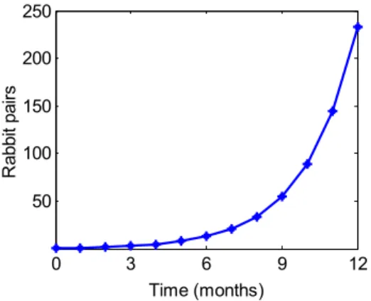
6. Fibonacci sequence growth curve of rabbit population ... 33
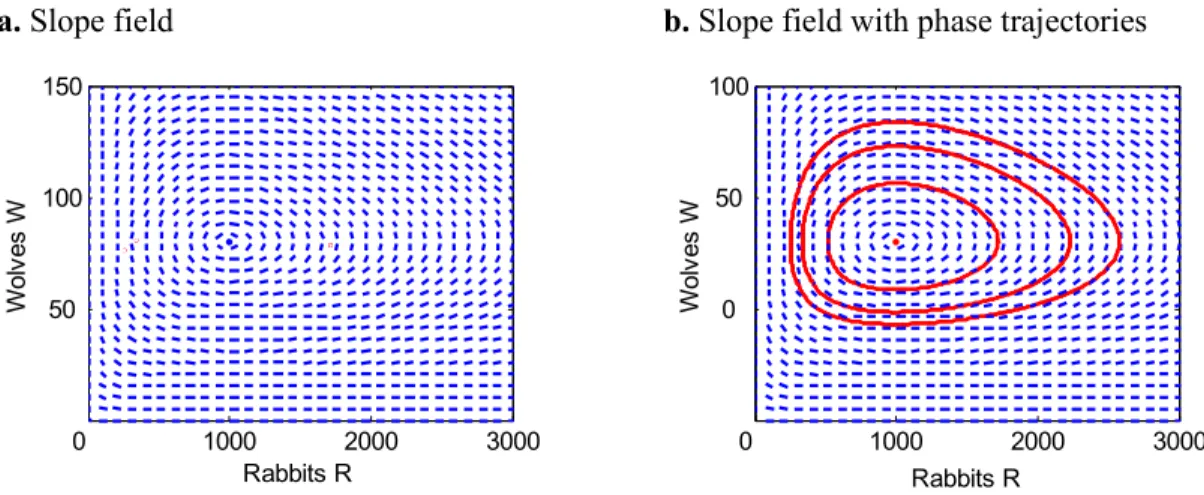
7. Qualitative plots (slope field and phase portrait) of the Lotka-Volterra system ... 37
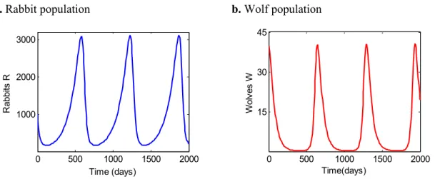
8. Simulations of the Lotka-Volterra system ... 38
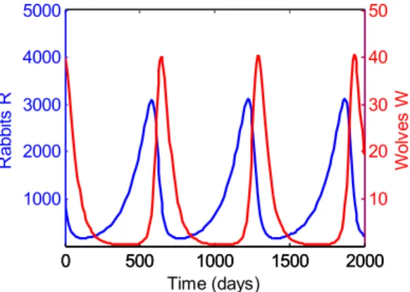
9. Solutions of Lotka-Volterra system superimposed ... 38
10. Ecological data of lynx-hare predator-prey system ... 39
11. Schematic diagram of the basic HIV immune system model ... 42
12. Schematic diagram of the initial proliferation process of HIV virions ... 43
13. Simulations of the basic HIV model for two regimes of R0 values: R0 1 and R0 1 ... 45
14. Schematic diagram of the Kirschner-Webb model ... 53
15. Simulations of the Kirschner-Webb model for three different regimes of the external viral source values: gV=5, 20, and increasing gV from 5 to 20 over time ... 54
16. Bifurcation diagram of Kirschner-Webb model created by varying gV from 5 to 20 ... 57
17. Two parameter bifurcation diagram for Kirschner-Webb model with growth rate of external viral source gV vs. number of virions produced from cell bursting N ... 58
18. Bifurcation diagram of Kirschner-Webb model created by varying
gV from 0 to 5 ... 61
19. Simulations of Kirschner-Webb model for early continuous and late continuous AZT monotherapy treatment ... 63
20. T cell counts for early and late treatment regimes superimposed ... 64
21. Simulations of the uninfected state for the Kirschner coinfection model ... 69
22. Simulations of the HIV infected state for the Kirschner coinfection model ... 70
23. Simulations of the TB infected state for the Kirschner coinfection model ... 71
24. Simulations of the HIV-TB coinfected state for the Kirschner coinfection model ... 72
25. Simulations of the primary and asymptomatic stage of HIV infection for new model ... 77
26. Simulations of the complete course of HIV infection for new model ... 79
27. Simulations of the HIV-TB coinfection for new model ... 80
28. Simulations of the simultaneous treatment of HIV and TB for new model ... 85
29. Simulation of premature cessation of TB treatment for new model ... 86
30. Simulations of treating TB then HIV at advanced stages of immune system collapse for new model ... 87
31. Simulations of treating TB then HIV at early stages of immune system collapse for new model ... 89
32. Simulations of treating HIV then TB for new model ... 91
Chapter 1
Introduction
HIV/AIDS represents a multifaceted challenge to society. Its burden is felt not only by the mortality rates associated with the disease, but also in terms of its social and economic impact. In order for humanity to come to terms with this scourge, there needs to be collaboration by the different groups of people who are involved in the fight against the disease. This need is most apparent in the scientific sphere. As an example the collaboration of mathematical and biological scientists has proved successful in revealing features of viral dynamics that were previously unknown. Mathematical models have been used to demonstrate the somewhat counter-intuitive fact that during the asymptomatic stage of HIV disease progression the viral turnover rate is approximately 10 billion HIV virions per day.
Such successes clearly encourage the development of mathematical models to yield insight into the quantitative and qualitative dynamics of disease progression. An area where this insight could potentially be useful is the problem of HIV-TB coinfection. Sub- Saharan Africa in particular has very high HIV-TB coinfection rates. TB is curable in HIV-positive individuals. In fact, if TB is not treated, the HIV disease progression increases in its rapidity and death soon results. Generally, when people are HIV-TB coinfected, their immune system is already well on its way to collapsing towards AIDS.
This means that the immune system is in a delicate state; therefore, death could result from either one of the two diseases. Even so, medical practitioners are reluctant to treat both diseases simultaneously due to the adverse compounded side-effects and undesirable drug-drug interactions of combining HIV and TB treatment. This problem is significant enough to warrant the running of medical trials in order to answer the question, when would the optimal point in time be to commence antiretroviral treatment in coinfected individuals? The START trials (STarting Anti-Retroviral Therapy) undertaken by the
Centre for the AIDS Programme of Research in South Africa (CAPRISA) in order to
Introduction
address the issues involving treatment of coinfection seek to ascertain whether the best time to start antiretroviral treatment for HIV-TB coinfected individuals is at the beginning, the peak, or after the completion of the TB treatment phase.
The main aim of the dissertation focuses on this HIV-TB treatment predicament. One might sense that the development of a mathematical model to simulate the treatment of HIV-TB coinfection could lead to some practical insight as to what the best strategy to treat for coinfection is likely to be. Since medical trials such as START are typically time intensive, a mathematical model could offer predictions to medical researchers while they wait for the trials to run its course. Furthermore, once the trials are completed, the model can be evaluated using the clinical data from the trials. Thus the central goal in undertaking this research was to develop a mathematical model to simulate the treatment of HIV-TB coinfection and to test the various strategies used to treat coinfection. By doing this one is able to offer some suggestions, in terms of the model’s results, as to what the optimal method of treating for coinfection is likely to be.
One should bear in mind that building and testing such a model requires an interdisciplinary approach. One needs firstly to be familiar with the biological aspects of the dynamics. Thereafter one needs to translate these biological dynamics into mathematical formalism by way of equations, which are then solved computationally in many cases. For this reason the contents of this dissertation can be viewed as having two distinct, but equally important, parts. The first part, Chapters 2 to 4, is devoted to describing the biological underpinnings of Human Immunodeficiency Virus (HIV), Tuberculosis (TB), the human immune system and the treatment therapies for HIV and TB. The second part, Chapters 5 to 8, deals with the mathematical modelling component of the research.
An introduction to the biological aspects of HIV-TB coinfection and its treatment was undertaken to familiarise the reader with the mechanisms of biological interactions of the system to be modelled. However, in attempting to understand the system one should not get bogged down by trying to understand the minute details of the system; rather one
Introduction
should familiarise oneself with those aspects of the system that would yield insight when translated into mathematical formalism. The above thought is astutely summarised by Albert Einstein [wikiquote.org],
“Things should be made as simple as possible, but not any simpler”
The discussion of the biological aspects of HIV and TB was done with this thought in mind. From a mathematical modeller’s point of view one needs only to understand the underlying mechanism by which the observed biological interactions between HIV, TB and the immune system take place. Once these interactions are translated into mathematical formalism they present themselves as nonlinear differential equations. As one finds later in the dissertation, even simple-looking nonlinear differential equations which have simple underlying mechanisms of interaction, can produce exceedingly complex dynamics. Happily for us the converse holds true as well in that the complex immunological dynamics which take place within the human body can be reproduced by simple underlying mechanisms. Therefore, as a mathematical modeller one need not attempt to explain the minute details of the biology, rather one need only understand the underlying mechanisms of interaction which are necessary to build a successful model, a model that yields insight into the immunological dynamics. The nitty-gritty of the biology should be left to the biologist.
However, with that said any person reading the biological account that follows in Chapters 2 to 4 will have a coherent and relevant understanding of the biology of HIV, TB and the immune system. Chapter 2 is dedicated to familiarising the reader with HIV, its transmission routes, molecular structure, characteristics of disease progression, replication cycle, drug therapies and historical accounts of the disease. Chapter 3 deals with describing the human immune system. The functioning and description of the immune system cells that are specifically involved during the pathogenesis of HIV and TB are described. Chapter 4 gives an account of Tuberculosis. A discussion is made of the epidemiological statistics, implications for coinfection with HIV, pathogenic mechanisms and the treatment of the disease.
Introduction
The second part of the thesis, Chapters 5 to 8, presents the mathematical modelling aspect of the research, where the goal is to create a mathematical model of HIV-TB coinfection with treatment. In order to achieve this objective the following strategy was pursued.
Initially the reader is familiarised with the mathematical modelling of biological systems by being introduced to various population models. Population models are relevant to modelling HIV since HIV models are merely in-host population models. This is followed by the review of various HIV models. Some of these HIV models included treatment mechanisms. The modelling of HIV-TB coinfection is then reviewed by the presentation of a previously developed HIV-TB coinfection model. All models reviewed – HIV, HIV with treatment and HIV-TB coinfection – made it possible to build a suite of models that each contained certain desirable mathematical features. Combining these different features separately and together culminated in the development of a final all-inclusive model of HIV-TB coinfection and its treatment.
The strategy outlined above to develop a mathematical model of HIV-TB coinfection is presented in Chapters 5 to 8. Chapter 5 introduces the reader to mathematical biological modelling by simulating and analysing well-known population and predator-prey models.
Chapter 6 reviews various HIV models. Model simulations, mathematical analyses and numerical bifurcation analyses are carried out. Mechanisms for simulating HIV treatment are added to the models. Treatment strategies and implication are discussed. Chapter 7 presents a model for HIV-TB coinfection. Simulations and discussions of the various permutations that the model displays are undertaken. The culmination of the dissertation is presented in Chapter 8. A new model is developed by using some of the mathematical structures found in the models that were previously presented. The model contains mechanisms that are able to simulate both the treatment of HIV and TB. Model simulations are carried out to evaluate the dynamics displayed by the model. Thereafter, the various treatment strategies used to treat HIV-TB coinfection are evaluated. The final chapter, Chapter 9, forms the conclusion of the dissertation. This chapter reviews the motivation for the dissertation, the research problems that were posed and the aims and objectives of the dissertation. A summary of the results from the model of HIV-TB coinfection with treatment is made. These results are then used to offer possible suggestions when treating for HIV-TB coinfection.
Chapter 2
Human Immunodeficiency Virus
2.1. Basic Virology
Viruses are pathogens that may cause death and disease to the living organisms that they infect. They are small particles that range in size from about 20 to 300 nano- metres [13, 29], and are probably the least complex of all life forms. In its basic form a virus consists of genetic material in the form of DNA or RNA. This is surrounded by a capsid, which is a casing composed of protein. Further to this it can also be encapsulated by a membrane consisting of a mixture of proteins and fatlike substances that serve to protect the viral genetic material [22]. Viruses are in fact so simple that they do not possess many of the characteristics used to define life. For example they cannot metabolise or reproduce on their own. In order to reproduce a virus must infect a living cell and use its host’s biological processes to replicate. Morbidity and mortality, which are associated with viral infection, are only side-effects of the virus’ primary reproductive objective.
The virus’ life cycle is typically categorised into a few key events. The replication cycle begins with the virus attaching itself to the host cell, thereafter inserting its genetic material into its host. The viral genetic material is then exposed by an uncoating process and viral proteins are produced. These newly produced viral proteins manipulate the host cell to replicate the viral genome. Yet more viral proteins are produced for the coat and membrane of the nascent virus. Finally the virus particles are assembled and the new virions are released from the host cell. The virions may undergo internal structural changes once they have budded from their host cell to form a mature virus capable of causing infection.
Human Immunodeficiency Virus
2.2. HIV: An Overview
Infection with Human Immunodeficiency Virus (HIV) results in Acquired Immunodeficiency Syndrome (AIDS), which is characterised by a failing immune system and susceptibility to opportunistic infections caused by fungi, bacteria, parasites and other viruses. Due to the collapsing state of the immune system the infected individual has an inability to fight off the constant assault of opportunistic infections, which finally results in death.
Human Immunodeficiency Virus belongs to a category of viruses known as retroviruses [49, 99]. Retroviruses have their genome in the form of RNA which has to be translated into DNA during its lifecycle. This is generally the reverse of what usually occurs during the biological processes found in nature, hence the prefix ‘retro’. Typically most biological processes proceed from DNA to RNA. In addition to being a retrovirus HIV is also a lentivirus [20, 99]. Lentiviruses are characterised by long incubation periods and long duration of illness. This has grave implications for the epidemiology of the disease since HIV-positive individuals can remain asymptomatic for many years and not know that they are infected, while spreading the disease to many others. HIV infection is spread by the transfer of infected bodily fluids such as blood, semen, vaginal fluid, or breast milk [6], which contain HIV present as free virus or as infected immune cells. Transmission can occur during any process that transfers infected bodily fluids from one individual to another. Typical routes of transmission are unprotected sexual intercourse, sharing of unsterilised hypodermic needles, usually amongst intravenous drug users, blood transfusions and from mother to child during breast feeding and child birth [124, 137].
2.3. HIV Virion Structure
The HIV genome encodes at least nine genes, these are - Gag, Pol, Env, Tat, Rev, Nef, Vif, Vpu and Vpr [99]. The first three genes are structural genes that encode for structural proteins used as building blocks, or are used in the construction of the building blocks, to
Human Immunodeficiency Virus
form the physical structure of new viral particles. The six remaining genes are regulatory genes, which encode for proteins that regulate the processes that the HIV virion performs, including viral replication by way of manipulation of the host cell’s biological processes.
Figure 1: Structure of HIV virion [USA. Fed. Gov.].
A pictorial representation of an HIV virion is shown in figure 1. It is approximately spherical and has a diameter of about 120 nanometers [82]. The HIV virion’s outer layer (viral envelope) is composed mainly of a bilayer of phospholipids [114]. Newly formed HIV virions are coated with the phospholipids bilayer as the viral capsid (core) emerges from the host cell during the budding process. The surface of the viral envelop is studded with the gp160 glycoprotein complex [18, 153, 154], which consists of a cap made from the glycoprotein gp120 and a stem made from the glycoprotein gp41. The surface glycoprotein complex enables the HIV virion to bind and subsequently fuse its capsid, including its genetic material, into its host cell [18, 51]. Just below the viral envelope is a matrix composed of the protein p17 [53, 67, 89]. The p17 protein performs important functions throughout the viral life-cycle, including anchoring the glycoprotein gp41 onto the surface of the virus, assisting in viral penetration, transporting the proviral integration complex across the nuclear envelope, and localizing the assembling virion to
Human Immunodeficiency Virus
the cell membrane [89]. Contained within the p17 matrix is the viral capsid, which is conical in shape and is composed of the protein p24 [1, 19]. The capsid contains the genetic material of HIV in the form of two copies of single-stranded RNA and enzymes such as reverse transcriptase and integrase, which are essential to the viral production process [3, 132].
2.4. HIV Replication Cycle
The replication cycle of HIV can typically be broken down into a number of steps. These steps are listed as: 1. Virion Binding and Capsid Insertion; 2. Reverse Transcription and Integration; 3. Transcription and Translation; 4. Assembly, Budding and Maturation.
2.4.1. Virion Binding and Capsid Insertion
Since HIV virions cannot replicate on their own they need to manipulate the biological processes of their host cells in order to do so. When an HIV virion comes into contact with a cell expressing CD4 on its cell surface, the gp120 glycoprotein spikes that protrude from the surface membrane of the HIV virion bind to the CD4 receptors [26, 32]. This binding induces a conformational change in the gp120/gp41 glycoprotein complex which allows the gp120 glycoprotein to bind to the chemokine coreceptors CXCR4 or CCR5 [36, 38, 51, 146]. This in turn induces an additional conformational change that
exposes the gp41 glycoprotein and facilitates the entry of the viral capsid into the host cell [18, 48, 78, 106].
2.4.2. Reverse Transcription and Integration
Once the HIV virion has penetrated the cytoplasm of its host cell an uncoating step leads to the release of the viral RNA from its core [15]. The reverse transcriptase enzyme then copies the viral RNA into DNA [124]. The viral DNA is then transported into the nucleus of the host where the integrase enzyme facilitates the splicing of the viral DNA into the
Human Immunodeficiency Virus
host cell’s DNA [21, 152]. The viral DNA that has been integrated into its host genome is known as a provirus.
2.4.3. Transcription and Translation
In the nucleus of the cell transcription of the cellular DNA into RNA, which are used as blueprints for protein building, takes place. At the same time RNA copies of the viral DNA, which has been spliced into the cellular DNA, are produced [58]. These copies are messenger RNA (mRNA) and travel to the cell’s cytoplasm [47], where the Env precursor polyprotein gp160 is synthesized in the endoplasmic reticulum with subsequent post-translational modification of the Env polyproteins occurring at the endoplasmic reticulum and Golgi apparatus to form the gp120/gp41 glycoprotein complex [132]. The Gag and Gag-Pol polyproteins are synthesized in the ribosomes. The Gag polyprotein is the precursor to the matrix, capsid and nucleocapsid proteins [47, 65]. The Gag-Pol polyprotein is the precursor to the matrix and capsid proteins, as well as the protease, reverse transcriptase and integrase enzymes [119]. The polyproteins are transported to the cell membrane where viral assembly takes place.
2.4.4. Assembly, Budding and Maturation
During or after transport of the Gag and Gag-Pol polyproteins to the cell membrane the Gag precursor recruits two copies of the single-stranded viral RNA genome, interacts with the Gag-Pol precursor and assembles into structures lining the inner face of the plasma membrane [47]. The Env glycoproteins, which form the viral envelope spikes, are also incorporated into the assembled structures. The assembled Gag protein complex, which encapsidates the two copies of the viral genome, buds from the plasma membrane, see figure 2, taking some of the membrane with it [132]. At this stage the particles are not yet infectious. They go through a maturation process during which the Gag and Gag-Pol
…
Human Immunodeficiency Virus
polyproteins are cleaved by the protease enzyme into the proteins and enzymes that make up a functional HIV virion and triggers the rearrangement of the cleavage products to form mature HIV virions capable of causing infection [59].
Figure 2: Electron microscope image of HIV virion budding [CDC].
2.5. HIV Disease Progression
The clinical course of HIV infection shown in Figure 3, leading ultimately to AIDS, varies between individuals in terms of viral load, symptoms and duration. However, there are four typical stages during disease progression that an HIV-positive individual experiences. These four stages were classified in 1990 by the World Health Organisation (WHO) [142] and are intended to be a means of establishing disease prognosis in a resource limited setting, such as one would typically find in Sub-Saharan Africa. These stages are classified as: Stage One – Primary Infection, Stage Two – Asymptomatic Infection, Stage Three – Symptomatic Infection, and Stage Four – AIDS.
The World Health Organisation revised its classification of the various HIV stages in 2005; however, both classification systems are similar. For the sake of simplicity, as is necessary for mathematical modelling purposes, a review is made of the initial classification system.
Human Immunodeficiency Virus
Figure 3: Graph of generalised HIV disease progression [Wikimedia Commons].
2.5.1. Primary Infection
Subsequently to initial infection with HIV viral replication commences in cells expressing CD4 receptors, primarily in CD4+ T cells. Individuals develop high viral loads that typically peak somewhere in the range 103–105 virions per mm3 of blood [124], resulting in high numbers of infectious virus and infected cells circulating in the peripheral blood. Within five to six weeks individuals may develop acute HIV syndrome, which is characterised by flu-like symptoms. This lasts for about a week, at which time the immune system mounts HIV specific immune responses, and the individual’s flu-like symptoms disappear. This is accompanied by a decrease in the viral load of about one to two orders of magnitude, which is largely due to HIV specific CD8+ T cell immune response; however, a limitation in the number of cells susceptible to HIV infection could also play a part [124, 127]. The CD8+ T cells ability to decrease viral load is an important factor since the rapidity of disease progression is correlated with the level (set point) the viral load settles down to shortly after primary infection [91]. The set point viral load is characterised by being relatively stable over prolonged periods of time due to approximately equal rates of production and clearance of HIV virions.
Human Immunodeficiency Virus
2.5.2. Asymptomatic Infection
The viral load settling to its set point value signals the onset of the asymptomatic phase.
This phase is characterised by clinical latency, shown in Figure 3, during which the HIV- positive individual remain free from any major symptoms. The typical duration of the asymptomatic phase is from 8 to 11 years [93, 129]. However, about 22% of individuals progress to AIDS within 5 years [96] and an estimated 8–15% of individuals will remain free from any AIDS defining illnesses for 20 years [95]. Even though HIV-positive individuals do not show AIDS related symptoms during this phase, the internal dynamics of the disease are not static in that there is a high turnover rate of HIV virions of the order 1010 virions per day [107], accompanied by a gradual decline in the CD4+ T cell count [132].
2.5.3. Symptomatic Infection
Characteristic of disease progression through the various stages of HIV is an increase in viral load accompanied by a decrease in CD4+ T cell count [73]. The increase in viral burden is temporally associated with the emergence of viral mutants displaying enhanced pathogenicity [25]. The failing immune system coupled with the high viral loads allows opportunistic infection, which would otherwise be controlled by the immune system, to appear. The diseases and symptoms which characterise WHO stage 3 are: unexplained weight loss of more than 10%, chronic diarrhoea for more than a month, prolonged fever (constant or intermittent) for more than a month, oral thrush, oral hairy leukoplakia, pulmonary tuberculosis and severe bacterial infections (i.e. pneumonia, pyomyositis). An individual in stage 3 of HIV infection would typically be bedridden for up to 50% of the day towards the end of this stage.
2.5.4. AIDS
The final stage of disease progression is characterised by the severe impairment of the
immune system with an associated CD4+ T cell count of less than 200 per mm3 of blood.
Human Immunodeficiency Virus
As a result a whole host of life-threatening opportunistic infections, c.f. WHO report [142], known as AIDS Related Complexes (ARCs) appear. Without antiretroviral therapy an individual with AIDS would live on average for 9.2 months [93]. The average survival time, using the present treatment strategies, of individuals diagnosed with AIDS, is in excess of 5 years [123].
2.6. HIV/AIDS Drugs and Treatment Strategies
Antiretrovirals, which have been approved to treat HIV, come primarily in two variants [124] – reverse transcriptase inhibitors and protease inhibitors. Protease inhibitors are designed to inhibit the activity of the HIV protease enzyme by preventing it from cleaving the Gag polyprotein into the proteins and enzymes that make up a functional HIV virion. Reverse transcriptase inhibitors are designed to inhibit the HIV reverse transcriptase enzyme from transcribing the viral RNA into DNA, thereby foiling an essential part of the HIV life-cycle. Reverse transcriptase inhibitors fall into two categories, nucleoside analogues or nonnucleoside analogues. Nucleoside analogues function as chain terminators. They are incorporated into the growing DNA molecule by mimicking the structure of the host cell’s DNA building blocks and thereby preventing its extension. Since they are structurally different from the host cells DNA when the viral DNA combine with these DNA building blocks they cannot integrate into the host cells DNA. A nonnucleoside analogue on the other hand binds to the HIV reverse transcriptase enzyme and disrupts the enzyme’s ability to transcribe the viral RNA into DNA.
In addition to the above-mentioned antiretroviral drugs, there are several drugs in the developmental stage that target other key aspects of the HIV life-cycle. Fusion inhibitors block an HIV virion’s ability to fuse with a candidate host cell’s membrane, thereby rendering it unable to enter and infect the host cell. Integrase inhibitors disrupt the activity of the HIV integrase enzyme which facilitates the integration of the viral DNA
…
Human Immunodeficiency Virus
into the host cell’s DNA. Entry inhibitors block an HIV virion’s ability to enter a candidate host cell by binding to the CCR5 chemokine coreceptor, a vital mechanism of host cell entry.
Current treatment strategy for HIV is termed HAART (Highly Active Antiretroviral Therapy). There is much debate about the optimal timing during disease progression when HAART should commence. However, HAART is typically commenced when an individual’s CD4+ T cell count is less than 200 per mm3 of blood or when an AIDS-related complex has been identified. HAART has several adverse side-effects;
however, this is outweighed by its ability to reduce morbidity and mortality. Nevertheless HAART cannot completely eradicate HIV so treatment is generally continued for the duration of the illness [124]. HAART consists of a combination of at least three drugs belonging to two different classes, typically two nucleoside analogue reverse transcriptase inhibitors and either a protease inhibitor or a nonnucleoside analogue reverse transcriptase inhibitor. By combining these antiretroviral drugs one is able to suppress viral replication to low levels, thereby decreasing viral load and giving the immune system an opportunity to recover. Equally importantly, by slowing down the HIV replication rate, the emergence of drug-resistant HIV mutants is at least delayed if not prevented [124].
2.7. Viral Mutations and Drug resistance
Viral mutations are a consequence of the high HIV virion turnover rate of the order 1010 virions per day [107] and the fact that the reverse transcription step, which transcribes the genomic viral RNA into proviral DNA, is highly error prone and lacks proof-reading capabilities [112, 117]. This results in the production of genetically heterogeneous HIV virions, characterised by the appearance of mutant strains of the disease. Genetically heterogeneous HIV virions, called quasispecies, can have up to 15% diversity between them in an HIV-infected individual [88]. Genetic variation is thought to be one of the primary factors which enable HIV to escape eradication by the immune system.
Human Immunodeficiency Virus
During antiretroviral therapy the selective forces to which the viral mutants are subjected allow the virus to develop drug resistance; moreover HIV drug-resistant strains can be transmitted from one individual to another [148]. HIV mutants, with varying degrees of resistance, have been found for all drugs that have been used, or are being considered for use, as anti-HIV therapy [23]. The emergence of anti-HIV drug resistance is the most common cause of treatment failure. Nevertheless treatment failure due to drug resistance can be reduced by ensuring strict compliance with the antiretroviral administration routine and by using a cocktail of multiple antiretroviral drugs simultaneously, in most cases three drugs, which slows the replication rate of the virus, thereby curtailing the mutation rate.
2.8. Origin of HIV
HIV seems to be a zoonotic infection crossing species from African chimpanzees to human beings [52, 63] establishing two different strains of the disease. HIV-1 and HIV-2 are genetically distinct and crossed from different chimpanzee subspecies [80]. HIV-1 is responsible for the global HIV pandemic; therefore all references to HIV in this dissertation pertain to HIV-1 unless otherwise explicitly stated. HIV-1 is closely related to a strain of Simian Immunodeficiency Virus, classified as SIVcpz, harboured by the Pan Troglodytes Troglodytes chimpanzees native to West Equatorial Africa [52]. The geographic location of origin of HIV-1 is most likely the Democratic Republic of Congo.
Infected individuals in that country have a constant and extremely high genetic diversity of HIV-1 stemming from the likelihood that the disease was present there for the longest period, having more time to mutate relative to anywhere else on the globe [134, 135].
Estimates of the timescale of SIVcpz crossing species into human beings, introducing the HIV-1 disease, place the event around 1930 [80, 121].
The HIV-2 strain is less virulent and not as easily transmissible as HIV-1 and is largely confined to West Africa [83]. Like HIV-1, however, HIV-2 is also closely related to a strain of Simian Immunodeficiency Virus, classified as SIVsm, harboured by the Sooty
Human Immunodeficiency Virus
Mangabey monkeys indigenous to West Africa [52]. A reconstruction of the HIV-2 lineage puts the likely location of SIVsm crossing species to establish the HIV-2 disease in the West African country of Guinea-Bissau around 1940; however, the disease only experienced rapid exponential growth, establishing itself as an epidemic, during the period 1955–1970 [111]. This period coincides with Guinea-Bissau’s war for independence which most likely created the social conditions necessary for the rapid spread of the disease [83].
2.9. History of HIV
The earliest cases of HIV on record are plasma samples taken in 1959 of a man in the Democratic Republic of Congo [155], tissue samples of an American teenager who died in Saint Louis in 1969 [103] and tissue samples of a Norwegian sailor who died around 1976 [72]. It was, however, only in 1981 that the disease was clinically diagnosed and its existence brought to the attention of the global medical community when a group of homosexual men in Los Angeles, San Francisco and New York were afflicted by rare diseases caused by the suppression of the immune system [101].
In 1969, when the disease was initially introduced into the United States of America, it was unable to take off. Possible reasons for this could be that the disease needed carriers who engaged in frequent sexual intercourse with large numbers of partners in order to establish itself as an epidemic, a condition that was likely met by the homosexual population that initially carried the disease. By 1982 there were over 800 cases of AIDS in the United States. It spread from the initial homosexual population to haemophiliacs, who received blood derived products from donors, intravenous drug users, and the heterosexual population.
With the disease rapidly spreading, the search was now on to identify its causative agent.
In 1983 Luc Montagnier and his team, working at the Pasteur Institute in Paris, identified the presence of a retrovirus in the lymph node tissue obtained from infected
Human Immunodeficiency Virus
individuals [4]. Soon afterwards Robert Gallo and his team, working at the National Institute of Health in Bethesda, Maryland, were also able to identify the retrovirus from infected patients [50, 122]. In addition the team at the NIH was able to multiply the retrovirus in continuous culture and was thereby able to develop a specific test for the disease [110]. This was a significant step which now gave health workers the ability to accurately diagnose the status of infected individuals and identify contaminated blood samples.
Since then there has been a wealth of research performed on the disease and numerous important features of the retrovirus’ structure and dynamics have been established.
Nevertheless there is still no vaccine or cure for the disease and there remains much research to be done in order to improve our understanding of the disease so that a vaccine or cure may be developed.
Chapter 3
Human Immune System
3.1. Immune System: An Overview
The immune system consists of organs, tissues and cells that function as an integrated unit to protect the body by overcoming infection due to pathogens such as viruses, bacteria, fungi and parasites. The immune system consists of the lymph vessels, lymph nodes, thymus, spleen, bone marrow and an elaborate network of interacting white blood cells [29]. At a fundamental level the immune system defence can either be adaptive or innate.
The adaptive immune system response is specific in that it adapts the immune response to maximise its effectiveness to eliminate a specific pathogen. After the immune system has eliminated the pathogen, it develops immunological memory whereby it is able to launch a quicker and more effective defence against the pathogen if it subsequently reappears.
The innate immune system response is nonspecific. It does not differentiate between the different types of pathogens that initiate the immune response. Thus it deals with all pathogens in a generic way. As a consequence of its generic manner, even for pathogens it has encountered before, the innate immune system does not develop immunological memory. Previous exposure to the pathogen would not be of any added advantage during future encounters.
Human Immune System
3.2. Immune System Cells
White blood cells are the major protagonist of the immune system’s defence against foreign substances (antigens). There are various subcategories amongst white blood cells that perform important functions in a healthy immune system. Foremost among these are the lymphocytes, which are made up of B cells and T cells, as well as the phagocytes, namely the macrophages.
3.2.1. B cells
B cells are produced in the bone marrow. They are able to recognise antigens that invade the body. B cells have antibody molecules on their cell surface that are able to identify localised regions, known as antigenic determinants, found on the surfaces of antigen molecules. The antigen binding site, found on specific regions on the antibody molecule, binds to an antigen if the antigenic determinant and the antigen binding site have complementary shapes and fit like a lock and key. The ability to combine the protein subunits that make up the antibody molecule in a vast number of different ways ensures that the antibody molecule binds to almost any antigen entering the body.
When the diverse array of B cells, each with differing antibody molecules on their cell surface, encounter an antigen only the B cell with the antibody molecule that best fits the antigenic determinant of the given antigen is activated to divide and form clones. This process, called clonal selection, ensures a high specificity for antibody-antigenic binding.
Adding further to the specificity of the process is somatic mutation, whereby antibodies with slightly differing shapes are produced and might have a greater binding affinity to the given antigen. Variants that bind better have a stronger signal to multiply, thus replicating more rapidly and effectively outcompeting the other B cells that do not have as high a specificity. This process, called affinity maturation, ensures optimal specificity of the antibody-antigen binding. The stronger replication signal that accompanies the dynamics of greater specificity also ensures that large numbers of antibodies are
…
Human Immune System
produced only when the body needs them. Furthermore in subsequent encounters with the given antigen the immune system is able to launch a faster and more effective attack due to some clones becoming long-lived memory cells.
B cells release their antibody molecules into the blood. The antibodies mark the pathogen for elimination by other immune cells such as, for example, phagocytes and the complement system of blood enzymes. The antibodies might even be able to neutralise the pathogen on their own by forming a coating so dense that it effectively prevents the pathogen from reproducing or invading target cells.
3.2.2. T cells
T cells are produced in the bone marrow, but undergo a maturation process in the thymus where they develop into functional immune cells that are differentiated according to the various tasks that each subcategory of T cells is required to carry out. The CD8+ T cells are the killer cells, while the CD4+ T cells are the helper cells. They are differentiated by the CD (cluster of differentiation) glycoprotein T cell receptor on their cell surface, either CD4 or CD8.
Vital to the proper functioning of the immune system is its ability to differentiate between self and nonself, ensuring it does not mount an immune response against its own cells and tissues by falsely identifying them as foreign, as is the case during autoimmune disease.
The task of differentiating between self and nonself lies with the T cells.
When a virus infects a T cell, it undergoes antigen processing whereby the cell processes the viral proteins into smaller fragments, known as peptides. Parts of the peptides are then bound on the cell surface to the Major Histocompatibility Complex (MHC) molecules.
T cell receptors bind to these MHC-peptide complexes; however, the interaction has a high degree of specificity and binding only occurs according to fit, analogously to the lock and key mechanism. During the T cell maturation process in the thymus, cellular peptides of immune system cells form MHC-peptide complexes that are presented to the
Human Immune System
T cells for binding. Those T cells that are adequately able to bind to the MHC-peptide complexes are allowed to survive. The remaining T cells die by apoptosis, a process of programmed self destruction of cells. This mechanism of identifying T cells that are able to bind to MHC-peptide complexes is known as positive selection. However, in order to ensure that the T cell do not mistake normal cellular peptides for pathogenic peptides, the T cells also undergo negative selection. During negative selection those T cells that bind only weakly with self MHC-peptide complexes are allowed to survive. The rest of the T cells, which bind too strongly with the self MHC-peptide complexes, also undergo apoptosis in order to avoid autoimmune disease. T cells that survive the positive and negative selection processes can mature to CD4+ or CD8+ T cells, which are differentiated by the class of MHC molecule on antigen presenting cells to which their receptors bind. CD8+ T cell receptors bind to MHC class one (MHC-1), while CD4+ T cell receptors bind to MHC class two (MHC-2).
3.2.2.1. CD8+ T cells
The CD8+ T cells are cytotoxic. If a cell is infected by a virus, the cell processes the viral proteins into antigenic peptide fragments and transports them to the cell surface where they are bound to MHC-1. The MHC-peptide complexes are presented on the cell surface. CD8+ T cells with the correct specificity are able to bind to the MHC-peptide complex and kill the infected cells by the release of perforins, proteins that cause death to the cell by puncturing its cellular membrane [14]. Once the CD8+ T cells are activated they undergo clonal expansion, producing more CD8+ T cells of the same specificity ready to kill the remaining virally infected cells. CD8+ T are also equipped with chemicals and defence mechanisms that can inhibit infected cells from replicating or infecting other cells.
3.2.2.2. CD4+ T cells
The CD4+ T cells cannot by themselves kill infected cells. Nevertheless they do have an important role to fulfil in terms of regulating and optimising the various aspects of
Human Immune System
cell-mediated immunity, for example CD4+ T cells are necessary for the proliferation of activated CD8+ T cells. They also fulfil a crucial role when establishing an antigen specific antibody immune response. Antigen Presenting Cells (APC), such as macrophages and B cells, internalise pathogens and present antigenic peptides bound to MHC-2 on their cell surface, which are then recognised by CD4+ T cells. CD4+ T cells of the correct specificity that are able to bind to the MHC-peptide complex ensure that the B cells are able to distinguish between self and nonself, while signalling to the B cells to increase its numbers via clonal expansion and produce more antibodies for the given antigen.
3.2.3. Macrophages
Macrophages are phagocytes that engulf and digest antibody-bound pathogens and other debris, such as dust, dead cells and tissue. Macrophages are derived from monocytes, white blood cells produced in the bone marrow that mature through a series of changes to become a macrophage. As is the case with B cells, macrophages are also antigen presenting cells. Subsequently to digesting an antigen they break the antigen down into smaller peptide fragments and present the antigenic peptides bound to MHC-2 on their cell surface. Circulating T cells are then able to recognise this MHC-peptide complex and become activated to mount a specific immune response against the antigen, which includes the stimulation of B cells to produce more antibodies for the given pathogen.
Antibody-coated pathogens are cleared more easily by the immune system. This antigen-specific immune response strengthens the ability of the immune system to overcome the pathogen.
Chapter 4
Tuberculosis
"We cannot win the battle against AIDS if we do not also fight TB. TB is too often a death sentence for people with AIDS. It does not have to be this way."
– Nelson Mandela, XV International AIDS conference, Thailand 2004
4.1. Tuberculosis: An Overview
Tuberculosis (TB) is a contagious disease which typically results from being infected with the bacteria Mycobacterium tuberculosis, which is a rod-shaped bacillus [29].
Figure 4: Electron microscope image of Mycobacterium tuberculosis [CDC].
TB is one of the oldest known diseases, with evidence of infection found in the vertebra of Stone Age Man and Egyptian mummies; as well as in records from ancient Greece, Rome, India, China and medieval Europe [55, 94, 98]. TB infection is spread through the air. It occurs by the inhalation of aerosol bacilli of its causative agent. TB commonly
infects an individual’s lungs. This form of TB is known as Pulmonary Tuberculosis. As a
Tuberculosis
result of active infection granulomas subsequently develop at infected sites in the lungs.
These granulomas consist predominantly of aggregates of T cells and TB infected macrophages [68]. Granulomas undergo cell death, referred to as necrosis. Lung cavitation can result from necrosis due to the destruction of lung tissue. TB infection is not only confined to the lungs. Infection in other organs and tissues does occur as a result of bacilli migration via infected dendritic cells and peripheral blood.
4.2. Tuberculosis Epidemiology
TB is a major public health problem in most of the developing world. In Sub-Saharan Africa the problem is aggravated by the fact that TB is the main opportunistic disease of HIV [34, 40, 61]. As data became available, the World Health Organisation was able to make estimates for HIV-TB epidemiology for 2000, then 2002 and recently for 2004.
Estimates for 2000 showed that TB infections attributable to HIV in WHO African member states was about 31%, with South Africa estimated to have had 2 million of the global total of 11 million HIV-TB coinfected individuals [27]. The 2002 estimates for HIV prevalence amongst adult TB patients revealed that Southern Africa faced grim prospects, with Zimbabwe (75%) and South Africa (60%) ranking amongst the worst affected [144]. Estimates for 2004 showed coinfection prevalence for the African continent increased to about 33%, with Southern African countries still faring poorly – Swaziland (81%), Botswana (77%), Lesotho (76), Zimbabwe (68%), Namibia (61%), South Africa (60%), Zambia (54%) and Malawi (52%) all being severely affected [145].
4.3. Tuberculosis-HIV Coinfection
Many people infected with HIV develop TB as the first manifestation of AIDS.
HIV-positive individuals do not have the internal immune system resources to keep TB in check. In fact they are more than 20 times likely to develop active TB than people who are HIV-negative as the suppression of the immune system worsens [115]. Studies have shown that the host’s immune response to the TB bacterium enhances HIV replication
Tuberculosis
and might accelerate the natural progression of the HIV infection [57, 97, 149]. In addition HIV can also accelerate disease progression in TB infection [31]. Therefore without treatment dually-infected individuals succumb to coinfection at a rapid rate.
4.4. Tuberculosis Pathogenesis
The etiological agent of TB is Mycobacterium Tuberculosis, first identified in 1882 by Robert Koch [79]. Mycobacterium Tuberculosis is an intracellular pathogen primarily infecting macrophages. TB infection begins when an uninfected individual inhales aerosol droplets containing Mycobacterium Tuberculosis. TB bacilli are expelled from the lungs of infected individuals when they cough. Once the bacilli are airborne they are inhaled by other individuals and typically get lodged in their alveoli, the tiny air sacs of the lung where gaseous exchange of oxygen and carbon dioxide take place [56]. Here the pathogen is engulfed and ingested by alveolar macrophages. Normally once pathogens have been phagocytosised by macrophages, they are transported to the macrophage’s lysosomes, where digestive enzymes break down and destroy the pathogen. However, it seems that mycobacteria may be able to block their delivery to the lysosomes enabling them to survive within the macrophages and start to replicate [68]. The ability of bacilli to evade the initial nonspecific immune response depends upon various factors. These include the state of the immune system, where diseases like HIV may inhibit immune responses, as well as the virulence factor of the mycobacteria [125]. The more virulent strains of TB are characterised by greater transmissibility and produce greater morbidity and mortality in infected individuals.
The replication of bacilli within the macrophages attracts blood monocytes that mature into macrophages and ingest the released bacteria. During this stage in disease progression symbiosis occurs between bacteria and macrophages, with neither population eliminating the other. Bacteria continue replicating while blood-derived macrophages continue arriving. Macrophages that have ingested the mycobacteria process and present mycobacterial antigens to T lymphocytes. Two to three weeks after infection cell-
Tuberculosis
mediated immunity develops with the arrival of antigen-specific T lymphocytes that intensifies the immune response and activates macrophages to kill the intracellular mycobacteria [133]. In addition to the activated cell-mediated response the immune system also initiates an inflammatory response. The inflammatory response serves to inhibit dissemination of the mycobacteria by encapsulating mycobacteria infected macrophages, activated macrophages, and T cells within granulomas. The granulomas subsequently develop central areas of necrosis which kill most of the mycobacteria;
however, the pathogen is almost never totally eradicated. Nevertheless, the vast majority of the TB-harbouring population, approximately 90% of TB infected individuals, are able adequately to contain the bacteria and do not progress to any symptoms of active TB [46]. In fact it is estimated that latent TB may be carried by a third of the global population [42]. From the remaining 10% of infected individuals the immune systems of approximately 5% are not able to adequately contain the bacteria and active TB results within a year of infection, with the other 5% going on to develop active TB at some later stage in life [139]. Immunosuppression, such as infection with HIV, greatly increases the risk of reactivation of latent TB amongst previously infected individuals. Under these conditions granulomas can become liquefied and are able to serve as a rich medium for the revived bacteria to replicate prodigiously [125].
4.5. Tuberculosis Treatment
The treatment of TB aims to cure patients of the disease, prevent death from active TB, prevent relapse of TB, decrease disease transmission and prevent development of acquired drug resistance [143]. In order to achieve these aims a combination of anti-TB drugs are used. The most widely used first-line treatment anti-TB drugs are Isoniazid, Rifampin, Pyrazinamide and Ethambutol. A typical treatment regimen involves taking Isoniazid, Rifampin, Pyrazinamide and Ethambutol for two months, then Isoniazid and Rifampin for the next four months. It is essential to maintain a strict adherence to the
…
Tuberculosis
drug intake regime as skipping a treatment or not completing the full course could result in a multidrug resistant strain of TB. Multidrug resistant TB takes longer to treat, treatment is more expensive, side-effects associated with the treatment are greater and treatment success rates are smaller.
In order to avoid such complications a TB control strategy has been developed from the collective best practices, clinical trials and programmatic operations of TB control over the past two decades [126]. This strategy, known as Directly Observed Treatment Short- course (DOTS), is widely regarded as being the most effective strategy for treating TB and is recommended by the World Health Organisation. The South African Department of Health has also adopted the DOTS strategy to treat TB by implementing its core features [126], which involve sputum smear microscopy to detect infectious cases among people attending health care facilities with symptoms of TB, standardised short-course anti-TB treatment with direct observation of treatment, an uninterrupted supply of TB drugs and a standardised recording and reporting system which allows assessment of treatment results.
4.5.1. Antituberculosis Drugs 4.5.1.1. Rifampicin
Rifampicin may be bacteriostatic, by inhibiting the multiplication of bacteria, or bactericidal, in that it kills bacteria. The bacteriostatic or bactericidal action of Rifampicin depends on the susceptibility of the bacterial species and the concentration of the drug. In the case of M. tuberculosis Rifampicin is bactericidal. In particular it possesses early bactericidal activity against active and semidormant bacterial populations, producing a sterilizing effect [71, 130]. Rifampicin inhibits the action of DNA-dependent RNA polymerase [17, 70], which is the enzyme that promotes the synthesis of RNA using a DNA or RNA template, thereby leading to the suppression of RNA synthesis and the subsequent translation to proteins in susceptible bacteria.
Tuberculosis
4.5.1.2. Ethambutol
Ethambutol is used in the initial phase of TB treatment primarily to prevent drug resistance to Rifampicin [7]. Ethambutol is generally considered to have a bacteriostatic effect on M. tuberculosis when used at typical doses during TB treatment [5]. The exact mechanism of interaction between Ethambutol and M. tuberculosis is still unknown;
however, several hypotheses have been advanced. The most prominent amongst these theories propose that ethambutol interferes with the biosynthesis of arabinogalactan, a constituent of the mycobacterial cell wall [37]. A further theory suggests that Ethambutol acts as a chelating agent, capable of forming bonds with metals, and disrupts and deactivates one of several metal-containing enzyme systems in the nucleic acid structures of mycobacteria [90].
4.5.1.3. Isoniaziad
Isoniazid is bacteriostatic against dormant and semidormant mycobacteria and bactericidal against rapidly dividing mycobacteria. Its primary use stems from its potent early bactericidal action against actively dividing mycobacteria. Isoniazid’s therapeutic mechanism results from its ability to inhibit the synthesis of mycolic acids, a major constituent of the mycobacterial cell wall [140]. Isoniazid inhibits the biosynthesis of mycolic acids by specifically targeting the enzyme InhA, which is essential to the biosynthesis process [118]. Mycolic acids form a lipid shell, which serves to determine permeability properties at the cell surface. They endow several unique characteristics to M. tubeculosis and are important to the growth, survival and pathogenicity of the mycobacteria [113].
4.5.1.4. Pyrazinamide
The concomitant use of Pyrazinamide with the other anti-TB drugs plays a unique role in shortening TB therapy from the previous 9–12 months to 6 months [151]. Pyrazinamide exerts its therapeutic effects by inhibiting the synthesis of fatty acids in M. tuberculosis
Tuberculosis
by disrupting the activity of the enzyme fatty acid synthetase I [156]. The drug is converted into its active form, pyrazinoic acid, by the M. tuberculosis enzyme pyrazinamidase. Pyrazinamidase, and consequently pyrazinamide, are only active at an acidic pH [150]. Once in its active form the drug shows bactericidal activity against M. tuberculosis, including bacilli within macrophages, presumably because of its acidic intracellular ph [5]. Its importance within the anti-TB drug regime stems from its sterilizing ability of dormant and semidormant tubercle bacilli, which are unaffected by other drugs [69, 151].
Chapter 5
Mathematical Biology
Mathematics can be applied to various systems in nature in order to gain qualitative and quantitative insight into the system. One can use mathematics to develop a model of the system, as has been the case in this dissertation where the aim was to develop a mathematical model of the interaction between HIV, TB and the human immune system, including the effects of drug therapy.
Many systems found in nature can be modelled by nonlinear coupled differential equations. Mathematical modelling is an iterative process in which one refines the model by successive iterations. The steps involved in modelling natural phenomena, as can be seen in Figure 5, are studying the phenomena, formulating a mathematical model of the phenomena, solving the model analytically or computationally, analysing and interpreting the solution, using the results to make predictions and gain insights about the phenomena and testing the validity of the insights and predictions.
Before a mathematical model is formulated one has to construct a verbal model of the underlying mechanisms of interaction of the system. One must then identify and understand the various parameters and variables that make up the system. Thereafter, one can combine the parameters and variables to form mathematical equations that follow certain rules, which according to our experience and knowledge, govern the system.
Often in order to make the equations mathematically tractable one has to simplify and generalise these rules. Thus in essence most mathematical models are only idealised approximations of the phenomena being studied. The model does not necessarily have to reproduce exactly what happens in nature. It needs only to reproduce the salient features of the system so that one is able to approximate its future behaviour, as well as make quantitative estimates of the different parameters that govern the system.
Mathematical Biology
After the initial steps of studying the system and building the mathematical model have been completed, the next step is to find solutions to the equations. In general one cannot solve the equations analytically; therefore one ha
![Figure 1: Structure of HIV virion [USA. Fed. Gov.].](https://thumb-ap.123doks.com/thumbv2/pubpdfnet/10706757.0/16.918.267.654.267.579/figure-1-structure-hiv-virion-usa-fed-gov.webp)
![Figure 2: Electron microscope image of HIV virion budding [CDC].](https://thumb-ap.123doks.com/thumbv2/pubpdfnet/10706757.0/19.918.230.690.258.564/figure-electron-microscope-image-hiv-virion-budding-cdc.webp)
![Figure 3: Graph of generalised HIV disease progression [Wikimedia Commons].](https://thumb-ap.123doks.com/thumbv2/pubpdfnet/10706757.0/20.918.139.786.110.468/figure-graph-generalised-hiv-disease-progression-wikimedia-commons.webp)
![Figure 4: Electron microscope image of Mycobacterium tuberculosis [CDC].](https://thumb-ap.123doks.com/thumbv2/pubpdfnet/10706757.0/32.918.335.587.597.808/figure-electron-microscope-image-of-mycobacterium-tuberculosis-cdc.webp)
![Figure 5: Flowchart of mathematical model development [128].](https://thumb-ap.123doks.com/thumbv2/pubpdfnet/10706757.0/40.918.240.680.423.666/figure-5-flowchart-mathematical-model-development-128.webp)
![Figure 10: Ecological data of lynx-hare predator-prey system. One finds that the coupled oscillations shown in solutions to the Lotka-Volterra system are indeed found in nature [147].](https://thumb-ap.123doks.com/thumbv2/pubpdfnet/10706757.0/48.918.241.671.248.450/figure-ecological-predator-coupled-oscillations-solutions-lotka-volterra.webp)
