IN SILICO IDENTIFICATION OF SELECTIVE NOVEL HITS AGAINST THE ACTIVE SITE OF WILD TYPE MYCOBACTERIUM
TUBERCULOSIS PYRAZINAMIDASE AND ITS MUTANTS
A thesis submitted in partial fulfilment of the requirements for the degree of
Master of Science in Bioinformatics and Computational Molecular Biology (Coursework and Thesis)
of
RHODES UNIVERSITY, SOUTH AFRICA Research Unit in Bioinformatics (RUBi)
DEPARTMENT OF BIOCHEMISTRY AND MICROBIOLOGY Faculty of Science
by
GOWO PRUDENCE
G20G2285
FEBRUARY 2021
DECLARATION
I, Prudence Gowo, declare that this mini thesis entitled, In silico identification of selective novel hits against the active site of wild typeMycobacterium tuberculosispyrazinamidase and its mutants, submitted to Rhodes University is solely my own research work. I have acknowledged all authors’
concepts and referenced direct quotations from their works. I also declare that this thesis has never been submitted to any different institution for whatever degree.
Signature ...
Date...
DEDICATION
To my best friend Loveness, you taught me everything, including achieving the impossible. I love you mom!
ACKNOWLEDGEMENTS
For its completion and success, I would like to acknowledge the supremacy of God, my ultimate Enabler; The One who started this journey and completed it. His love and grace never fails me ! For her unrivalled love and support in every way, my heartfelt gratitude goes to my mom, Mrs Loveness Muisa. You made sure I went through, you are irreplaceable!
I also want to acknowledge the unpararelled work and guidance of my supervisor Professor Özlem Tastan Bishop, may God bless you. I would like to recognise the work of my project mentor Mrs Rita Boateng; her patience, guidance and encouragement was second to none. You were the power.
I will also want to offer my invaluable gratitude to the NRF bursary for the financial support, all my lecturers for the coursework and guidance. I am deeply grateful.
To this clique;
Dr. “bro” Kenny Chiwarawara, Loving sis Chipo Magwaba, Dr. “sis” Nomzamo Dube, Project partner Thomas Kenyon,
Marvel, Lillian Mbaisi, Mingi and all my classmates;
You were my social and academic pillars, the fire that kept pushing me and my absolute cheerleaders. I appreciate you, your presence, encouragement, love and dedication to my success.
Thank you.
ABSTRACT
The World Health Organization declared Tuberculosis a global health emergency and has set a goal to eradicate it by 2035. However, effective treatment and control of the disease is being hindered by the emerging Multi-Drug Resistant and Extensively Drug Resistant strains on the most effective first line prodrug, Pyrazinamide (PZA). Studies have shown that the main cause of PZA resistance is due to mutations in the pncA gene that codes for the target protein Pyrazinamidase (PZase).
Therefore, this study aimed to identify novel drug compounds that bind to the active site of wild type PZase and study the dynamics of these potential anti-TB drugs in the mutant systems of PZase.
This approach will aid in identifying drugs that may be repurposed for TB therapy and/or designed to counteract PZA resistance. This was achieved by screening 2089 DrugBank compounds against the whole wild type (WT) PZase protein in molecular docking using AutoDOCK4.2. Compound screening based on docking binding energy, hydrogen bonds, molecular weight and active site proximity identified 47 compounds meeting all the set selection criteria. The stability of these compounds were analysed in Molecular Dynamic (MD) simulations and were further studied in PZase mutant systems of A3P, A134V, A146V, D8G, D49A, D49G, D63G, H51P, H137R, L85R, L116R, Q10P, R140S, T61P, V139M and Y103S. Generally, mutant-ligand systems displayed little deviation from the WT systems. The compound systems remained compact, with less fluctuations and more hydrogen bond interactions throughout the simulation (DB00255, DB00655, DB00672, DB00782, DB00977, DB01196, DB04573, DB06414, DB08981, DB11181, DB11760, DB13867, DB13952). From this research study, potential drugs that may be repurposed for TB therapy were identified. Majority of these drugs are currently used in the treatment of hypertension, menopause disorders and inflammation. To further understand the mutant-ligand dynamic systems, calculations such as Dynamic Residue Network (DRN) may be done. Also, the bioactivity of these drugs on Mycobacterium tuberculosis may be studied in wet laboratory, to understand their clinical impart in
TABLE OF CONTENT
DECLARATION...I ACKNOWLEDGEMENTS...III ABSTRACT...IV TABLE OF CONTENT...V LIST OF FIGURES...XI
CHAPTER ONE...1
1. LITERATURE REVIEW...1
1.1 TUBERCULOSIS...1
1.1.1 Introduction...1
1.1.2Tubercle bacilli...3
1.1.3 Diagnosis of TB...4
1.1.4 Drug resistance...4
1.1.5 TB treatment...6
1.2 PYRAZINAMIDE (PZA)...8
1.2.1 Mechanism of PZA...9
1.2.2 Alternatives to PZA...12
1.3 PYRAZINAMIDASE...13
1.3.1 Structure and mechanism...13
1.4 PYRAZINAMIDE RESISTANCE...16
1.5 PZase-PZA COMPLEX AND RESISTANT MUTATIONS...18
1.6 DRUG REPURPOSING...20
1.6.1 Advantages and limitations of drug repurposing...21
1.7 PROBLEM STATEMENT...22
1.9 AIM...23
1.10 OBJECTIVES...23
2. STRUCTURE BASED VIRTUAL SCREENING...24
2.1 CHAPTER OVERVIEW...24
2.2 INTRODUCTION...24
2.2.1 Virtual screening...25
2.2.2 Molecular docking...27
2.2.2.1 Molecular docking algorithms...27
2.2.2.2 AutoDOCK4...28
2.3 METHODOLOGY...29
2.3.1 Data retrieval...30
2.3.2 PZase protonation...31
2.3.3Protein and compound preparation...31
2.3.5 Initial docking validation...33
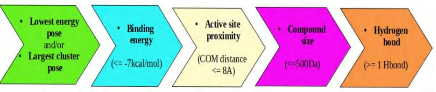
2.3.6 Criteria for compound selection...34
2.4 RESULTS AND DISCUSSION...35
2.4.1 Docking validation...35
2.4.2 Blind docking screening...36
2.4.2.1 Blind docking outcome...38
2.4.3 Protein-ligand interactions...41
2.4.4 Pharmacology of the hit compounds...45
2.5 CONCLUSION...48
3. WILD TYPE - MOLECULAR DYNAMICS...50
3.1. CHAPTER OVERVIEW...50
3.2 INTRODUCTION...51
3.2.1 Protein dynamics...51
3.2.2 Simulation parameters...52
3.2.3 Force fields...53
3.2.3.1 GROMACS...53
3.2.3.2 AMBER...54
3.2.4 Advantages and limitations of molecular dynamics...54
3.3 METHODOLOGY...55
3.3.2 MD simulation runs...56
3.3.2.1 Protein and ligand preparation...56
3.3.2.2 Energy minimization...57
3.3.2.3 Equilibration...57
3.3.2.4 MD simulation...57
3.3.3 Post MD trajectory analysis...57
3.3.3.1 RMSD...58
3.3.3.2 RMSF...58
3.3.3.3 Radius of gyration...59
3.3.3.4 Hydrogen bonding profiling...59
3.3.3.5 VMD visualization...59
3.4 RESULTS AND DISCUSSION...59
3.4.1 Initial MD simulation...60
3.4.2 Root Mean Square Deviation...61
3.4.3 Root Mean Square Fluctuation...63
3.4.4 Radius of gyration...64
3.4.5 Hydrogen bonds...66
3.4.6 Drug use and target site...68
3.6 CONCLUSION...70
4.1. CHAPTER OVERVIEW...72
4.2 INTRODUCTION...72
4.2.1 Mutations...72
4.2.2 Pyrazinamidase mutations...73
4.2.3 Mutation study...74
4.3 METHODOLOGY...75
4.3.1 Mutants preparation...76
4.3.2 MD preparation and analysis...78
4.4 RESULTS AND DISCUSSION...78
4.4.1 Root Mean Square Deviation...79
4.4.1.1 Ligand RMSD...79
4.4.1.2 Backbone RMSD...82
4.4.2 Radius of gyration...84
4.4.2.1 Active site radius of gyration...84
4.4.2.2 Whole system gyration...86
4.4.3 Hydrogen bonds...88
4.4.4 RMSF...92
4.5 CONCLUSION...95
5 CONCLUSION AND FUTURE WORK...96
5.2 FUTURE WORK...98 REFERENCES...99 APPENDICES...111
LIST OF FIGURES
Figure 1.1: Estimated TB incidence cases for 2019 (image from WHO, 2020)...2 Figure 1.2: Schematic representation of pyrazinamide mechanism in Mycobacteria tuberculosis (mechanism generated from Zhang et al., 2013)...11 Figure 1.3: Crystal structure of M. tb PZase (PDB ID:3PL1)...14 Figure 1.4: a) Catalytic triad of M. tb pyrazinamidase with metal binding site residues and active site residues b) Catalytic function of PZase on nicotinamide and pyrazinamide to produce nicotinic acid and pyrazonoic acid ...15 Figure 2.1 Overall molecular dynamics steps and tools used...30 Figure 2.2: Molecular docking box size covering whole protein as used in blind docking. a)
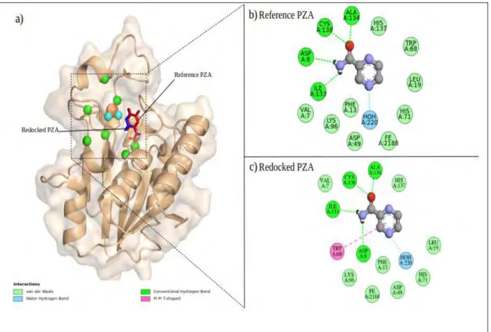
Visualized grid box dimensions viewed in AutoDOCK Tools b) The spacing, x,y,z centers and dimension parameters...33 Figure 2.3:Docking validation of PZA on wild type PZase a) Visualization of superimposed
redocked PZA structures. The 2D interactions of the redocked PZA against PZAse b) from the reference study by Sheik Amamuddyet al,2020 and c) from this study, as visualized in Discovery Studio...36 Figure 2.4: Systematic order summarizing the flow of blind docking compound selection. Ligand filtering was done in 5 stages to extract and identify the best docked DrugBank compounds...37 Figure 2.5: Blind docking screening of the 2089 DrugBank compounds against wild type PZase a) Lowest energy and highest cluster ligand poses b) Hit compounds (93 ligands) on the active pocket of PZase with the active site residues highlighted in green...38 Figure 2.6: Characteristic heatmaps of the selected 47 compounds in reference to the control PZA. a) Docking binding energies below -7 kcal/mol. b) Euclidean distances within 8Å. c) The number of hydrogen bonds formed between ligand and protein. d) The calculated compound molecular weight values...40 Figure 2.7: Discovery Studio 2D visualization of the 47 best docked DrugBank compounds against PZase. The colour key represents the different bonds formed within each complex. The ligand is represented as balls and sticks while the receptor residues are disc shaped...44 Figure 3.1: Overall summary of the performed Molecular Dynamics simulation methods. Three key steps are Force-field inferring, Molecular dynamics run and analysis obtained results...55 Figure 3.2: Violin plots for Ligand RMSD of the last 10ns of the 20ns simulation period. The control PZA in blue, selected stable compounds in yellow and rejected compounds in red...61
Figure 3.3: Violin plots of a) Ligand RMSD and b) Protein backbone RMSD of the selected 47 compounds...62 Figure 3.4: RMSF analysis. a)A heat map showing the local residue fluctuations during the 150ns simulation period across all hit compounds. b) Mapped fluctuating regions on the protein, green
Figure 3.5: Radius of Gyration a) for the protein and b) active site residues within 8Å of PZA in the catalytic cleft...65 Figure 3.6: Number of hydrogen bonds throughout the 150ns simulation period...67 Figure 3.7: Summary of Hydrogen bonds formed throughout the 150ns simulation...68
Figure 4.1: Summary of the performed molecular dynamics simulation on the mutant systems. The first step was introduction of mutations followed by the general MD steps of inferring force-field, running molecular dynamic simulations and analysis of obtained results...75 Figure 4.2: Representation of positions on the mutated amino acid residues in PZase, based on the center of mass of the docked PZA ligand...77 Figure: 4.3: Ligand RMSD violin plots of the hit compounds across mutants. The WT systems are highlighted in blue while mutant ligand systems are in yellow...81 Figure: 4.4: Backbone RMSD violin plots for 150 ns of the hit compounds across mutants. The WT systems are highlighted in blue, mutant systems ligands in yellow...84 Figure 4.5: Active site radius of gyration violin plots for the MD simulation of 150 ns on the hit compounds across PZase mutants. The WT systems are highlighted in blue while mutant systems are in yellow...86 Figure 4.6: Whole protein system radius of gyration for PZase WT and mutant systems. The WT systems are highlighted in blue while mutant systems are in yellow...88 Figure 4.7: Distribution of hydrogen bonds through out 150 ns MD simulation. The light colour represent fewer bonds while a dark colour shows more hydrogen bonds at a specific time...90 Figure 4.8: Occupancy of hydrogen bonds through out the 150ns simulation period. The percentage value is highlighted in each box...92 Figure 4.9: RMSF heat maps of local residue fluctuations during the 150 ns MD simulation period in 13 hit compounds across 16 PZase mutations. ...94 Figure 4.10: Mapped fluctuating regions on mutant PZase protein structure...94
Figure 5.1: Overall flow of compound screening from data retrieval to molecular docking and molecular dynamic simulations of the WT and mutant PZase...97
LIST OF TABLES
Table 1.1: Summary of the common drugs used in TB treatment (Zumlaet al.,2013; Zhang and Yew, 2015; Zhouet al.,2017; WHO, 2019; Barozi, 2020)...7
Table 2.1: Summary of general clinical uses of the selected 47 hit compounds. The compound names and uses are from the DrugBank online database
(www.DrugBank.ca)...46
Table 3.1: Summary of the uses and target sites of 13 identifiied stable DrugBank compounds in WT PZase dynamic simulations...69
Table 4.1: Selected mutations from the previous group study by Sheik Amamuddyet al(2020) where PZA-bound protein complexes had an estimated exit time point of less than 50ns...76
Table 4.2.: Selected mutants from Sheik Amamuddyet al(2020) from group 1-3. Mutants were selected based on systems where ligand appeared stable prior to release...77
LIST OF ABBREVIATIONS
AIDS Acquired Immune Deficiency Syndrome
BCG Baccille Calmette-Guerin
CHPC Center for High Performance Computing
FAS1 Fatty Acid Synthase1
GROMACS GROningen MAchine for Chemical Simulations
HIV Human Immunodeficiency Virus
ID Identification
LAM Lipoarabinomannan
LB Ligand-based
MBS Metal Binding Site
MD Molecular Dynamics
MDR-TB Multidrug Resistant TB
Mtb Mycobacterium tuberculosis
M.tbC Mycobacterium tuberculosis Complex
PDB Protein Data Bank
POA Pyrazinoic Acid
PROSA Protein Structure Analysis
PZA Pyrazinamide
PZase Pyrazinamidase
Rg Radius of Gyration
RMSD Root Mean Square Deviation
RMSF Root Mean Square Fluctuation
rpsA 30S Ribosomal Protein S1
SB Structure-based
TB Tuberculosis
WHO World Heatlh Organization
WT Wild Type
XDR-TB Extensively Drug Resistant tuberculosis
2D Two- dimensional
3D Three-dimensional
TABLE OF AMINO ACIDS
Full name Three letter code One letter code
Alanine Ala A
Arginine Arg R
Asparagine Asn N
Aspartic acid Asp D
Cysteine Cys C
Glutamic acid Glu E
Glutamine Gln Q
Glycine Gly G
Histidine His H
Isoleucine Ile I
Leucine Leu L
Lysine Lys K
Methionine Met M
Phenylalanine Phe F
Proline Pro P
Serine Ser S
Threonine Thr T
Tryptophan Trp W
Tyrosine Tyr Y
Valine Val V
CHAPTER ONE
1. LITERATURE REVIEW
1.1 TUBERCULOSIS 1.1.1 Introduction
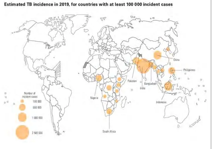
Tuberculosis (TB) was declared a global health emergency in 1993 by the World Health Organization (WHO) (Floyd et al., 2018), a disease claiming approximately 1.7 billion infections and 10 million sicknesses each year (WHO 2019). According to Singh et al.,(2018), a third of the world’s population has been infected with TB and there is a documented case of TB infection in almost every country. WHO reported TB-related deaths of up to 1.4 million in 2019 out of the 10 million infections.Majority of the infections were reported from Africa, South-East Asia and West Pacific regions (WHO, 2020). South Africa has been reported to be among the eight high burden countries (India, China, Indonesia, Philippines, Pakistan, Nigeria, Bangladesh) that contribute to two thirds of the global infections (Figure 1.1, WHO 2020). It is also the only high burden country having zoonotic TB cases (Mycobacterium bovis) (WHO, 2020).
A target has been set by WHO to eradicate TB by 2035 with the acknowledgment that better diagnostic methods, preventative and therapeutic measures have to be employed (CryPTIC, 2018).
The ultimate goal is guided by reducing deaths and incidence rates by 90% and 80% respectively between 2015 and 2030 (WHO 2019; Floydet al.,2018). However, WHO (2020) reports that due to the current COVID-19 pandemic, the extra pressure on health services may slow or reverse the progress done on TB eradication. Although lockdown measures and physical distancing policies
imposed across all nations may reduce the incidences of TB transmission and infection, this may be an offset of worsening TB therapy outcomes, having longer infectiousness periods and poverty (WHO, 2020). Already, the high burden countries have reported massive reduction in the number of new cases within the first few months of COVID-19 lockdown. The high burden countries reported 25–30% lower TB cases in India, Indonesia and the Philippines within the first six months of 2020 compared to 2019 while South Africa had a decrease of up to 50% (WHO, 2020).
Figure 1. 1: Estimated TB incidence cases for 2019 (image from WHO, 2020)
The main population at risk is adults with underlying conditions such as diabetes, HIV/AIDS or malnutrition (WHO, 2020). Generally, there is a double risk in males than female (WHO, 2019), and according to Jimenez-Corona et al., 2006 this difference is mainly due to men being more
exposed to local transmission in crowded working areas, poor ventilated and imprisonment shelters.
Mens’ excessive use of tobacco and alcohol also increase the rate at which latent TB progresses to active disease and symptoms such as coughing may be mistaken for tobacco use while one is already symptomatic (Jimenez-Corona et al., 2006). However, McQuaid et al., (2020) states that males and females have the same risk of Multi Drug Resistant (MDR) TB.
TB mainly affects the lungs (pulmonary TB), however, other organs such as the brain, joints, central nervous system, lymph and circulatory system may also be affected. An infected person shows respiratory symptoms such as emaciation, low fever, night sweats among many other symptoms (Zhouet al., 2017). It is mostly spread when an infected person expels the bacteria into the air through coughing or sneezing (Delogu et al., 2013; WHO, 2020). Failure of an individual’s innate immune defense mechanism to eliminate the bacteria leads to its replication and spreads to other organs and tissues. The cell-mediated response attempts to control the bacterial replication resulting in latent TB (dormantbacilli) with no symptoms or signs of the disease which can last for days or even years. A decrease in cell mediated response mechanism leads to manifestation of the disease and thus active TB (Deloguet al.,2013). This process makes up the stages of TB which are exposure, latent, and active TB. Patients with latent TB are at risk of reactivation of the disease, which is one major problem in controlling TB globally (Smithet al.,2004).
1.1.2Tubercle bacilli
The causative agent of TB was discovered by Robert Koch in 1882 as a bacillus species called Mycobacterium tubercle bacilli (M. tb) (WHO, 2020). M. tbis a small, rod-shaped, aerobic bacillus bacterium with a slow reproductive cycle of 24 to 48 hours under optimal conditions (Deloguet al,
2013; Muller, 2016). It has a thick cell wall made up of mycolic acids and waxy components on its inner and outer layers that prevent harmful agents and antibiotics from entering into the cell (Zhang et al., 2003). The synthesis of these membrane components are the target sites of the effective anti- TB drugs such as isoniziad and ethambutol (Muller, 2016). The cell also has a deficiency of pyrazinoic acid (POA) efflux pumps which make it susceptible to POA derivative drugs like pyrazinamide (Zhang et al., 2003). During a disease process, M. tb can be found in micro- environments that are acidic, have nutrient deficiency and low or high oxygen content. All these different environmental conditions lead to the development of heterogeneous bacterial populations that are either non-replicating or growing with different levels of susceptibility to anti-TB drugs (Zhanget al.,2012).
1.1.3 Diagnosis of TB
Different methods have been developed to detected TB infection with each technique having different detection approach. TB infection can be identified using a microscope to visualize sputum smears using the Ziehl-Neelsen technique, chest X-rays, molecular tests such as Gene-Xpert, immunology-based technique like TB LAM test and cultured based methods (Lawn et al, 2017;
Brogeret al.,2019; WHO, 2020). Multiple line probe assays tests for drug resistance have also been developed, however, culture-based assays are the standard susceptibility tests frequently used (Floydet al.,2018).
1.1.4 Drug resistance
Effective treatment and eradication of TB is being hindered by the emergence of Drug Resistant Tuberculosis (DR-TB) such as MDR-TB and Extensively Drug Resistant (XDR-TB). MDR-TB is
defined as resistance ofM. tbto at least isoniazid and rifampicin, while XDR-TB is the resistance to both first line and second line injectable TB drugs including fluoquinolones (Zhang and Yew, 2015).
Resistance against newly discovered drugs targeting both growing and non-growing M. tb (bedaquiline, pretomanid, delamanid) has been reported (Zhang and Yew, 2015). Studies revealing the vital factors of TB virulence including the unique components of its cell membrane contributing to virulence and persistence have also been done (Smithet al,2004).
Zhang and Yew (2015) stated that chromosomal gene mutations and protein modifications account for the two types of drug resistance in M. tb. These are genetic and phenotypic resistance respectively. Mutations on the genes encoding the proteins that are targeted by the present anti-TB drugs is the major cause of M. tb resistant strains which might occur as a result of sub-optimal physician prescription, failure of patient compliance and bacterial efflux pump (Zhang and Yew, 2015; WHO 2020; WHO, 2020). The resistant gene may also be passed from one individual to the other (Zhang and Yew, 2015) and the majority of MDR-TB and XDR-TB cases are due to the transmitted Beijing genotype in China, Europe and Africa (Zhang and Yew 2015). Zhanget al(2012) also states that persister bacteria, with the potential and ability to survive antibiotic stress, are one of the main causes of prolonged TB treatments and drug resistance. Multiple clinical experiments have demonstrated that persisters that can be found in lesions, sputum or adipose tissue, are the problem cause in TB relapse and drug resistance (Connollyet al.,2007; Zhanget al.,2012).
1.1.5 TB treatment
Statistics conducted before drugs for TB treatment became available reported that 70% of the people diagnosed smear positive with pulmonary TB died within 10 years and overall 40% of the people that tested positive for all forms of clinical TB died (Floyd et al., 2018). A decrease in incidence and mortality rate was obtained from around the 1940s after the introduction of effective anti-TB drugs, resulting in TB being regarded as a disease of the past. However, it has remained an infectious disease responsible for the highest number of deaths globally (Floyd et al.,2018, WHO, 2020).
Currently, the available TB vaccine, Baccille Calmette-Guerin (BCG), is only effective in preventing severe forms of TB infections in children and is restricted to HIV negative children. No vaccine has been synthesized for adults (Floyd et al., 2018, WHO, 2020). The currently used first line drugs for drug-susceptible TB are isoniziad (INH), rifampicin (RMP), ethambutol (EMB) and pyrazinamide (PZA) (Zhang and Yew, 2015; Zhou et al., 2017; WHO, 2019). These drugs are prescribed over a period of 6 months with a minimum success rate of 85% (WHO, 2020). Generally, RMP interferes with RNA synthesis and binds to rpoB forming a hydroxyl radical. On the other hand, INH attacks the enolyl acyl carrier protein reductase enzyme and inhibits mycolic acid synthesis in cell wall while EMB targets arabinosyl transferase, resulting in no synthesis of cell wall arabinogalactan (Zhang and Yew 2015). Table 1.1 gives a summary of some drugs used in TB treatment.
Table 2.1: Summary of the common drugs used in TB treatment (Zumla et al., 2013; Zhang and Yew, 2015; Zhouet al.,2017; WHO, 2019; Barozi, 2020).
Group Drug name
First line drugs Isoniziad, Rifampicin, Ethambutol, Pyrazinamide
Injectable drugs Kanamycin, Aminkacin, Streptomycin, Capreomycin
Fluoroquinolones Moxifloxacin, Levofloxacin, Gatifloxacin Second -line drugs Ethionamide, Terrizidone, Para-amino salicylic
acid, Prothionamide
Drugs with unclear efficacy Amoxicillin, linezolid, clofazimine
Majority of the developed antibiotic drugs are based on their effect on growing bacteria with little or no activity on persister bacteria (Cogan, 2006). Since one of the main causes of different anti-TB drug susceptibility is due to persisters, drugs with mechanisms that target persisters will greatly improve treatment of TB (Zhang et al., 2012). Apart from developing drugs that target persisters, manipulating the host immune system by enhancing and utilizing its defense mechanism with vaccines and immuno-modulating agents, stimulating innate and adaptive immunity may assist in preventing and quickening recovery from the disease (Bishopet al.,2001).
Some anti-TB drugs like rifamycins and fluoroquinolones primarily target growing bacteria but also have limited activity on non-growing persisters. The few identified persister-active compounds are not readily bioavailable and have high toxicity, thus need further studies for optimization (Cogan, 2006). In 1944, Bigger proposed an intermittent drug dosing approach to allow persisters to grow in the absence of antibiotics and become susceptible to drugs (Bigger, 1944; Lewis, 2012), however, his model was discovered to be practical in vitro as complex conditions are encountered in vivo
(Cogan, 2006). The development of pyrazinamide drug that targets persisters by disrupting vital processes needed for their survival in stressful conditions has greatly improved the treatment regimens in TB (Bishopet al.,2001; Cogan, 2006).
1.2 PYRAZINAMIDE (PZA)
Pyrazinamide is a pro-drug used in the therapy of TB alongside other first line drugs. It is also used to prevent relapse of TB and is incorporated with second line drugs in treating drug susceptible and resistant M. tb (Juniad et al., 2018; Sheik Amamuddy et al., 2020). The drug pyrazinamide (PZA) was chemically synthesized in 1936 by Dalmer and Walter, and was later discovered as an anti-TB drug in 1952 based on the effects of its analog, nicotinamide, on mycobacteria in animal models (Zhang and Mitchison, 2003; Zhang et al.,2014). It was initially used as a second line TB drug due to its high hepatic toxicity effects which were caused by the high dosages (3.0 g daily) and prolonged treatment periods. Further studies later discovered lower dosage concentration (1.5 to 2 g daily) with effective sterilizing effects and in synergy with other TB drugs (Zhang et al., 2014). It has a molecular weight of 123.1, melting point of 188-189 0C and molecular formula C5H5N3O (Zhang and Mitchison, 2003; Zhang et al., 2015). PZA has poor solubility in organic solvents and dissolves in water (15 mg/ml) at room temperature while its active derivative pyrazinoic acid (POA) readily dissolves in organic solvents like dimethyl sulfoxide (DMSO) (Zhang and Mitchison, 2003).
Pyrazinamide has been reported to have excellent sterilizing bactericidal effectin vivoagainstM. tb while having no notable activity in vitro (Peterson et al., 2015; Gopal et al., 2016). Its activity in vitrocan be induced in the presence of efflux pump inhibitors, in mild acidic conditions or in media with nutrient deficiency, anaerobic conditions and molecules that alter energy metabolism. It has a bactericidal effect on semi-dormanttubercle bacilliin a pH range of (4.8-5.0) (Zhang and Mitchison,
2003). According to Juniad et al (2018), the use of PZA in TB treatment cuts the therapy time by 33% from the standard 9 to 12 months to 6 months. Additionally, its removal from TB therapy reduces other drugs’ efficiency to destroy bacterial cells (Bishop et al., 2001). However, the 6 months therapy period is still long enough to facilitate drug resistance development and patient noncompliance, thus the goal is to develop drugs for TB treatment within 2 months or less (Gopalet al.,2016).
Pyrazinamide in humans and murine models have been reported to be effective in the first two months of TB treatment in combination with the first line drugs (Zhang and Mitchison, 2003;
Ahmad et al., 2013). However, Ahmad et al (2013) states that in second line regimen of murine models, PZA contributes sterilizing activity beyond two months when incorporated with streptomycin and isoniazid which also resonates with his obtained study with moxifloxacin and levofloxacin. Failure of PZA activity beyond the first two months may be due to overlapping sterilizing effect with rifamycins or antagonistic effects with isoniazid (Ahmad et al., 2013; Zhang et al., 2014). Apart from the good antibacterial effect exhibited by PZA, it has negative side effects of damaging the liver, therefore alternative compounds with trivial side effects need to be identified (Zhouet al.,2017).
1.2.1 Mechanism of PZA
In spite of the significant role played by PZA in TB treatment and its inclusion in all TB drug combinations, its mechanism is the least understood among all anti-TB drugs (Zhang et al., 2013;
Gapolet al., 2016; Sheik Amamuddyet al., 2020). Its role as a persister drug has attracted a lot of attention in trying to understand its mode of action in TB treatment and also in developing drugs for other persistent infections (Cagon, 2006).
Different mechanisms of PZA onM. tbhave been suggested (Lamontet al., 2020). Multiple studies revealed that the sterilizing activity of PZA depends on the acidic environment in the lesion caused by inflammation, explaining its use only in the first two months of therapy (Zimhony et al., 2000;
Shi et al., 2011; Zhang et al., 2013) while further studies demonstrated the independence of PZA bactericidal effect in nearly neutral and alkaline conditions with critical accumulation concentrations of POA (Dillionet al., 2014).
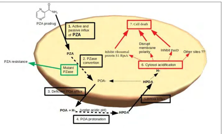
The accepted model for PZA mechanism involves the acidification of cytoplasmic bacilli by POA mediated proton shuttling (Figure 1.2) (Zhang et al., 2013; Peterson et al., 2015). PZA is activated only in acidic conditions (Bishopet al., 2001), and at this low pH, POA becomes toxic and inhibits the growth of M. tb (Juniad et al., 2018). PZA enters into M. tb by passive diffusion (Zhang and Mitchison, 2003; Junaidet al.,2018) and possibly by active transport (Zhang and Mitchison, 2003).
Initially, POA is formed as a charged anionic form with no bactericidal effect in a neutral cytoplasmic environment, however, in acidic conditions, POA is excreted by a weak efflux pump and is converted to uncharged protonated POA (HPOA) which accumulates in the cell and eventually kills the cell (Zhang et al., 2003; Zhang and Mitchison, 2015; Juniad et al., 2017).
Protonated POA acidifies the cell cytoplasm and affects bacterial cell activity by inhibiting functioning of vital enzymes such as ribosomal protein S1 rpsA and panD which are involved in translation, co-enzyme A synthesis and charges on the cell membrane thus affecting membrane transportation (Zhanget al.,2013; Zhang and Yew, 2015).
Several target sites and pathways for TB treatment have been proposed and studied (Shiet al.,2014, Gopalet al.,2020; Smithet al.,2004). Zimhonyet al(2000) suggested fatty acid synthase1 (FAS1) as a molecular target site of PZA, however these findings were later refuted by Boshoff et al., (2002). Studies by Shi et al (2011) identified 30S ribosomal protein S1 (rpsA) as a target site for PZA/POA which inhibits trans-translation process. However, Personne (2014) have shown that strains with a defective trans-translation pathway were still susceptible to PZA and Dillion (2017) also concluded that PZA is independent ofrpsAand trans-translation.
Figure 1.2: Schematic representation of pyrazinamide mechanism in Mycobacteria tuberculosis (mechanism generated from Zhanget al., 2013; Petersonet al,2015; Junaidet al., 2018; Zhang and Mitchison, 2003)
rspA and panD sites have been targeted as potential drug sites for PZA/POA (Zhang et al., 2003;
Zhang and Yew, 2015; Gopal et al., 2020). The study by Shi et al (2014) suggested panD as the target site for PZA/POA. A recent study by Gopal et al(2020) supported Shi et al(2014) findings and revealed the mechanism of POA as a weak panD inhibitor that triggers degradation of the
enzyme by caseinolytic protease. Degradation of this enzyme blocks the synthesis of co-enzyme A, an enzyme involved in energy metabolism reactions. Gopal studies in 2016 and 2017 have illustrated that the use of PZA/POA in TB treatments reduces the levels of co-enzyme A. This site however, is not the exclusive target site of the PZA drug as conditional susceptibility was observed in strains without the panD region (Dillion et al., 2017). The mechanism of PZA remains elusive and further studies are still to be done (Sheik Amamuddyet al.,2020).
1.2.2 Alternatives to PZA
PZA derivatives can be studied for their activity against M. tb. Some synthesized PZA derivatives were studied by Zhou et al (2017), who discovered N-(3-thiomorpholinopropyl)pyrazine-2- carboxamide as a potential compound, however further analysis is needed. One of the PZA derivatives is morphazinamide (MZA) which has a similar impact on M. tb in both acidic and neutral pH conditions. It is converted to PZA, formaldehyde and morphiline in bacterial cells.
However, its use in animal TB treatment is inferior to PZA regardless of its high activity in in vitro models thus considered less useful in TB treatment (Zhang et al., 2003). A synthetic analogue of PZA, 5-chloro-pyrazinamide, is also active against M. tb. However, its mode of action is independent of the enzyme pyrazinamidase and has no effect on M. tb in mouse models thus makes it different to PZA (Zhang et al., 2003; Zhanget al., 2014). Studies have also shown that esters of POA have anti-TB properties similar to the mechanism of PZA in vitro studies, but have failed to show significant activity againstM. tbin vivo (Zhanget al.,2003). Therefore, PZA remains the best prodrug for TB treatment regardless of the emerging PZA-resistant M. tb strains. However, identification of an alternative drug that mimics the mechanism of PZA with little to no side effects to counteract drug resistance is crucial for progress in TB eradication.
1.3 PYRAZINAMIDASE
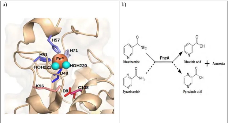
Pyrazinamidase (PZase) has been identified as the enzyme responsible for the conversion of prodrug PZA into its active form pyrazinoic acid (POA) (Juniad et al., 2018). This enzyme is present in microorganisms including M. tb, Saccharomyces cerevisiae, Acinetobacter baumanii, Escherichia coli and Pyrococcus (Rasool et al., 2019). The overall structure of M. tb PZase is similar to the crystal structures of A. baumanii and P. horikoshi, however, with crucial variations (Petrellaet al.,2011).P. horikoshiiPZase was shown to contain Zn2+ion in its crystal structure (Du et al.,2001) whileA.baumaniiPZase had Zn2+ and Fe2+ions in the ratio 1:1 (Fyfeet al.,2009) and M. tb PZase contains Fe2+(Petrella et al.,2011). PZase is located in the cytoplasm and is encoded by the pncA gene of M. tb (Zhang et al., 2003). According to Zhang et al., (2003), PZase is also responsible for the conversion of nicotinamide to its acidic form nicotinic acid, which is used in making nicotinamide adenine dinucleotide (NAD) in bacterial species (Zhanget al.,2003).
1.3.1 Structure and mechanism
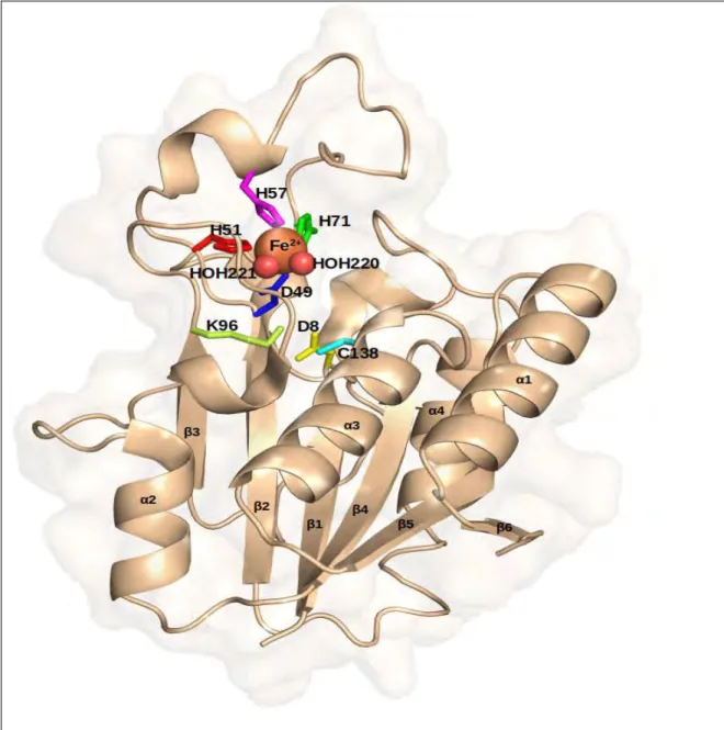
M.tbPZase is a metallo-enzyme with an amidase activity, made up of alpha helices surrounding six parallel beta-sheets (Juniadet al.,2018; Sheik Amamuddyet al.,2020) (Figure 1.3) with a substrate binding cavity of approximately 7Å wide and 10 Å deep (Petrella et al., 2011). Its metal binding site (MBS) consists of an iron (Fe2+ion) coordinated by two water molecules (H2O 220, H2O 221), three histidine and one aspartate residues (H51, H57, H71, D49) in a distorted tetragonal bipyramidal arrangement (Figure 1.4a) (Juniad et al., 2018; Petrella et al., 2011). These residues leave a space on the cavity for PZA binding. It has also been reported that M. tb PZase might contain low amounts of manganese and zinc or may contain them in the metal binding site (Petrella et al.,2011) and some few metal ions can competitively bind as co-factors altering the structure and function of the enzyme (Rasoolet al.,2019).
Figure 1.3: Crystal structure of M. tb PZase (PDB ID:3PL1) showing the alpha helices, loops and six beta pleated sheets in wheat. The metal ion (Fe2+) is represented as an orange sphere while water molecules are red spheres. The active site and metal binding site amino acid residues are represented as sticks. The structure was generated in PyMOL.
The active site residues Asp8, Lys96 and Cys138 (Figure 1.4a) located at the end of β strand 1, β strand 3 and at the N-terminal of alpha helix 3 respectively, make up a catalytic triad (Juniadet al., 2018; Petrellaet al.,2011). According to Petrellaet al(2011), the key residue Cys138 is involved in the nucleophillic attack of PZA while Asp8 and Lys96 act as the activating base and stabilizer of the
acylenzyme. The addition-elimination mechanism found inA. baumanii also applies inM. tbwhich results in the release of ammonia and the formation of acyl-enzyme intermediate after a nucleophilic attack on PZA carbonyl carbon by the thiolate form of Cys138 supported by Asp8. In the final hydrolytic step, the formed Acyl-enzyme intermediate is hydrolysed by water (H2O 202) (Petrella et al., 2011). Figure 1.4b shows the general activity of PZase/pncA on PZA and nicotinamide as explained above.
a) b)
Figure 1.4:a)Catalytic triad ofM. tbpyrazinamidase with metal binding site residues in blues (H51, H57, H71, Asp49) and active site residues in reds (Asp8, Lys96, Cys138). The metal ion (orange) and water molecules (H2O 220/221) in cyan spheres b) Primary function of Pzase/pncA on nicotinamide and pyrazinamide to produce nicotinic acid and pyrazonoic acid with ammonia as a by-product respectively (active site residues visualized in PyMOL, equation generated from Zhang et al., 2013).
The protein has 52-70 residues that form a loop lid controlling access to the active pocket, balanced by H51 and H71 residues (Sheik Amamuddyet al.,2020). On the extended part of this loop, there is a conserved residue found inM. tb,A. baumanii andP. horikoshispecies (Trp68 residue), which is located above the catalytic site and delineates binding site sides (Petrella et al., 2011). The active
site also has a cis-peptide bond between Ile33 and Ala134 residues that draws the nitrogen atom of Ala134 towards the center, forming an oxyanion hole with nitrogen from Cys138 that is occupied by a water molecule (H2O 221) (Petrellaet al.,2011).
1.4 PYRAZINAMIDE RESISTANCE
Current studies have reported some resistance in the use of PZA prodrug (Juniadet al.,2018). This can be accounted for by the loss of PZase enzyme activity (Zhang and Mitchson, 2003; Kim et al., 2012; Zhang and Yew, 2015). A recent study by Jumaet al (2019) in Tanzania revealed that 50% of patients with MDR-TB and 21.3% with drug sensitive TB also had PZA resistance. There have also been reports on increasing rate of PZA resistance cases (Sheen et al., 2020). Determination of resistance of M. tb to PZA is difficult to interpret and analyze using acidic media approaches thus sequencing of the gene pncAis often done to identify mutations that are related to PZA resistance (Petrella et al., 2011). A study by CRyPTIC (2018) showed that correct sequencing of the M. tb isolates on pncA gene predicted PZA resistance with 96.8% specificity and 91,3% sensitivity and susceptibility, accommodating other factors as causes for PZA resistance.
Strains of M. tb that are resistant to PZA have been found in isolates lacking pncA mutations (Simons et al.,2013). These results supported the results obtained by Sreevatsan et al.,1997; Kim et al.,2012; Shiet al., 2014, whose analysis also showed that approximately 0% to 30% (depending on geographical region) of M. tb isolates show PZase inactivity in the absence of pncA mutation thus suggesting alternative mechanisms for PZA resistance. Multiple studies have been done in attempt to understand the mechanism of PZA resistance (Bishopet al.,2001; Shiet al.,2011; Zhang et al., 2014; Shi et al., 2014; Palmer and Kishony, 2014; Gopal et al., 2016; Dillon et al., 2017).
Analyses of genetic sequences of PZA-resistant strains reveal no PZA-associated prominent clustering mutations apart from those identified in prodrug activating pyrazinamide (Gopal et al., 2016). Although the resistance of PZA can be encoded by three genes (pncA, rpsA andpanD), pncA gene contributes up to 72-99% of the mutations (Juniad et al., 2018). Resistance of this drug according to Junaid and colleagues is due to scattered mutations in the coding and promoter region of thepncAgene (Kimet al.,2012; Juniadet al.,2018). The most identified type of mutations in the pncAgene are missense and nonsense mutations which cause amino acid substitution or nucleotide insertions or deletions (Zhang and Mitchison, 2003).
Mutations on the pncAgene are commonly found on three regions 3-17, 61-85 and 132-142 which have an effect on the folding of the active site (Zhang et al.,2003; Sheik Amamuddyet al., 2020).
These mutations are responsible for 54% of PZA resistant strains (Sheik Amamuddy etal.,2020). A study on various PZase species by Lemaitre et al., 1999 showed that these three regions are highly conserved therefore alteration on these regions affect the structural or catalytic function of the enzyme. This study also suggested Cys138, Ala134, Thr135, Trp68 and Asp8 as the key residues for PZA hydrolysis and stated that mutations on these specific residues or close to these residues causes active site modifications leading to the loss of PZase activity and PZA resistance (Lemaitre et al., 1999). Apart from acquired PZA resistance through mutations, natural PZA resistance also occurs in some mycobacteria such asM. bovis that have a point mutation on thepncAgene at nucleotide 169 from Cytosine to Guanine which replaces Histidine (H57) in M. tb with Aspartic acid in M. bovis.
The M. kansasii and M. smegmatis species are also naturally resistant to PZA due to the weak activity of PZase enzyme and highly active POA efflux pump respectively (Zhanget al., 2003).
1.5 PZase-PZA COMPLEX AND RESISTANT MUTATIONS
The mechanism of resistance to the drug PZA remains unclear and is yet to be determined (Sheik Amamuddy et al., 2020). Due to the increase in M. tb PZA resistant strains, there is need for new approaches to identify novel drug target sites and new drugs for TB treatment. Previous studies have been focusing on the wet laboratory experiments such as PZA susceptibility tests (Yoonet al., 2014; Morlocket al.,2017). Computational methods can aid in analyzing the effect of mutagenicity on PZase function (Rasoolet al.,2019).
PZase-PZA complex can be analysed by computational methods. Recent studies using computational approaches have been focusing on the effect of mutations on the metal ion and PZA binding (Sheen et al., 2012; Kadem-Maaref et al., 2017; Khan et al., 2018; Rasool et al., 2019;
Sheik Amamuddyet al, 2020). Mutations that have been identified to have major effects on PZase activity include Ala3 to Gly17, Thr61to Leu85 and Gly132 to Thr142 (Juniad et al.,2017). Juniad et al(2017) however stated that mutations on other sites might also have an effect on the solubility, structure and function of the protein. Previous studies have also demonstrated that mutations on the metal binding site lowers the binding affinity of the co-factor ion which is crucial for the activity of PZase (Sheenet al.,2012; Rasoolet al.,2019;).
According to Petrella et al., 2011, mutations on pncA gene not only affect catalytic functioning of the protein, but also affect the thermal stability and folding properties of the protein. Their study on mutating residues that make up the catalytic triad (Asp8Glu, Cys138Ala, and Lys96Gln), substrate binding site (Phe13Leu and Trp68Leu), metal binding site (His51Ala, Asp49Gly, and His57Asp) and the oxyanion hole formation (Ala134Val) resulted in none or low PZase activity. These results
demonstrated the importance of specific residues for PZase activity and also support that mutations have a great effect on the integrity of the 3D structure and activity. Khanet al(2018) study showed that the mutations Leu19Arg, Arg140His and Glu144Lys on PZase caused changes in the protein stability, flexibility, activity and in the binding pocket size when analyzed using molecular dynamics simulations.
A study by Rasool et al., 2019based on the Density Functional Theory (DFT) approach reported that mutagenicity on PZase is detrimental to its activity and results in weak binding of its co-factor metal (iron) and the prodrug PZA. The study revealed that the mutations Asp12Gly, Asp12Ala, Thr135Pro and Asp136Gly weaken the binding of PZA as these mutations occur close to the active site. Junaidet al (2018) investigated the effect of mutating Asn11 to Lys, Pro69 to Thr and Asp126 to Asn on the pncA gene. Their study showed that these mutations resulted in an increase in fluctuations in the mutant protein compared to the wild type, indicated by a weakened binding of PZA to the active site and an alteration in the active site volume which in turn altered the binding of PZA to PZase.
A study by Kadem-Maaref et al (2017), analyzing the effect of different metals on PZase function using DFT model revealed that cobalt and nickel are more active than iron and can effectively replace it as a co-factor while magnesium, zinc and copper decreases its activity. These results resonate with those obtained by (Sheen et al.,2012 and Rasool et al., 2019). Sheik Amamuddy et al (2020) study investigated the mechanism of PZA resistance by studying the unbinding events of PZA on the WT and mutant Pzase. Their study revealed that mutations on MBS residues caused iron ion delocalization which led to the opening of the lid and unbinding of PZA.
1.6 DRUG REPURPOSING
Despite the advancement in technology and knowledge on human disease states, the traditional development of new drugs is substantially expensive and time consuming (Rudrapal et al., 2020).
Therefore, the use of already discovered drugs (drug repurposing or repositioning) is being adopted for lower costs and shorter timelines to treat common and rare diseases. Drug repurposing is a strategy that identifies new alternative uses on drugs that have been approved, discontinued, abandoned or under experimental investigations to target other medical conditions (Elder and Tindall, 2020; Khan et al., 2020). The approach generally follow three steps of i) identifying a potential molecule for the given state, ii) preclinical assessment of drug effects and iii) efficacy evaluation in clinical trials (Pushpakom et al., 2019). According to Rudrapal et al., (2020) approximately 30% of US Food and Drug Administration approved drugs and vaccines are as a result of repositioned drugs. Some of the common effective drugs from repositioning are minoxidil, aspirin, valproic acid including sildenafil (Viagra) which was initially developed for hypertension and angina pectoris treatment but has been repurposed to treat erectile dysfunction (Pushpakom et al.,2019; Elder and Tindall, 2020; Khanet al.,2020; Rudrapalet al.,2020).
Various computational approaches like signature matching, genome-wide association and molecular docking and/or experimental approaches like phenotypic screening and binding-target assays may be used to identify repositioning opportunities (Pushpakomet al.,2019; Rudrapalet al., 2020). The experimental-based approach is a protein target-based and cell-based screening method of original drugs for new pharmacological effects. The in silico approach is based on molecular interactions between protein and drug molecules through virtual screening of drug databases using computational biology tools (Rudrapal et al., 2020). In this study, the computational molecular
docking approach will be used to screen multiple drugs against target protein PZase (conventional docking).
1.6.1 Advantages and limitations of drug repurposing
Advantages of drug repurposing include low chances of failure as the drug would have been approved and successfully tested in preclinical models and humans thus also implies less time and less investment in preclinical tests (Pushpakom et al., 2019; Khan et al., 2020). According to Rudrapalet al (2020), the average traditional approach requires 10-16 years to develop a new drug while 3-12 years are required to design a drug through drug repurposing. Repurposed drugs may also reveal off-target or on-target effects, exposing new target sites and pathways that can be further exploited as potential drug-target sites (Pushpakomet al.,2019).
Successful drug repurposing has been achieved through both computational and experimental approaches, with fast screening on large data using computational methods compared to experimental methods. However, there are barriers hindering the wide use and success rate of these techniques. The challenges faced include legal and intellectual property barriers and organizational hurdles which require collaborations from pharmaceutical companies, biotechnology firms and academic communities.
1.7 PROBLEM STATEMENT
Tuberculosis has been declared a global health emergency by the WHO with South Africa being reported to be among the high burden countries that contribute two thirds to the overall TB incidences. The World Health Organization has set a target to eradicate TB by 2035 aiming to reduce deaths and incidence rates by 90% and 80% respectively between 2015 and 2030 (WHO, 2020). The emergence of MDR-TB and XDR-TB M. tb strains has become a major public health problem threatening the progress made in TB treatment worldwide. In order to accomplish the goal set, intense research and development of new novel drugs and drug targets has to be done. The prodrug PZA with bactericidal effect on semi-dormant mycobacterium tuberculosis has been identified as a critical drug needed in all TB treatment combinations, reducing therapy time from 9 months to 6 months (Gopal et al., 2016; Juniad et al., 2018). However, various mechanisms contributing to PZA drug resistance have been reported (Zhang and Mitchson, 2003; Kim et al., 2012; Zhang and Yew, 2015) with previous studies focusing on wet laboratory experiments and in silico analysis on the effect of mutations on active site and metal binding site of pyrazinamidase (Sheen et al., 2012; Kadem-Maaref et al., 2017; Khan et al., 2018; Rasool et al., 2019; Sheik Amamuddy et al., 2020). Exploration on alternative compounds to PZA has been partially conducted in wet laboratory research (Zhang et al., 2003; Zhang et al., 2014; Zhou et al., 2017) with no in silico approaches to identify compounds against PZase active site. This study aims to identify novel selective compounds against the active site of M. tb PZase and analyse their behaviour in the presence of mutations. The identification of scaffolds or selective novel compounds against M. tb PZase might lead to the design of more effective drugs, for cure and eradication of TB.
1.8 HYPOTHESIS
The study hypothesizes that DrugBank database have compounds that can selectively bind to the active site of M. tb PZase, mimicking the behavior of PZA prodrug. Therefore, computational techniques of virtual screening and dynamic simulations may be employed for the identification of novel compounds for drug repurposing in TB.
1.9 AIM
The study aimed to virtually screen DrugBank compounds against the active site of M.tbPZase and analyze the effect of point mutations on the identified hit compounds. This is so as to identify scaffolds that may lead to the development of effective TB drugs.
1.10 OBJECTIVES
1. To identify potential DrugBank compounds that selectively bind to PZase active site by performingin silico molecular docking studies.
2. To perform Molecular Dynamics (MD) calculations on wildtype PZase to identify stable protein-drug complexes.
3. To introduce mutations on PZase-DrugBank complexes and study the effect of the mutations on the complexes.
CHAPTER TWO
2. STRUCTURE BASED VIRTUAL SCREENING
2.1 CHAPTER OVERVIEW
Compounds that bind on to the active or allosteric site of a target protein can induce conformational changes and either inhibit or promote the protein’s functionality. The aim of this chapter (Chapter 2) was to identify potential DrugBank (Wishartet al, 2018) compounds that can selectively bind onto the active site of wild type PZase with better binding characteristics compared to the control Pyrazinamide. The entire protein surface was subjected to two thousand and eighty-nine (2089) DrugBank compounds through molecular docking using AutoDOCK 4.2 (Morris et al., 2009). The protein and PZA ligand structures were prepared using AutoDOCK4 Tools (Morris et al., 2009).
Pyrazinamide was used as a control and the docking parameters were validated by redocking PZA onto the wildtype PZase and comparing its pose and interactions with those obtained by the previous group study of Sheik Amamuddy et al., (2020). The best hit compounds were selected based on lowest binding energy, active site proximity, low molecular weight and presence of hydrogen bond interactions before being subjected for further analysis. This chapter provides a brief introductory description on high throughput virtual screening, applied docking methodology, discussion of the results obtained and summarized conclusion.
2.2 INTRODUCTION
Proteins interact with other molecules and their functionality in biological processes occur through recognition of other molecules. Identification of the small molecules protein-target site provides insight on the underlying molecular modes of actions, giving information on their pharmaceutical
effects (Wanget al., 2012). The first target-substrate binding was reported by Fischer in 1894, who interpreted using the lock and key analogue, based on the protein-substrate complementary shape.
However, his interpretation did not explain allosteric modulation and non-competitive binding which lead to other binding models such as the induced-fit by Koshland in 1958 being proposed (Salmaso and Moro, 2018).
2.2.1 Virtual screening
Computational methods have been applied in drug discovery processes since 1980s, leading to the establishment of (Computer Aided Drug Design) CADD techniques which improved from analysis of a rigid ligand-target binding to flexible ligand-protein complexes (Salmaso and Moro, 2018;
Nguyen et al., 2019). These techniques are developed primarily for virtual screening hit/lead optimization as well for the designing of novel compounds (Kitchenet al.,2004; Salmaso and Moro, 2018). Virtual screening, which aims to increase the novel compounds hit rate and reduce experimentally tested compounds, accomplishes its goal by screening a large data set of compounds in search for binding capacity for a targeted molecule.
The CADD techniques are grouped as Ligand-based (LB) or Structure-based (SB) methods. The Ligand-based method only depends on information about the similarity of known ligands (Sliwoski et al., 2014). In this study, the Structure-based method was used, which depend on the crystal structure of the target molecule (obtained by NMR, Xray crystalography or from homology modelling) and on the fact that binding to the target structure may be optimized since ligand-target binding is influenced by structure complementarity (Sliwoski et al., 2014; Salmaso and Moro, 2018). The SB technique has made prominent inhibitors of HIV-1 reverse transcriptase (Vadivelan
et al., 2011), heat shock proteins (Doddareddy et al., 2011) and Plasmodium parasite (Chaudhary and Prasad, 2014) among many other therapy fields.
The availability of databases that consist of chemical compounds from natural and/or synthetic origin have enabled easier identification and screening on potential hit compounds. One of the most comprehensive source of small biological molecules is the CheMBL database that is compiled from publications and other chemical databases like PubChem. Generally, only approximately 11 000 compounds are readily available from pure natural products (Kinghornet al,2019). Several natural product (NP) databases have been developed, however, majority of these databases are specialized including databases such as Super Natural II (Banerjee et al., 2015), one of the largest online database that mainly consist of purchasable compounds and NPAtlas (Van Santen et al., 2019) database focusing on microbial natural products. Some databases like AnalytiCon provides over 2000 semi-synthetic compounds (Kinghorn et al, 2019) while other databases like PubChem (Kim et al., 2016) and ZINC (Irwin et al., 2012) consist of millions of mainly commercially available small and large synthetic molecules. The ZINC database is widely accepted as a meta-database of readily purchasable compounds (Kinghornet al.,2019).
Recently, a free and open generalistic NP database that inco-operates and curates data from various databases (composed of 401,624 compounds), COlleCtion of Open Natural ProdUcTs (COCONUT) has been designed for diverse and advanced searches for NPs (Sorokina et al, 2021). In Africa, some key present natural compound databases are SANCDB, a South African Natural Compound Database that is made up of highly curated natural compounds from plants and marine habitats (Hatherley et al., 2015) and NANPDB, a Northern African Natural Database (Ntie-Kang et al.,
2017). In this study, DrugBank database compounds were screened in search of orthosteric compounds to the WT PZase using molecular docking technique. DrugBank contains information on millions of drugs and their target sites, their clinical and drug repurposing trials (Wishart et al, 2018).
2.2.2 Molecular docking
In order to identify novel ligand compounds, prediction and interpretation of ligand binding modes is vital. A well developed technique that accomplishes this is molecular docking, which predicts and identifies the best ligand orientation to a protein molecule counterpart. It is an important technique because it reduces time and cost to design novel pharmaceutical drugs. The main goals of molecular docking are prediction of pose, virtual screening and estimation of binding affinity (Guedes et al., 2014). The first algorithm for molecular docking was developed by Kuntzet al(1982), which was a fully rigid docking technique. According to (Salmaso and Moro, 2018), docking methods can be grouped based on the molecules degree of flexibility (Pagadalaet al.,2017).
2.2.2.1 Molecular docking algorithms
Molecular docking process has two distinct steps which are orientation sampling and scoring function. The sampling process searches conformational space while the scoring function associates the bound conformation to the global minimum energy. One type of sampling method is rigid docking, in which both proteins and ligands are treated as rigid molecules. This is based on the key- lock model and is mainly done in protein-protein docking where there are infinite conformational changes to sample. Semi-flexible docking however considers the ligand as flexible and protein as rigid. This samples the conformational changes of the ligand while maintaining the protein
conformation (Salmaso and Moro, 2018). The third method is flexible docking, which treats both ligand and protein molecules as flexible entities (Salmaso and Moro, 2018). This method is computationally expensive and thus techniques with a balance in accuracy and speed are preferred.
Majority of the algorithms consider the protein rigid, that is, allow the bond to rotate yet prohibiting bond angle and lengths (Guedeset al.,2014).
The goal of the scoring function is to determine poses in the sampling engine and distinguish correct binding poses from non-binding modes (Salmaso and Moro, 2018). The scoring functions are divided into three main groups which are Empirical (such as GlideScore and LUDI), Knowledge based (DrugScore and GOLD/ASP) and Force-field based (AutoDOCK) scoring functions (Salmaso and Moro, 2018). The force-field based programs approximate the systems energy by calculating the bonded and non-bonded components as by Lennard-Jones and Coulomb function while the Knowledge-based are based on that the ligand-protein interactions are correlated with favourable interactions (Salmaso and Moro, 2018). Other scoring functions employ algorithms from more than one group to develop a multi-phase approach with better scoring, an example is AutoDOCK4 which is a semi-empirical function (Hueyet al.,2007). In this study, AutoDOCK4 was used to dock all the ligand molecules to the wild type PZase protein.
2.2.2.2 AutoDOCK4
AutoDOCK is an automated molecular docking program that is computationally characterized by its use of one CPU (Central Processing Unit) core during execution (Santos-Martins et al.,2019). Its semi-empirical scoring function is made up of a force-field that calculates hydrogen bonding, repulsion, desolvation and electrostatics (Morris et al., 2009). The program has two main
algorithms, which are Autogrid4 and AutoDOCK4 (Morris et al., 2009; Lokesh and Kannbiran, 2016).
In AutoDOCK, intermolecular (ligand and target) interactions are calculated based on the search parameters defined by the user. These intermolecular interactions are calculated using the Autogrid4 program that creates energy maps for all the amino acids in the defined search area (Santos-Martins et al., 2019). AutoDOCK4 then calculates the interactions of the ligand to the amino acids using AMBER force field and linear regression approaches. It provides an option to treat the target as rigid or flexible while automatically determining the ligand’s flexibility on rotatable and non- rotatable bonds (Lokesh and Kannbiran, 2016).
Ligand poses are generated using the Lamarckian Genetic Algorithm (LGA) which employs a global and local search in genetic algorithms (Santos-Martins et al., 2019). Execution for ligand pose search is terminated when either the given score evaluations or GA generations are met. The ligand pose results are then clustered based on root mean square deviation (RMSD), where generally, if the first cluster has at least 20% of the ligand poses, the search process is considered a success (Santos-Martinset al.,2019). The binding error found in AutoDOCK4 is approximately 2.5 kcal/mol (Morriset al.,2009; Lokesh and Kannbiran, 2016).
2.3 METHODOLOGY
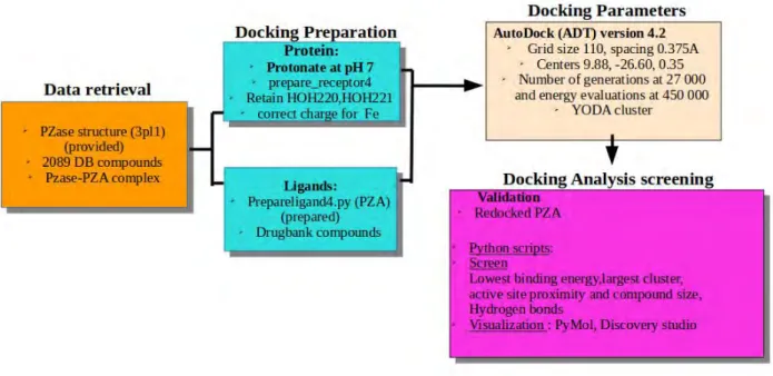
The flow chart below (Figure 2.1) shows the overall procedure applied to successfully dock and select novel hit compounds. The key steps taken include data retrieval, protein-ligand docking
preparations, setting docking parameters, screening and identifying successfully docked orthosteric compounds.
Figure 2.1: Overall applied molecular docking steps, tools and techniques. The docking simulation was perfomed using AutoDOCK 4.2 algorithm. Arrows show the flow of the procedure.
2.3.1 Data retrieval
Due to no available co-crystallized PZase-PZA complex structure in Protein Data Bank (PDB), homology modelling technique was applied to generate a complex structure as described in the study by Sheik Amamuddy et al., (2020). The PZase-PZA complex was provided by the previous group (Sheik Amamuddyet al.,2020). The protein’s PDB ID is 3PL1 (Petrellaet al.,2011). It is an X-ray diffractioned structure with no co-crystallized molecules obtained at 2.20Å resolution having an R-Value Free of 0.240. A data-set of minimized 2089 DrugBank compounds were also provided by Sheik Amamuddyet al.,(2020), who prepared using an in house built script incorporating RDkit tool (Landrum, 2006).
2.3.2 PZase protonation
According to Zhang et al., (2008), PZase is active at a pH of 7.0, therefore in this study, the
modelled structure was protonated at pH 7 using the webserver
(https://server.poissonboltzmann.org/pdb2pqr) (Dolinsky et al, 2004). The protonation states were assigned using PROPKA with the pH set at 7.0. AMBER force field was used as the output naming scheme. Additional selected options were to avoid rebuild of atoms close to existing ones, optimize hydrogen bonding, create an APBS input file and to keep chain IDs in the output file. One key option was not to remove waters from the output file, since there are two crucial water molecules involved in stabilizing the metal binding site. The created .pqr file was edited by adding Iron and renaming the water molecules.
2.3.3Protein and compound preparation
The protein was prepared for docking using the Python command (prepare_receptor4.py -r ‘name of receptor’) in AutoDOCKTools/Utilities24 by adding hydrogens and gasteiger charges. The Gasteiger-Huckel method computed the assigning of charges. Non polar hydrogens were mergerd and AutoDOCK atom types were assigned. The output modified file was saved as a .pdbqt file.
Since the waters on position 220 and 221 and the Iron metal are required for the functioning of the protein, the water molecules were retained and the metal ion was concatenated to the output file with its charge manually edited to +2.000. The control ligand, PZA was prepared using the Python command (prepare_ligand4.py -l ‘name of ligands’) which added gasteiger charges, hydrogens and torsions, merged non-polar hydrogens, detected aromatic carbons and rotatable bonds and saved the modified output file in a .pdbqt format. All the 2089 DrugBank compounds were prepared in a
similar manner to PZA and they were provided by the previous group (Sheik Amamuddy et al., 2020).
2.3.4 Docking parameters
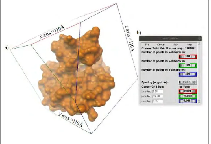
Blind docking was done using AutoDOCK (ADT) version 4.2 on all 2089 DrugBank compounds and PZA (as a control) against the PZase enzyme in a Linux based cluster, YODA. The parameters were set in ADT (version 1.5.6). Grid box size was set to cover the whole protein with 110Å in all x,y,z directions (Figure 2.2 a). The grid center box spacing was set to 0.375Å while the x,y,z centers were set to 9.88, -26.60 and 0.35 respectively (Figure 2.2 b). LGA was used to search for 100 conformations with the maximum number of generations at 27 000 and energy evaluations at 450 000. The semi empirical scoring algorithm calculated the interaction energies. All compounds were docked using the above mentioned parameters and tools. The best poses were selected based on largest cluster and lowest energy, hydrogen bonds and center of mass distance (active site proximity).
Figure 2.2: Molecular docking box size covering whole protein as used in blind docking. a) Visualized grid box dimensions viewed in AutoDOCK Tools. The protein is represented as an orange surface while its center is marked by a yellow line. b) The spacing, x,y,z centers and dimension parameters assigned to obtain the required box size. All x,y,z dimensions are colour co- ordinated as red, green and blue respectively.
2.3.5 Initial docking validation
In order to validate the set parameters and the ability of AutoDOCK 4.2 to reproduce the correct poses, the prepared PZA ligand was docked on to PZase using the above mentioned docking parameters. Due to the absence of a co-crystallized PZA-PZase complex, the obtained pose and interactions were compared to those obtained by Sheik Amamuddy et al.,(2020). Ligand pose and residues interactions were visualized in BIOVIA Discovery Studio Visualizer and PyMOL.