A TAXONOMIC STUDY OF SELECTED REPRESENTATIVES OF
SIPHONOSTOMATOIDA (COPEPODA) FROM OSTEICHTHYES IN COASTAL WATERS OFF SOUTHERN AFRICA
by
MAKWENA MELITA SEBONE
DISSERTATION
Submitted in fulfilment of the requirements for the degree of
MASTER OF SCIENCE in
ZOOLOGY in the
FACULTY OF SCIENCE & AGRICULTURE (School of Molecular & Life Sciences)
at the
UNIVERSITY OF LIMPOPO
SUPERVISOR: PROFESSOR S.M. DIPPENAAR
2023
i DECLARATION
I declare that the dissertation hereby submitted to the University of Limpopo, for the degree of Masters of Science in Zoology has not previously been submitted by me for a degree at this or any other university; that it is my work in design and execution, and that all material contained herein has been duly acknowledged.
____________________________ ___________________
Sebone MM (Miss) Date
19/11/2022
ii ABSTRACT
Currently Copepoda consists of 14 600 species of which 2 275 species are members of the Siphonostomatoida. Siphonostomatoida consists of 40 families, with 17 families symbiotic on fish. Sphyriidae has 44 accepted species in eight reported genera, of which four genera infect teleosts and the remaining four infect elasmobranchs. Adult females undergo transformation through loss of locomotory appendages to suit their mesoparasitic lifestyle and develop outgrowths on the cephalothorax or neck for attachment to the host. To date, only 176 marine siphonostomatoid species have been reported from South African waters, with only nine sphyriid species.
Sphyriids previously collected from marine bony fish off the east, south and west coasts of southern Africa and preserved in 70% ethanol were studied. Specimens were examined with stereo- and compound microscopes and identified using published literature. Selected specimens were stained in lactic acid with added lignin pink, appendages were dissected and illustrated with the aid of a drawing tube.
Selected specimens were also studied through scanning electron microscopy.
The examined specimens were identified as species of Sphyrion and Lophoura. Re- descriptions were done for all valid Sphyrion species females (S. laevigatum, S. lumpi and S. quadricornis) and new descriptions for the males of S. laevigatum and S.
quadricornis. Post-metamorphosis females of Sphyrion species can be differentiated by the shape of cephalothorax, length of the neck in relation to the length of the trunk and the length of posterior processes in relation to the trunk length, while males are mostly very similar. New information is provided regarding the appendages of S.
laevigatum and S. quadricornis. The appendages of the three species bear close resemblance to one another. Additionally, an identification key for the post- metamorphosis females of Sphyrion species is provided.
iii
Re-descriptions were done for five female Lophoura species (L. caparti, L. cornuta, L.
cf edwardsi, L. tetraloba and Lophoura sp.) and a new description of the male of L.
tetraloba. Differences between young and post-metamorphosis females of L. cf edwardsi and L. tetraloba were observed in the width of the holdfast organ processes and the length of porous peduncle and stalks of the posterior processes which appear to grow with age. The difference between the young and adult male of L. tetraloba lies in the lengths of the cephalothorax in relation to the trunk length and segmentation visible on the trunk of the young male but not adult male. The post-metamorphosis females of Lophoura species can be differentiated by the shape and number of processes on the holdfast organ, in combination with the cephalothorax length in relation to the neck length, neck length in relation to the trunk length, shape of the trunk, and the length and structure of the posterior processes. An identification key was drawn up for all species of the Lophoura post-metamorphosis females.
An attempt was made to provide the COI barcodes for all the species of Sphyrion and five species of Lophoura. These would have confirmed the species identification of morphologically variable species e.g. S. laevigatum and S. lumpi and also provide an estimation of the interspecific divergence amongst the different species. Additionally, it would have assisted in distinguishing between L. tetraloba and L. cf edwardsi and provided an estimation of the amount of sequence divergence between the two genera. Unfortunately sequencing of apparently successfully amplified products was unsuccessful probably due to low DNA quality which possibly degraded due to collection methods used for the fish hosts and parasites and prolonged preservation of specimens.
This study provides new host records i.e. Coelorinchus simorhynchus, Coelorinchus trunovi and Saurida undosquamis for Sphyrion quadricornis off South Africa which is also a new geographical record. Allocyttus verrucosus, Coelorinchus simorhynchus, Coelorinchus trunovi, Mesovagus antipodum and Ventrifossa nasuta are also new host records for S. lumpi. Additionally, Epigonus denticulatus and Bassanago albescens are new host records for Lophoura caparti and L. cornuta respectively off
iv
South Africa, which is a new geographical record for both species. Furthermore, Coelorinchus fasciatus and Lucigadus ori are new host records for Lophoura tetraloba and L. cf edwardsi off South Africa, which is also a new geographic record for both species. Thus, the results of the study improve the current knowledge of the marine siphonostomatoid biodiversity off South Africa as well as their distribution and infected hosts.
v ACKNOWLEDGEMENTS
▪ My deepest gratitude to University of Limpopo, and Department of Biodiversity for giving me this opportunity, and provision of a great research lab, with necessary equipments, and also provision of funding for conferences.
▪ My appreciation to the NRF for funding this research.
▪ My heartfelt thanks to my dearest supervisor Prof SM Dippenaar for her dedicated support, encouragements, and motivations. A very incredible mentor who taught me a lot and shared with me every opportunity to enhance my academic expertise. I can’t thank you enough for being patient with me even when I was incompetent at times.
▪ My sincere thanks to Mr Mangena (Sporo) for being the best, dedicated mentor and patiently helping me out with molecular work, with extreme positivity.
Thanks for all the encouragements and motivations.
▪ Thanks to Ms Lebepe (Madame) for helping with proposal writing, her encouragements, and also for assisting in collection of specimens.
▪ Thanks to Mr Mokumo for checking on me with words of encouragement and sharing his M.Sc. experiences.
▪ Thanks to Bea Jordaan, for her contributions in collection of the specimens, and sharing all the best and hilarious memories from the field trips.
▪ Thanks to Dr Anine Jordaan (North West University) for help with Scanning Electron Microscopy.
▪ My heartfelt thanks to my beloved parents for persistent support throughout.
▪ My deepest thanks to all my siblings (Neza, Moloko, Koki, Lebo, Lorraine and Charmaine) and my nephews (Phuti and Moloko) for being there for me in ways that words can’t describe. You have always been my pillar of strength.
▪ My deepest Thanks to my best friend Mulalo for the support and motivations provided throughout.
▪ My gratitude to postgraduate students from the Department of Biodiversity, including my friend Thabelo for all the support and motivations.
▪ My gratitude to Sean Fennessy of the Oceanographic Research Institute (ORI) for fishes obtained during the east coast surveys of KZN marine waters, in 2006
vi
and the KZN bight Ecosystem Functioning Project in 2010 funded by the ACEP African Coelacanth Ecosystem Programme.
▪ My gratitude to D Vaughan (now Central Queensland University) and R Leslie (then DAFF) for collection of specimens during demersal cruises on board the Africana.
▪ I acknowledge PARSA and WAC for the opportunity to attend international conferences.
▪ I would like to always thank God for guidance.
vii RESEARCH OUTPUTS
Publications
Dippenaar SM, Sebone MM. 2022. Morphology of three Sphyrion (Copepoda:
Siphonostomatoida: Sphyriidae) species infecting teleost fishes off South Africa with the first description of males of two species. Diversity 14(11): 929.
https://doi.org/10.3390/d14110929
Sebone MM, Dippenaar SM. (in preparation). Reports and re-descriptions of selected Lophoura species (Copepoda: Siphonostomatoida: Sphyriidae) off South Africa.
Oral presentations
Sebone MM, Dippenaar SM. Sphyriidae females infecting marine teleosts off the South African coast. Virtual International Conference on Copepoda (e-ICOC) 25 – 30 July 2022.
Sebone MM, Dippenaar SM. The study of Sphyrion females infecting marine Osteichthyes off the South African coasts. 4th ICPOW & 50th Annual PARSA Conference, Kruger National Park, South Africa. 11 – 15 September 2022.
viii TABLE OF CONTENTS
DECLARATION i
ABSTRACT ii
ACKNOWLEDGEMENTS v
RESEARCH OUTPUTS vii
TABLE OF CONTENTS viii
LIST OF FIGURES x
LIST OF TABLES xvii
CHAPTER 1: General Introduction 1
1.1. Copepoda Milne Edwards, 1840 1
1.2. Copepods as fish symbionts 2
1.3. Siphonostomatoida Burmeister 1835 2
1.4. Family Sphyriidae Wilson C.B., 1919 4
1.5. Osteichthyes 5
1.6. Coastal waters and their lifeforms 5
1.7. Biological taxonomy 6
1.8. Purpose of the study 6
1.8.1. Aim 6
1.8.2. Objectives 7
1.9. Significance of the study 7
CHAPTER 2: Methodology 9
2.1. Sampling 9
2.2. Data collection 9
2.2.1. Morphological data 9
2.2.2. Molecular data 10
2.3. Data analysis 12
2.3.1. Cladistics analysis 12
CHAPTER 3: Genus Sphyrion Cuvier, 1830 16
3.1. Introduction 16
3.2. Materials and Methods 17
3.3. Results 17
3.3.1. Descriptions of Sphyrion species 17
ix
3.3.2. Key to the adult females of Sphyrion species (according to current
study): 26
3.4. Discussion 26
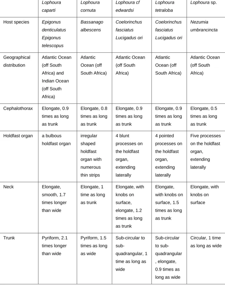
CHAPTER 4: Genus Lophoura Kölliker in Gegenbaur, Kölliker & Müller, 1853 47
4.1. Introduction 47
4.2. Material and methods 51
4.3. Results 51
4.3.1. Descriptions of Lophoura species 51
4.3.2. Identification key to all the adult female Lophoura species 66
4.3.3. Cladistic analysis 68
4.4. Discussion 70
CHAPTER 5: Molecular systematics 95
5.1. Introduction 95
5.2. Material and methods 96
5.3. Results 96
5.4. Discussion 97
CHAPTER 6: General discussion and conclusions 100
REFERENCES 108
x LIST OF FIGURES
1. Figure 1.1: A South African map showing the positions of the coasts, oceans, currents and ecoregions (adapted from Roux et al. 2013 and modified) ... 8 2. Figure 3.1: Sphyrion laevigatum (Quoy & Gaimard, 1824) post-metamorphosis female. A. general habitus, cephalothorax ventral view, trunk dorsal view; B.
cephalothorax, posteroventral view; C. cephalothorax, anteroventral view; D.
cephalothorax, cephalic region with appendages; E. antenna; F. maxillule; G.
maxilliped. Scale bars: A – D = 1 mm and E – G = 10 µm. (ap – antennary processes, a1 – antennule, a2 – antenna, mx1 – maxillule, mp – maxillary processes, mxp – maxilliped) ... 31 3. Figure 3.2: Sphyrion laevigatum (Quoy & Gaimard, 1824) post-metamorphosis female. A. general habitus, cephalothorax ventral view, trunk dorsal view; B.
general habitus, cephalothorax posterodorsal view, trunk ventral view.
Scanning electron micrographs: C. cephalic region, ventral; D. cephalic region, posteroventral view; E. antennule; F. antenna. Scale bars: A, B, D = 1 mm; C = 500 µm; B; E = 10 µm; F = 100 µm. (ap – antennary processes, a1 – antennule, a2 – antenna, mt – mouth tube, mx1 – maxillule, mp – maxillary processes, mgp – maxillary gland pore, mxp – maxilliped). ... 32 4. Figure 3.3: Sphyrion laevigatum (Quoy & Gaimard, 1824) post-metamorphosis female. Scanning electron micrographs: A. maxillule; B. mouth tube; C. labium tubercle; D. maxillary processes with maxillipeds posteromedially; E. maxillary gland pore; F. maxillipeds. Scale bars: A, E = 50 µm; B = 300 µm; C = 10 µm;
D = 500 µm; F = 100 µm. (lbr – labrum, lbm – labium). ... 33 5. Figure 3.4: Sphyrion laevigatum (Quoy & Gaimard, 1824) male. A. general habitus, lateral view; B. trunk; C. antennule; D. Antenna; E. maxillule; F.
mandible; G. maxilla; H. maxilliped. Scale bars: A – H = 10 µm. ... 34 6. Figure 3.5: Sphyrion laevigatum (Quoy & Gaimard, 1824) post-metamorphosis female. A. general habitus, cephalothorax ventral view, trunk dorsal view; B.
general habitus, cephalothorax anterodorsal view, trunk ventral view; C.
cephalothorax, ventral view. Scale bars: A – C = 1 mm. ... 35 7. Figure 3.6: Sphyrion laevigatum (Quoy & Gaimard, 1824) post-metamorphosis female. A. general habitus, cephalothorax ventral view, trunk dorsal view; B.
xi
general habitus, cephalothorax dorsal view, trunk ventral view; C.
cephalothorax, region with appendages; D. antennule; E. antenna; F. maxillule;
G. maxilliped. Scale bars: A – C = 1 mm and E – G = 10 µm. (ap – antennary processes, a1 – antennule, a2– antenna, mx1 – maxillule, mp – maxillary processes, mxp – maxilliped). ... 36 8. Figure 3.7: Sphyrion lumpi (Krøyer, 1845) post-metamorphosis female. A.
general habitus, cephalothorax lateral view, trunk ventral view; B. general habitus, cephalothorax ventral view, trunk lateral view; C. general habitus, cephalothorax dorsal view, trunk dorsal view; D. cephalothorax, ventral view;
E. cephalothorax, cephalic region with appendages; F. antennule. Scale bars:
A – E = 1 mm and F = 10 µm. (ap – antennary processes, a1 – antennule, a2– antenna, mx1 – maxillule, mp – maxillary processes, mxp – maxilliped). ... 37 9. Figure 3.8: Sphyrion lumpi (Krøyer, 1845) post-metamorphosis female. A.
antenna; B. labium tubercle; C. maxillule; D. maxilliped. Scale bars: A – D = 10 µm. ... 38 10. Figure 3.9: Sphyrion lumpi (Krøyer, 1845) post-metamorphosis female. A.
general habitus, cephalothorax dorsal view, trunk dorsal view; B. general habitus, cephalothorax ventral view, trunk ventrolateral view; C. general habitus, cephalothorax dorsal view, trunk ventral view; D. general habitus, cephalothorax ventral view, trunk dorsal view. Scanning electron micrographs:
E. cephalic region, ventrolateral view; F. cephalic region; E. Scale bars: A – E
= 1 mm; F = 300 µm. (ap – antennary processes, a1 – antennule, a2 – antenna, mt – mouth tube, mx1 – maxillule, mp – maxillary processes, mgp – maxillary gland pore, mxp – maxilliped). ... 39 11. Figure 3.10: Sphyrion lumpi (Krøyer, 1845) post-metamorphosis female.
Scanning electron micrographs: A. antennule; B. antenna; C. mouth tube with labrum tubercle; C. labium tubercle; E. maxillule; F. maxillipeds. Scale bars: A, D = 10 µm; B = 50 µm; C, F = 100 µm; E = 20 µm. (lbr – labrum). ... 40 12. Figure 3.11: Sphyrion lumpi (Krøyer, 1845) post-metamorphosis female. A.
general habitus, cephalothorax lateral view, trunk ventral view; B. general habitus, cephalothorax lateral view, trunk dorsal view; C. cephalothorax, anterior view; D. cephalothorax, ventral view. Scale bars: A – D = 1 mm. ... 41 13. Figure 3.12: Sphyrion lumpi (Krøyer, 1845) post-metamorphosis female. A.
cephalothorax, ventral view; B. cephalic area; C. cephalothorax, dorsal view;
xii
D. antennule; E. antenna; F. antenna; G. maxillule; H. maxilliped. Scale bars:
A – C =1 mm, D – H = 10 µm. (ap – antennary processes, a2– antenna, mp – maxillary processes, mxp – maxilliped). ... 42 14. Figure 3.13: Sphyrion quadricornis Gaevskaya & Kovalenva, 1984 post-
metamorphosis female. A. general habitus, cephalothorax ventral view, trunk dorsal view; B. cephalothorax, ventral view; C. cephalothorax, dorsal view; D, cephalothorax, cephalic region with appendages; E. antennule; F. antenna; G.
labium tubercle; H. maxillule; I. maxilliped. Scale bars: A – D = 1 mm and E – I
= 10 µm. (ap – antennary processes, a1 – antennule, a2– antenna, mx1 – maxillule, mp – maxillary processes, mxp – maxilliped). ... 43 15. Figure 3.14: Sphyrion quadricornis Gaevskaya & Kovalenva, 1984 post-
metamorphosis female. A. general habitus, cephalothorax dorsal view, trunk ventral view; B. general habitus, cephalothorax ventral view, trunk dorsal view.
Scanning electron micrographs: C. cephalic area, ventral view; D. cephalic area, ventral view; E. antennule; F. antenna. Scale bars: A – C = 1 mm; D = 300 µm; E = 10 µm; F = 30 µm. (ap – antennary processes, a1 – antennule, a2 – antenna, mt – mouth tube, mx1 – maxillule, mxp – maxilliped, mp – maxillary processes, mgp – maxillary gland pore). ... 44 16. Figure 3.15: Sphyrion quadricornis Gaevskaya & Kovalenva, 1984 post-
metamorphosis female. Scanning electron micrographs: A. maxillule; B.
maxillary gland pore; C. mouth tube; D. labrum tubercle; E. labium tubercles; F.
maxilliped. Scale bars: A, D = 20 µm; B = 50 µm; C, F = 100 µm; E = 30 µm.
(ap – antennary processes, a1 – antennule, a2 – antenna, mt – mouth tube, mx1 – maxillule, mxp – maxilliped, mp – maxillary processes, mgp – maxillary gland pore). ... 45 17. Figure 3.16: Sphyrion quadricornis Gaevskaya & Kovaleva, 1984 male. A.
general habitus, lateral view; B. abdomen with caudal rami; C. antennule; D.
antenna; E. maxillule; F. mandible; G. maxilla; H. maxilliped; I. maxilliped. Scale bars: A – I = 10 µm. ... 46 18. Figure 4.1: Lophoura tetraloba Ho & Kim I.H., 1989 juvenile female. A. general habitus, trunk ventral view, cephalothorax lateral view; B. trunk, lateral view.
Lophoura edwardsi Kölliker, 1853, juvenile female. C. general habitus, trunk ventrolateral view, cephalothorax lateral view; D. trunk, dorsal view. Scale bars:
A – C = 1 mm ... 76
xiii
19. Figure 4.2: Lophoura tetraloba Ho & Kim I.H., 1989 post-metamorphosis female. A. general habitus, trunk ventral view, cephalothorax dorsolateral view;
B. general habitus, dorsal view, cephalothorax ventrolateral view; C. general habitus, trunk dorsal view, cephalothorax ventral view; D. general habitus, trunk ventral view, cephalothorax dorsolateral view; E. general habitus, dorsal view, cephalothorax lateral view. Scale bars: A – E = 1 mm. ... 77 20. Figure 4.3: Lophoura tetraloba Ho & Kim I.H., 1989 post-metamorphosis female. A. general habitus, trunk dorsal view, cephalothorax ventral view; B.
general habitus, trunk ventral view, cephalothorax dorsal view; C.
cephalothorax, anterodorsal view; D. cephalothorax, anterodorsal view; E.
abdomen; F. maxilliped. Scale bars: A – E = 1 mm and F = 10 µm. (mp – maxillary process, ap – antennary process). ... 78 21. Figure 4.4: Lophoura tetraloba Ho & Kim I.H., 1989 post-metamorphosis female. Scanning electron micrographs: A. cephalothorax, cephalic region, anterior view; B. cephalothorax anterolateral view; C. cephalothorax, cephalic region; D. antennule; E. antenna; F. mouth tube. Scale bars: A – B = 500 µm;
C = 300 µm; D – E = 10 µm; F = 50 µm. (ap – antennary process, a1 – antennule, a2 – antenna, mt – mouth tube, lbr – labrum, mx1 – maxillule, mp – maxillary processes, mgp – maxillary gland pore, cg – circular groove). ... 79 22. Figure 4.5: Lophoura tetraloba Ho & Kim I.H., 1989 post-metamorphosis female. Scanning electron micrographs: A. labium tubercle; B. maxillary process; C. maxillary pore; D. maxilliped. Scale bars: A = 10 µm; B = 500 µm;
C = 20 µm; D = 30 µm. (mxp – maxilliped). ... 80 23. Figure 4.6. Lophoura tetraloba Ho & Kim I.H., 1989 male. A. male amongst posterior processes of female; B. general habitus, lateral view; C. mouth tube;
D. caudal rami, lateral view. Scale bars: B = 50 µm; C = 20 µm; D = 10 µm. 81 24. Figure 4.7. Lophoura tetraloba Ho & Kim I.H., 1989 male. A. adult male general habitus, lateral view; B. juvenile male general habitus, lateral view; C. caudal rami, ventrolateral view; D. antennule; E. antenna; F. maxillule. Scale bars: A = 1 mm; B = 50 µm; C – F = 10 µm. ... 82 25. Figure 4.8. Lophoura tetraloba Ho & Kim I.H., 1989 male. A. mouth tube; B.
mandible; C. maxilla; D. maxilliped. Scale bars: A – B = 5 µm; C – D = 10 µm.
... 83
xiv
26. Figure 4.9: Lophoura cf edwardsi Kölliker, 1853 post-metamorphosis female.
A. general habitus, trunk dorsal view, cephalothorax lateral view; B. general habitus, trunk ventral view, cephalothorax lateral view; C. general habitus, trunk lateral view, cephalothorax dorsal view. Scale bars: A – C = 1 mm. ... 84 27. Figure 4.10: Lophoura cf edwardsi Kölliker, 1853 post-metamorphosis female.
A. general habitus, trunk dorsal view, cephalothorax ventral view; B.
cephalothorax, dorsolateral view; C. cephalothorax, anterior view; D, abdomen, dorsal view; E. antennule; F. antenna; G. maxilliped. Scale bars: A – D = 1 mm and E – G = 10 µm. (mp – maxillary process, ap – antennary process). ... 85 28. Figure 4.11: Lophoura cf edwardsi Kölliker, 1853 post-metamorphosis female.
Scanning electron micrographs: A. cephalothorax anterior view; B.
cephalothorax anterolateral view; C. antennule; D. antenna; E. cephalothorax, ventral view; F. maxilliped. Scale bars: A – B = 500 µm; C – D = 10 µm; E = 500 µm; F= 20 µm. (ap – antennary process, a1 – antennule, a2 – antenna, mp – maxillary process, mgp – maxillary gland pore, mxp – maxilliped, cg – circular groove). ... 86 29. Figure 4.12: Lophoura caparti (Nuñes-Ruivo, 1962) post-metamorphosis female. A. general habitus, trunk ventral view, cephalothorax dorsal view. B.
holdfast organ; C. abdomen, dorsal view. Scale bars: A – C = 1 mm ... 87 30. Figure 4.13: Lophoura caparti (Nuñes-Ruivo, 1962) post-metamorphosis female. A. general habitus, trunk dorsal view, cephalothorax ventral view; B.
cephalothorax, ventral view; C. cephalothorax, dorsal view; D. abdomen; E.
antennule; F. antenna; G. maxilliped. Scale bars: A – D = 1 mm and E – G = 10 µm. (mp – maxillary process, ap – antennary process). ... 88 31. Figure 4.14: Lophoura caparti (Nuñes-Ruivo, 1962) post-metamorphosis female. A. cephalic region, anterior view; B. antennule; C. antenna; D. maxillule;
E. maxilliped. Scale bars: B, C and E = 5 µm and D = 20 µm. (ap – antennary process, a1 – antennule, a2 – antenna, mp – maxillary process, mx1 – maxillule). ... 89 32. Figure 4.15: Lophoura cornuta (Wilson C.B., 1919) post-metamorphosis female. A. general habitus, trunk lateral view, cephalothorax lateral view; B.
holdfast organ, lateral view; C. abdomen, lateral view. Scale bars: A – C = 1 mm. ... 90
xv
33. Figure 4.16: Lophoura cornuta (Wilson C.B., 1919) post-metamorphosis female. A. general habitus, trunk ventrolateral view, cephalothorax lateral view;
B. general habitus, trunk ventrolateral view, cephalothorax lateral view; C.
cephalothorax, lateral view; D. cephalothorax, anterior view; E. trunk, ventral view. Scale bars: A – E = 1 mm. (mp – maxillary process, ap – antennary process). ... 91 34. Figure 4.17: Lophoura sp. post-metamorphosis female. A. cephalothorax and holdfast organ, posterolateral view; B. cephalothorax and holdfast organ, anterolateral view; C. trunk, ventral view; D. trunk, ventral view; E. abdomen, dorsal view; F. posterior processes. Scale bars: A – F = 1 mm ... 92 35. Figure 4.18: Lophoura sp. post-metamorphosis female. A. cephalothorax and holdfast organ, posterolateral view; B. cephalothorax, anterolateral view; C.
cephalothorax, anterior view; D. trunk, ventral view; E. abdomen, dorsal view.
Scale bars: A – E = 1 mm. (mp – maxillary process, ap – antennary process).
... 93 36. Figure 4.19: A 50% majority rule consensus tree of two most parsimonious trees (TL= 42, CI = 0.5238, HI = 0.4762, RI = 0.6078, RCI = 0.3184) estimating the phylogenetic relationships among Lophoura species (ingroup species), with Tripaphylus elongatus as an outgroup species. A – L represent nodes, while character transformations are marked on the branches. ... 94 37. Figure 5.1: Examples of PCR products visualized with 1.5% agarose gel electrophoresis. A. PCR products of Sphyrion and Lophoura species (Ladder, S. quadricornis, S. lumpi, S. laevigatum, L. tetraloba, L. edwardsi, L. caparti, positive control and negative control respectively); B. PCR products of Sphyrion and Lophoura species (Ladder, S. quadricornis, S. lumpi, S. laevigatum, L.
tetraloba, L. edwardsi, L. edwardsi, L. caparti, positive control and negative control respectively); C. PCR products of Sphyrion and Lophoura species (Ladder, S. quadricornis, S. lumpi, L. edwardsi, L. caparti, positive control and negative control respectively); D. PCR products of Sphyrion and Lophoura species (S. quadricornis, S. lumpi, S. lumpi, S. laevigatum, L. tetraloba, L.
edwardsi, L. caparti, positive control and negative control respectively); E. PCR products of Sphyrion species (S. lumpi, S. lumpi, S. laevigatum, positive control and negative control respectively); F. PCR products of Sphyrion and Lophoura species (S. quadricornis, S. lumpi, S. laevigatum, L. tetraloba, L. edwardsi, L.
xvi
caparti, positive control and negative control respectively). (L = ladder, fb = faint band, vb = visible band, pd = primer dimer). ... 99
xvii LIST OF TABLES
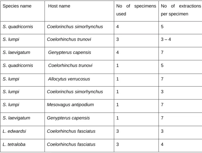
1. Table 2.1: Specimens used for DNA extractions with their species names and host names. ... 11 2. Table 2.2: Primers used in PCR amplification of a part of mitochondrial gene
(COI), with their sequences and product size anticipated... 12 3. Table 2.3: Characters with assigned character states used to compile a
character matrix for Lophoura species (according to present study and Benz et al. (2006)). ... 13 4. Table 2.4: Character matrix compiled for Lophoura species with Tripaphylus
elongatus (Wilson, 1932) as the outgroup taxon (compiled from Wilson 1919, 1935; Nuñes Ruivo 1962; Hewitt 1964; Szidat 1971; Kensley and Grindley 1973; Stadler 1978; Kabata 1979; Ho 1985; Hogans and Dadswell 1985; Ho and Kim 1989; Boxshall 1989, 2000; Castro-Romero and Gonzalez 2009;
Gómez et al. 2010; Dippenaar 2018)... 14 5. Table 3.1: Comparisons between the morphology of S. laevigatum female individuals. ... 20 6. Table 3.2: Comparison of general habitus of different Sphyrion species
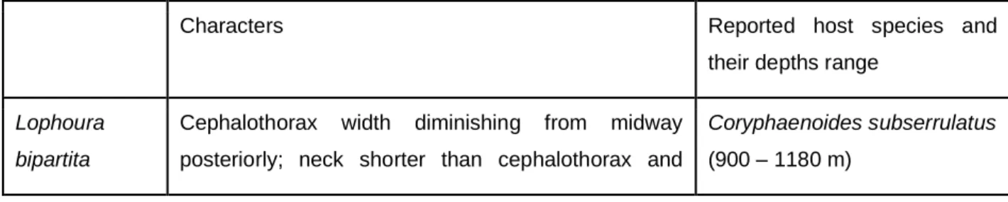
(according to the current study). ... 29 7. Table 4.1: A summary of the distinguishing features of Lophoura species based on the morphology of the transformed post-metamorphosis females (compiled from Wilson 1919, 1935; Nuñes Ruivo 1962; Hewitt 1964; Szidat 1971; Kensley and Grindley 1973; Stadler 1978; Kabata 1979; Ho 1985; Hogans and Dadswell 1985; Ho and Kim 1989; Boxshall 1989, 2000; Castro-Romero and Gonzalez 2009; Gómez et al. 2010) with their reported host species and the depths at which the hosts occur (Froese and Pauly 2022). ... 48 8. Table 4.2: Comparisons of Lophoura species, with reported hosts and
distribution in the current study. ... 65 9. Table 4.3: The consistency index (ci), retention index (ri) and rescaled
consistency index (rci) values calculated for each character used in the cladistic analysis of all Lophoura species with Tripaphylus elongatus as the outgroup taxon. ... 69
xviii
10. Table 5.1: Representatives of Sphyriidae for which DNA extractions were done, the host species from which they were collected, DNA concentration, UV absorption at 260 nm (A260), UV absorption at 280 nm (A280), ratio 260/280 and ratio 260/230... 96 11. Table 6.1: Reported host species of Lophoura and Sphyrion species with geographic location (compiled from Wilson 1919, 1935; Nuñes Ruivo 1962;
Hewitt 1964; Szidat 1971; Kensley and Grindley 1973; Kabata 1979; Ho 1985;
Hogans and Dadswell 1985; Ho and Kim 1989; Boxshall 1989; Ho 1992; Moran
& Piasecki 1994; Boxshall 2000; Castro-Romero and Gonzalez 2009; Gómez et al. 2010; Kazachenko 2017; Froese and Pauly 2022; Walter and Boxshall 2022; Dippenaar and Sebone 2022; and the current study). ... 101
1 CHAPTER 1: General Introduction
1.1. Copepoda Milne Edwards, 1840
This class consists of typically small aquatic crustaceans within the phylum Arthropoda. Copepoda exhibits high morphological diversity with abundant species (Boxshall and Halsey 2004). Currently, it consists of 10 orders (Platycopioida Fosshagen, 1985; Calanoida G. O. Sars, 1903; Misophrioida Gurney, 1933;
Canuelloida Khodami, Vaun MacArthur, Blanco-Bercial & Martinez Arbizu, 2017;
Gelyelloida Huys, 1988; Harpacticoida G. O. Sars, 1903; Mormonilloida Boxshall, 1979; Cyclopoida Burmeister, 1834; Siphonostomatoida Burmeister, 1835 and Monstrilloida Sars, 1901) with 14 600 species reported worldwide (Walter and Boxshall 2022). The members are morphologically diverse, ranging from free-living to symbiotic copepods inhabiting a wide variety of habitats, ranging from freshwater to marine and hypersaline waters (Boxshall and Halsey 2004; Eyun 2017).
Copepods are uniquely known by their broad, paddle-like swimming legs. They are typically small creatures with body lengths varying from about 0.2 mm to 2.8 mm.
However, some parasitic copepods are large, with a body length of about 250 mm, such as members of the Pennellidae Burmeister, 1835 (Boxshall and Halsey 2004). A copepod’s body is divided into the cephalosome, metasome and urosome. The cephalosome and urosome have 10 appendages each, used mostly for feeding and movement but also for attachment in symbiotic species (Huys and Boxshall 1991).
Copepods are of major importance to living systems. The aquatic food chain requires copepods which feed on phytoplankton and provide food for fish and other planktivorous organisms. Freshwater copepods feed on, amongst others, mosquito larvae, thus biologically controlling malaria. They also serve as intermediate hosts to other parasites, such as guinea worm and tapeworm (Boxshall and Halsey 2004).
Some copepods can impact fisheries by parasitizing economically important fish. Free- living copepods can be biological indicators of climate change and symbiotic copepods
2
can be used as biological tags while also providing information (e.g. geographical, diet, phylogenetic, etc.) about their hosts (Goater et al. 2014).
1.2. Copepods as fish symbionts
Of the ten Copepoda orders, only two have species that are fish symbionts, namely Siphonostomatoida and Cyclopoida (Huys and Boxshall 1991; Khodami et al. 2017).
These symbiotic copepods are mostly ectoparasitic, since they inhabit the host’s external body surface such as eye orbit, oral cavity, gill cavity and nasal slits (Huys and Boxshall 1991). They use appendages such as the antennae, maxillae and/or maxillipeds for attachment, with exception of the family Lernaeopodidae Milne Edwards, 1840, which embed maxillary arms (bulla) into the host tissue for firm attachment. Mesoparasites include members of the families Sphyriidae Wilson C.B., 1919 and Pennellidae Burmeister, 1835 since they embed the anterior part of their bodies into the host tissues, aided by cephalic holdfasts for attachment (Kabata 1979;
Boxshall and Halsey 2004). Endoparasitic copepods occur within the family Philichthyidae Vogt, 1877 with members inhabiting the skull bones (e.g. Colobomatus mylionus Fukui, 1965) and sensory canals of the lateral line system (e.g. Colobomatus pupa Izawa, 1974) (Madinabeitia et al. 2012, Madinabeitia and Iwasaki 2013).
1.3. Siphonostomatoida Burmeister 1835
The order Siphonostomatoida is a large order within the superorder Podoplea Giesbrecht, 1882 (Huys and Boxshall 1991), which consists of 40 families with 2 275 species (Walter and Boxshall 2022). Approximately 75% of the siphonostomatoid members are fish symbionts (Dippenaar 2004, 2016). Siphonostomatoids are mainly marine, although there are few symbionts of freshwater fish. This order is known for unresolved interfamilial relationships due to scanty knowledge of the representatives (Dippenaar 2009).
In addition to the 163 Siphonostomatoida from the checklist by Nunkoo (2019), 13 additional species (Lernaeopoda mustelli Thomson G.M., 1890 and Lernaeopoda bidiscalis Kane, 1892 (see Dippenaar 2020b); Neoalbionella izawai Dippenaar, 2020 (see Dippenaar 2020a); Naobranchia cygniformis Hesse, 1863 (see Dippenaar and
3
Sebone 2021); Pseudocharopinus malleus (Rudolphi in Nordmann, 1832) (see Dippenaar 2019); Caligus lineatus Hayes, Christison, Vaughan, Smit & Boxshall, 2021; Caligus tumulus Hayes, Christison, Vaughan, Smit & Boxshall, 2021; Caligus longipedis Bassett-Smith, 1898; Lepeophtheirus spinifer Kirtisinghe, 1937;
Lepeophtheirus acutus Heegaard, 1943; Caligus tenuis (van Beneden, 1852); Caligus rufimaculatus Wilson, 1905 (see Hayes et al. 2021); and Alella igillimpethu Erasmus, Hadfield, Wepener & Smit, 2022 (see Erasmus et al. 2022)) have been reported from marine fish of South Africa. Thus, only 176 marine symbiotic siphonostomatoids were reported from South Africa in comparison to the 2 275 (Walter and Boxshall 2022) marine siphonostomatoids reported worldwide. This is most likely due to limited studies examining the hosts for possible symbiotic copepods since there is a shortage of taxonomists studying marine siphonostomatoids in South Africa (Smit and Hadfield 2015; Dippenaar 2016). Without sufficient studies, there is a possibility that some species, including siphonostomatoids, may become extinct (with their hosts) prior to description or report which may lead to lack of knowledge about the biodiversity of siphonostomatoids in South Africa but also worldwide (Woodcock 2002; Narendran 2008; Dippenaar 2016).
Members of order Siphonostomatoida possess a tubular mouth resembling a siphon, which is formed by the fused labium and labrum (Kabata 1979). There is sexual dimorphism in members of this order, with the extreme being reached in families such as Lernaeopodidae and Sphyriidae where males are dwarf and attach to large-bodied females (Kabata 1979; Huys and Boxshall 1991).
The lack of sufficient knowledge to identify and classify some members of the Siphonostomatoida is clear considering for example the family Lernaeopodidae Milne Edwards, 1840 with about 22 renamed genera and 522 renamed species, while Pennellidae has about 17 renamed genera and 116 renamed species and Sphyriidae Wilson C.B., 1919 has about seven renamed genera and 30 renamed species.
Additionally, about 22 marine lernaeopodids and about 20 marine pennellids have incomplete descriptions for further taxonomic classification (Walter and Boxshall 2022).
4 1.4. Family Sphyriidae Wilson C.B., 1919
Species found in this family are mesoparasites of deep sea and open ocean fish (Huys and Boxshall 1991). The body of the adult females are transformed and the locomotory appendages lost or reduced after attaching to the host. Thus, the body can be divided into the head (cephalothorax), neck and genito-abdominal trunk (Kabata 1979). In cases where locomotion is required on the host’s body surface, the female uses the maxillae and maxillipeds to aid the movement (Boxshall and Halsey 2004). These appendages are used for prehension but once the female becomes attached to a suitable spot on the host, it develops holdfast protuberances on the cephalothorax margins or neck which is embedded into the host tissue, and it becomes a mesoparasite. The cephalothorax protuberances aid with firm attachment and eventually the maxillae and/or maxillipeds may disappear (Kabata 1979). The males continue to use the second maxillae and maxillipeds both for locomotion and prehension to the adult female. The sphyriid dwarf males bear a close resemblance to the lernaeopodid males (Kabata 1979; Huys and Boxshall 1991).
Sphyriidae currently consists of eight genera (i.e. Sphyrion Cuvier, 1830; Lophoura Kölliker in Gegenbaur, Kölliker & Müller, 1853; Tripaphylus Richiardi in Anonymous, 1878; Opimia Wilson C.B., 1908; Periplexis Wilson C.B., 1919; Paeonocanthus Kabata, 1965; Norkus Dojiri & Deets, 1988; Driocephalus Raibaut, 1999) including 44 species (Walter and Boxshall 2022). The genera are distinguished based on the morphology of the cephalothorax, posterior processes (simple and cylindrical vs subdivided and complex) and a neck with or without holdfast outgrowths (Kabata 1979).
Sphyriids are parasites of both elasmobranchs and teleosts (Boxshall and Halsey 2004), with only four genera (Paeonocanthus, Periplexis, Lophoura and Sphyrion) found on teleosts (Gómez et al. 2010). To date, only three genera and nine species (Lophoura elongata Kensley & Grindley 1973 from Histiobranchus bathybius (Günther, 1877); Sphyrion laevigatum (Quoy & Gaimard, 1824) from Atractoscion aequidens (Cuvier, 1830), Coelorinchus fasciatus (Günther, 1878) and Genypterus capensis (Smith, 1847) (Barnard 1955; Payne 1986); Sphyrion lumpi (Krøyer, 1845) from Antimora rostrata (Günther, 1878), Psychrolutes inermis (Vaillant, 1888) and Psychrolutes macrocephalus (Gilchrist, 1904) (Barnard 1955; Kensley and Grindley
5
1973); Tripaphylus elongatus (Wilson C.B., 1932) from Carcharhinus obscurus (LeSueur, 1818) (Dippenaar and Jordaan 2007; Dippenaar 2018); Tripaphylus benzi Dippenaar, 2018 from Mustelus palumbes Smith, 1957 (Dippenaar 2018); Tripaphylus hoi Dippenaar, 2018 from Mustelus palumbes Smith, 1957; Tripaphylus beatricae Dippenaar, 2018 from Mustelus mustelus (Linnaeus, 1758) (Dippenaar 2018);
Tripaphylus lewisi Dippenaar, 2018 from Hemipristis elongata (Klunzinger, 1871) (Dippenaar 2018); and Tripaphylus vaissierei (Delamare Deboutteville & Nuñes- Ruivo, 1954) from Sphyrna lewini (Griffith & Smith, 1834) (Dippenaar and Jordaan 2007; Dippenaar 2018)) were reported from South African waters.
1.5. Osteichthyes
Osteichthyes contributes 96% of fish species worldwide. The representatives are highly diverse, and their adaptation extends to extreme habitat conditions.
Osteichthyes consists mostly of Actinopterygii (ray-finned fish) and Sarcopterygii (lobe-finned fish). The Actinopterygii has a bony skeleton, fins with spines, a cartilaginous skull (partly calcified), and a pair of gill openings enveloped in the operculum. Sarcopterygii members possess fleshy lobes which join fins to the body (Smith and Heemstra 1988). Fishes of the Osteichthyes differ from those of the Chondrichthyes by possessing bony skeletons rather than cartilaginous skeletons and also the presence of a swim bladder to improve buoyancy (Perry 2007). Currently, there are 1 810 Osteichthyes species reported from South African marine waters (Froese and Pauly 2022). Although there is a high diversity of bony fish, there is currently no copepodologist actively researching the symbiotic siphonostomatoids infecting Osteichthyes in South Africa. Most available studies of South African siphonostomatoids were reported from representatives of Chondrichthyes (Dippenaar 2016).
1.6. Coastal waters and their lifeforms
The South African coast ranks the second longest in Africa with about 3 650 km (Rautenbach et al. 2019). It is composed of three ecoregions (KwaZulu-Natal, Agulhas bank and Namaqua) with the cold Benguela current of the Atlantic Ocean along the west coast (Fig. 1.1) and the warm Agulhas current of the Indian ocean along the east coast (Fig. 1.1). The east coast has lower biodiversity compared to the west and the
6
south coasts, due to less nutrients in warm water whereas the west and south coast are enriched with abundant biodiversity (Griffiths et al. 2010). The South African marine waters provide habitats to more than 12 000 species, with about 2 013 known fish species (Froese and Pauly 2022).
1.7. Biological taxonomy
Taxonomy is the science of studying and classifying biotic organisms into respective taxa (Boxshall 2020). There are two methods widely used to identify invertebrates, i.e.
classical and DNA-based taxonomy. Classical taxonomy includes a close observation of external traits and morphometric measurements. It has been used from ancient times before technological advancements (Wheeler 2008). DNA-based taxonomy includes the amplification of DNA fragments through polymerase chain reaction (PCR) to determine species-specific genetic variation, interrelationships within different taxa and systematics between taxa (Pfenninger et al. 2006). The integration of both classical and DNA-based taxonomy yields the best results for taxonomic purposes (Wheeler 2008) of species and overcomes the disadvantages arising when each method is used individually.
Biological taxonomy contributes to background knowledge about an organism, including the habitat preference, host association, geographical distribution, morphology and biology (Boxshall 2020). Taxonomy helps in discovering the number of species existing in the environment and gives an idea of local fauna and flora, thus helping in distinguishing endemic species (Frankham et al. 2002) and making biodiversity estimations.
1.8. Purpose of the study 1.8.1. Aim
The aim of this study was to study and report on selected representatives of Sphyriidae collected from marine bony fish in coastal waters off southern Africa and to attempt to clarify any existing confusion regarding their taxonomy and systematics.
7 1.8.2. Objectives
The objectives of this study were:
i. To identify all the collected specimens of the family Sphyriidae collected from marine bony fish to species level by studying the morphology through dissecting appendages, illustrating and completely describing each species.
ii. To compare the identified species with those in the literature and to attempt to clarify the existing uncertainty regarding the number of valid species where necessary.
iii. To support classical taxonomy by DNA-based taxonomy of the identified species.
1.9. Significance of the study
This study will provide additional information to the limited knowledge regarding the biodiversity of marine siphonostomatoids occurring off South Africa. It may also add to our knowledge of the hosts being infected by these collected sphyriid species as well as information regarding the host specificity of the selected species. An important contribution will be made to current knowledge by the attempt to resolve the current valid species discrepancies and providing re-descriptions and illustrations of collected species. The knowledge of our world’s symbiotic copepods can help the present and upcoming generations to better understand the world of copepods and their interactions with fish.
8
Figure 1.1: A South African map showing the positions of the coasts, oceans, currents and ecoregions (adapted from Roux et al. 2013 and modified)
9 CHAPTER 2: Methodology
2.1. Sampling
Specimens previously collected from marine bony fish off the east, south and west coasts of southern Africa, and preserved in 70% ethanol, were examined. These specimens were collected between 1991 and 2016 from fish caught as by-catch during demersal cruises, including hake assessment demersal cruises off the south and west coasts of South Africa on board the Department of Agriculture, Forestry and Fisheries (DAFF) research vessel R/V (Africana) as well as during demersal survey trawls off the South African east coast (Indian Ocean) (ACEP African Coelacanth Ecosystem Programme). The fish hosts were identified by researchers on board the vessels. The fish hosts examined from the west coast include Allocyttus verrucosus (Gilchrist, 1906); Coelorinchus simorhynchus Iwamoto & Anderson, 1994; Nezumia umbracincta Iwamoto & Anderson, 1994; Lucigadus ori (Smith, 1968); Coelorinchus fasciatus (Gunther, 1878); Epigonus denticulatus Dieuzeide, 1950; and Genypterus capensis (Smith, 1847); whereas the fish hosts from the east coast include Coelorinchus trunovi Iwamoto & Anderson, 1994; Epigonus telescopus (Risso, 1810); and Ventrifossa nasuta (Smith, 1935); and the fish hosts from south coast include Mesovagus antipodum (Hubbs & Iwamoto, 1977) and Bassanago albescens (Barnard, 1923). The host names were confirmed through Froese & Pauly (2022) and Fricke et al. (2022).
2.2. Data collection
2.2.1. Morphological data
The identification of collected specimens was performed using morphological characteristics. The collected specimens were stained in lactic acid with a small amount of dissolved lignin pink. The external morphology of collected specimens was studied using the wooden slide technique (Humes and Gooding 1964), through stereo- and compound microscopes and drawings done with the aid of drawing tubes. Studied specimens were dissected, and all appendages were drawn. Measurements were done using a 2 mm stage micrometer. Morphological terminology used is mostly according to Kabata (1979) and Huys and Boxshall (1991). Additionally, a series of
10
photographs of sections of selected specimens were taken using an imaging software Olympus Stream Essentials 2.2 and merged into one picture (since the size of specimens was too big to be captured in a single photo). Photographs of appendages of somes specimens were taken using an imaging software Olympus LCmicro 2.2.
Morphological features were used to classify the species into the respective genera using relevant literature (Wilson 1919, 1935; Nigrelli and Firth 1939; Barnard 1955;
Nuñes Ruivo 1962; Hewitt 1964; Szidat 1971; Kensley and Grindley 1973; Kabata 1979; Gaevskaya and Kovaleva 1984; Ho 1985; Hogans and Dadswell 1985; Payne 1986; Ho and Kim 1989; Boxshall 1989; Moran and Piasecki 1994; Boxshall 2000;
Castro-Romero and Gonzalez 2009; Gómez et al. 2010; Kazachenko 2017).
Scanning Electron Microscopy
Cephalic areas of the specimens were dissected off the cephalothoraces and dehydrated through ethanol series (70% – 80% – 90% – 100% – 100%) with each over 30 minutes intervals whereafter the samples were transferred into hexamethyldisilazane for at least 30 minutes and then transferred into a petri dish to dry completely. Samples were mounted onto stubs, sputter-coated with carbon and gold palladium. Scanning electron microscopy observations were carried out through a FEI Quanta 250 FEG SEM.
2.2.2. Molecular data
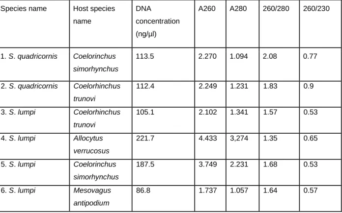
Specimens were rehydrated from 70% ethanol to distilled water by gradually decreasing the ethanol concentration. Genomic DNA was isolated from trunk tissues of specimens of Sphyrion laevigatum, Sphyrion lumpi, Sphyrion quadricornis, Lophoura edwardsi, Lophoura caparti and Lophoura tetraloba (see Table 2.1) using proteinase K digestion and the Qiagen genomic purification kit, following the instruction of the manufacturer. Extracted DNA concentrations, and UV absorption at 260 nm (A260), UV absorption at 280 nm (A280), ratio 260/280 and ratio 260/230 were measured using a Nanodrop. The recommended DNA concentration for successful PCR is 50 ng/μl and thus attempts to increase the concentration of extractions with lower concentrations included multiple extractions made from an individual which were combined and incubated at 56°C (dry bath) for 1 hour to evaporate. Thereafter the concentration of DNA was measured again and if still less than 50 ng/μl the
11
concentration step was repeated until the desired concentration was reached.
Samples consisting of DNA concentrations ranging from 76.8 ng/μl to 221.7 ng/μl were used to attempt to amplify part of the COI (cytochrome oxidase I) gene. Each polymerase chain reaction (PCR) was carried out in 20 μl volumes, containing 10 μM of both forward (cop-COI-1498F, LCO and LCO_t1) and reverse (HCO and HCO_t1) primers (see Table 2.2), 10 μl OneTaq® 2X Master Mix, 50 ng/μl of template DNA and distilled water to add up the volume. The PCR reactions were performed with the following conditions: initial denaturation at 95°C for 4 min, followed by 30 cycles of denaturation at 94°C for 1 min, annealing at 45°C – 48°C for 2 min (see Table 2.3), and extension at 70°C for 3 min. The final extension was carried out at 72°C for 10 min and cooling at 4°C for 30 min. The amplicons were separated on a 1.5% agarose gel stained with ethidium bromide, running at 150 V. PCR products were sent for sequencing to Inqaba Biotech laboratory.
Table 2.1: Specimens used for DNA extractions with their species names and host names.
Species name Host name No of specimens
used
No of extractions per specimen
S. quadricornis Coelorinchus simorhynchus 4 5
S. lumpi Coelorhinchus trunovi 3 3 – 4
S. laevigatum Genypterus capensis 4 7
S. quadricornis Coelorhinchus trunovi 1 5
S. lumpi Allocytus verrucosus 1 7
S. lumpi Coelorinchus simorhynchus 1 3
S. lumpi Mesovagus antipodium 1 7
S. laevigatum Genypterus capensis 1 7
L. edwardsi Coelorhinchus fasciatus 3 3
L. tetraloba Coelorhinchus fasciatus 3 4
12
L. tetraloba Lucigadus ori 1 4
L. caparti Epigonus denticulatus 2 6
L. caparti Epigonus denticulatus 1 6
Table 2.2: Primers used in PCR amplification of a part of mitochondrial gene (COI), with their sequences and product size anticipated.
Name of
primer Sequence Annealing
temperature
Anticipated
gene size Reference
LCO 2198 5’-GGTCAACAAATCATAAAGATATTGG-3’ 45 °C 670 bp Folmer et al. 1994 HCO
1490 5’-TAAACTTCAGGGTGACCAAAAAATCA-3’ 45 °C, 48 °C 670 bp Folmer et al. 1994
LCO 2198_t1
5’-
TGTAAAACGACGGCCAGTGGTCAACAAATCA TAAAGATATTGG-3’
45 °C 670 bp Messing
1983
HCO 1490_t1
5’-
CAGGAAACAGCTATGACTAAACTTCAGGGTG ACCAA
AAAATCA-3’
45 °C 670 bp Messing
1983
cop-COI-
1498F AAYCATAAAGAYATYGGDAC 48 °C 670bp Bucklin et
al. 2010
2.3. Data analysis
2.3.1. Cladistics analysis
Cladistic analysis was performed for all Lophoura species based on morphological characteristics, to determine evolutionary relationships among them. Tripaphylus elongatus (Wilson, 1932), one of members of family Sphyriidae, was used as the outgroup. Outgroup taxon helps to determine polarity of character states using the
13
outgroup comparison method. Thirteen morphological characters (Table 2.3) were identified (according to Benz et al. 2006) to use in compiling a character matrix for the 19 Lophoura species (see Tables 2.4). Parsimony analysis was performed with PAUP version 4.0b10 (Swofford 1985), using a heuristic search with tree-bisection reconnection (TBR) branch swapping technique to find the most parsimonious tree(s).
The homoplasy index, retention index, consistency index and rescaled consistency index were used to measure the goodness of fit of the characters on the tree. In cases where there is more than one most parsimonious tree, the strict consensus (only display nodes that concur in all most parsimonious trees) and 50% majority rule consensus (display nodes that occur in 50% of most parsimonious trees) trees were determined.
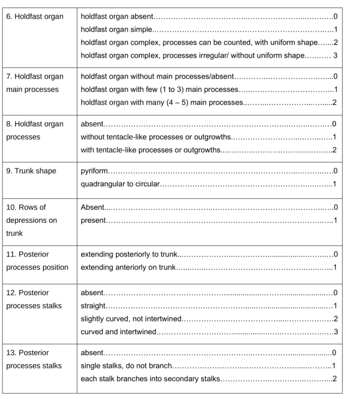
Table 2.3: Characters with assigned character states used to compile a character matrix for Lophoura species (according to present study and Benz et al. (2006)).
Characters States
1. Cephalothorax circular……….……….………..……...…….0 longitudinally elongated, cylindrical...……… ………….…….……..1
2. Cephalothorax length vs neck length
cephalothorax length shorter than neck length.………..…….0 cephalothorax length longer than neck length.………..…...…1
3. Cephalothorax vs trunk length
cephalothorax length shorter than trunk….………….………….…………...…..0 cephalothorax length longer than trunk…….……….…………..…….……1
4. Cephalothorax width
cephalothorax without longitudinal part.……….………0 longitudinal part of the cephalothorax with the same width throughout…..…..1 longitudinal part of the cephalothorax with different width..…………...……….2 5. Neck length in
relation to trunk length
longer than the trunk……….….………...0 as long as trunk………..……….…..1 shorter than the trunk……….………....…..2
14
6. Holdfast organ holdfast organ absent………..………...…….….0 holdfast organ simple.……….…..1 holdfast organ complex, processes can be counted, with uniform shape.…...2 holdfast organ complex, processes irregular/ without uniform shape.….…… 3 7. Holdfast organ
main processes
holdfast organ without main processes/absent….………..……….…....0 holdfast organ with few (1 to 3) main processes…...………...1 holdfast organ with many (4 – 5) main processes..……...………...…...2 8. Holdfast organ
processes
absent………..……..…….…0 without tentacle-like processes or outgrowths.….………..……...…..1 with tentacle-like processes or outgrowths....………...………....……...2
9. Trunk shape pyriform……….………...………..….0
quadrangular to circular………..…..…...1 10. Rows of
depressions on trunk
Absent...………..………..…..0 present……….…….………...……….…..1
11. Posterior processes position
extending posteriorly to trunk...………...…………...……..…0 extending anteriorly on trunk…....…...………..…..…...1
12. Posterior processes stalks
absent………...………...…0 straight……….………...…...…1 slightly curved, not intertwined.………..………....……….2 curved and intertwined.………...….……….…3 13. Posterior
processes stalks
absent……….……..………...0 single stalks, do not branch………..………....………..1 each stalk branches into secondary stalks………..…………..………...2
Table 2.4: Character matrix compiled for Lophoura species with Tripaphylus elongatus (Wilson, 1932) as the outgroup taxon (compiled from Wilson 1919, 1935; Nuñes Ruivo 1962; Hewitt 1964; Szidat 1971; Kensley and Grindley 1973; Stadler 1978; Kabata 1979; Ho 1985; Hogans and Dadswell 1985; Ho and Kim 1989; Boxshall 1989, 2000;
Castro-Romero and Gonzalez 2009; Gómez et al. 2010; Dippenaar 2018).
Character 1 2 3 4 5 6 7 8 9 10 11 12 13
15
L. bipartita 1 1 1 2 0 2 1 2 1 1 1 3 1 L. bouvieri 1 0 0 1 0 1 1 1 1 1 0 1 1 L. brevicollum 1 0 0 2 2 2 2 1 1 1 0 1 2 L. caparti 1 0 1 1 0 1 1 1 0 0 0 1 2 L. cardusa 1 0 1 1 2 3 0 2 0 1 0 1 1 L. cornuta 1 0 0 1 2 2 2 2 0 1 0 1 1 L. edwardsi 1 0 0 1 0 2 2 1 1 1 0 1 1 L. elongata 1 1 1 1 0 2 2 1 0 1 0 1 1 L. gracilis 1 1 1 2 1 2 2 1 0 1 0 1 2 L. laticervix 1 0 0 1 2 2 1 1 0 1 0 1 1 L. magna 1 0 0 1 2 3 1 2 0 1 0 1 1 L. pentaloba 1 0 0 1 2 2 2 1 0 1 1 3 1 L. simplex 1 0 0 1 0 0 0 0 0 1 0 1 1 L. szidati 1 0 1 2 0 1 1 1 0 1 0 2 1 L. tetraloba 1 0 0 1 0 2 2 1 1 1 0 1 1 L. tetraphylla 1 0 0 1 2 2 2 1 1 1 0 2 1 L. tripartita 1 0 1 1 0 2 1 1 0 1 0 1 2 L. unilobulata 1 0 0 1 0 1 1 1 0 1 0 1 2 L. ventricula 1 0 0 1 0 2 2 2 0 1 1 3 1 T. elongatus 0 0 0 0 0 0 0 0 0 0 0 0 0
16 CHAPTER 3: Genus Sphyrion Cuvier, 1830
3.1. Introduction
Sphyrion is one of the genera belonging to family Sphyriidae, which Krøyer (1863) previously placed under the Pennellidae (see Kabata 1979). Currently there are only three accepted species, namely Sphyrion laevigatum (Quoy & Gaimard, 1824), Sphyrion lumpi (Krøyer, 1845) and Sphyrion quadricornis Gaevskaya & Kovaleva, 1984 (Walter and Boxshall 2022). Similar to other sphyriid females, Sphyrion adult females possess highly modified bodies.
Post-metamorphosis females possess transversely elongated (hammer-shaped) cephalothoraces of shapes and sizes varying from one species to the other, attached to a cylindrical neck. The posterior part of the neck expands into the dorsoventrally flattened trunk with suborbicular or pyriform shape, bearing a small abdomen posteriorly. The abdomen accommodates branched and grape-like posterior processes, and long egg sacs, with multiseriate eggs. Males attach to the female body (on the posterior processes) resemble lernaeopodid males with the body indistinctly divided into the cephalothorax and compact trunk (Wilson 1919; Kabata 1979).
Sphyrion species are cosmopolitan and infect a wide range of host species (Ho 1992;
Moran & Piasecki 1994; Kazachenko 2017). To date, S. lumpi has been reported from 30 fish host species (i.e. Antimora rostrata (Günther, 1878); Anarhichas denticulatus Krøyer, 1845; Anarhichas lupus Linnaeus, 1758; Boreogadus saida (Lepechin, 1774);
Cyclopterus lumpus Linnaeus, 1758; Coelorinchus fasciatus (Günther, 1878);
Coelorinchus marinii Hubbs, 1934; Coryphaenoides rupestris Gunnerus, 1765;
Coryphaenoides armatus (Hector, 1875); Gaidropsarus ensis (Reinhardt, 1837);
Gadus morhua Linnaeus, 1758; Glyptocephalus cynoglossus (Linnaeus, 1758);
Laemonema laureysi Poll, 1953; Macrourus berglax Lacepède, 1801; Merluccius bilinearis (Mitchill, 1814); Molva dypterygia (Pennant, 1784); Psychrolutes inermis (Vaillant, 1888); Reinhardtius hippoglossoides (Walbaum, 1792); Sebastes norvegicus (Ascanius, 1772); Sebastes mentella Travin, 1951; Sebastes viviparus Krøyer, 1845; Macrourus holotrachys Günther, 1878; Merluccius merluccius (Linnaeus, 1758); Psychrolutes macrocephalus (Gilchrist, 1904); Sebastes fasciatus
17
Storer, 1854; Sebastes flammeus (Jordan & Starks, 1904); Solea solea (Linnaeus, 1758); Pagellus bogaraveo (Brünnich, 1768); Urophycis tenuis (Mitchill, 1814)) (Ho 1992, Alves et. al 2013, Walter and Boxshall 2022) belonging to 15 families, whereas S. laevigatum has been reported from six fish host species (i.e. Coelorinchus fasciatus (Günther, 1878); Genypterus blacodes (Forster, 1801); Genypterus capensis (Smith, 1847); Merluccius australis (Hutton, 1872); Merluccius hubbsi Marini, 1933;
Merluccius polli Cadenat, 1950) belonging to three families (Ho 1992, Walter and Boxshall 2022) and S. quadricornis has been reported from four fish host species (i.e.
Coelorinchus braueri Barnard, 1925; Coelorinchus innotabilis McCulloch, 1907;
Coelorinchus fasciatus (Günther, 1878); Macrourus berglax Lacepède, 1801) belonging to Macrouridae (Ho 1992, Walter and Boxshall 2022).
3.2. Materials and Methods Refer to Chapter 2.
3.3. Results
3.3.1. Descriptions of Sphyrion species
3.3.1.1. a. Sphyrion laevigatum (Quoy & Gaimard, 1824) Host: Genypterus capensis (Smith, 1847)
Locality: South coast of South Africa (Atlantic Ocean)
Material examined: 4♀♀ and 1♂ from G. capensis off the south coast (Atlantic Ocean) Material collected: 30♀♀ and 3♂♂ all from G. capensis off the south and west coast (Atlantic Ocean)
Description of post-metamorphosis female (Figs. 3.1 – 3.3)
Body length from tip of cephalothorax to the tip of the abdomen 27.6 mm (n = 15; 22.3 – 34.3 mm), cephalothorax length 7.9 mm (n = 15; 6.3 – 9.8 mm), width 20.4 mm (n = 15; 15.4 – 25.9 mm); neck length 9.1 mm (n = 15; 7 – 14 mm), width 2 mm (n = 15;
1.4 – 2.8 mm); trunk length 10.7 mm (n = 15; 8.4 – 12.6 mm), width 14.9 mm (n = 15;
11.3 – 19.6 mm); posterior process length 13.4 mm (n = 15; 8.4 – 17.5 mm), width
18
14.5 mm (n = 15; 11.5 – 22.4 mm); egg-sac length 36.6 mm (n = 3; 35 – 39.2 mm), width 1 mm (n = 10; 0.8 – 1.8 mm).
Cephalothorax (Figs. 3.1A – C, 3.2A – B) transversely elongated, bearing 8 smooth protuberances, 1 pair of enlarged protuberances laterally, 2 pairs of protuberances off different sizes anteriorly and 1 pair of protuberances posteriorly; dorsal surface smooth; ventral surface (Figs. 3.1A – C, 3.2A) with two pairs of enlarged processes medially, i.e., antennary and maxillary processes respectively (Figs. 3.1D, 3.2C – D).
Neck (Figs. 3.1A, 3.2A – B) elongated, cylindrical. Trunk (Figs. 3.1A, 3.2A – B) dorsoventrally flattened, sub-circular, wider than long with rudimentary abdomen.
Posterior processes (Figs. 3.1A, 3.2A – B) divided numerously into profusely branched structures. Egg sacs (Figs. 3.1A,