Depending on the type, intensity and duration of the force, and the particulars of the cell, the force can cause various types of responses in the subject cells. Both biochemical and physiological reactions are observed, including apoptosis and purely mechanical destruction of the cell.
Introduction
This review examines the types of hydrodynamic forces found in bioprocessing equipment and the impact of those forces on cells. Methods are provided for quantifying the magnitude of the specific forces, and response thresholds are noted for common cell types grown in free suspension, supported on microcarriers and anchored to stationary surfaces.
Shear Forces in Process Equipment
The magnitude of the fluid mechanical forces is often expressed as shear stress, τ, or shear rate, γ. Attempts have been made to characterize an average shear rate or a maximum shear rate in various types of bioreactors and process flow devices, as discussed next for the more common cases.
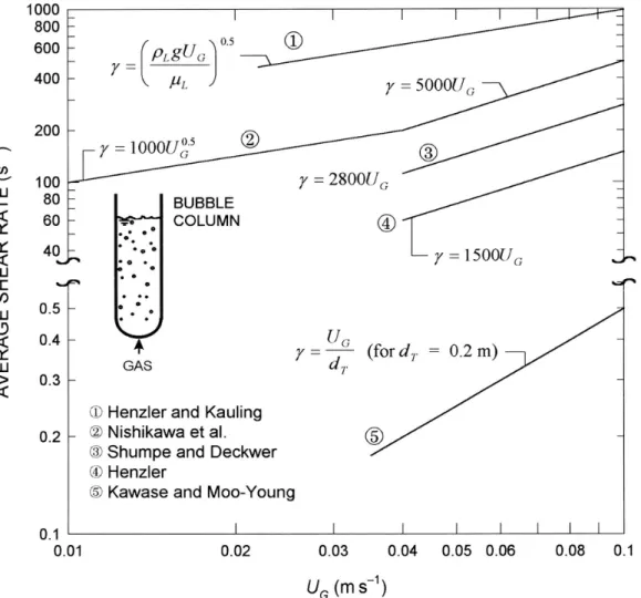
Gas-Agitated Bioreactors
Importantly, these findings regarding the axial variation of mean wall shear velocity (γw) do not agree with Eq. Based on measurements of one-dimensional energy spectra in the center of the riser,44 tur-.
Mechanically Stirred Vessels
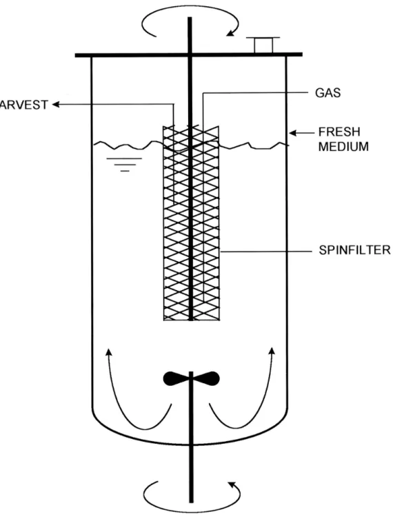
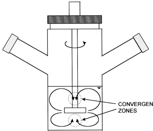
The reactor achieved complete gas-liquid separation and there was no gas in the downcomer. In perfusion culture of suspended animal cells, "spin filters" (Figure 5) or rotating wire mesh cylinders are sometimes used to retain cells in the bioreactor.
Pipework and Flow Channels
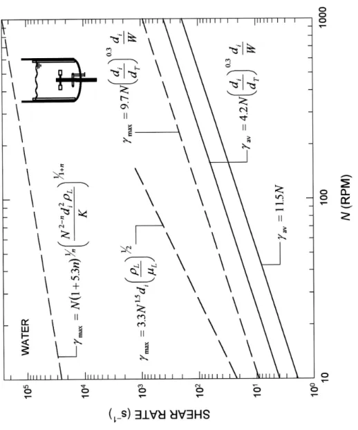
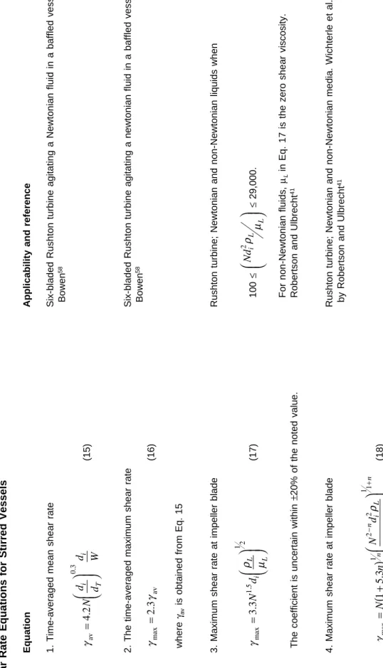
The coefficient is uncertain within ±20% of the quoted value.Robertson and Ulbrecht41 4. Maximum shear rate at fan blade Rushton turbine; Newtonian and non-Newtonian media. Comparison of the different shear rate correlations for water in a standard stirred tank stirred with a 0.1-m-diameter 6-blade Rushton turbine.
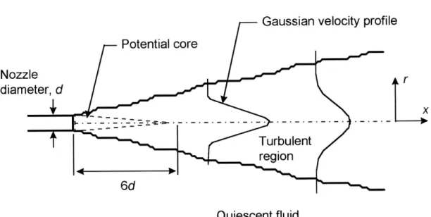
Turbulent Jets
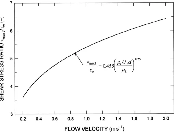
In a stable submerged turbulent jet, the maximum shear stress occurs in the direction of discharge, six to seven nozzle diameters downstream of the orifice. If the nozzle discharge velocity is the same as in the pipe, the maximum shear stress at the pipe wall is always less than in the jet.
Shear Phenomena in Isotropic Turbulence
Methods for calculating the specific energy dissipation rate in stirred vessels have been discussed elsewhere.60,69 The average energy dissipation rate in a non-aerated vessel is given as. The energy dissipation rate in the presence of aeration is generally lower than in the equivalent non-gassed condition. The maximum energy dissipation occurs near the impellers and this maximum value can be calculated using the equation.
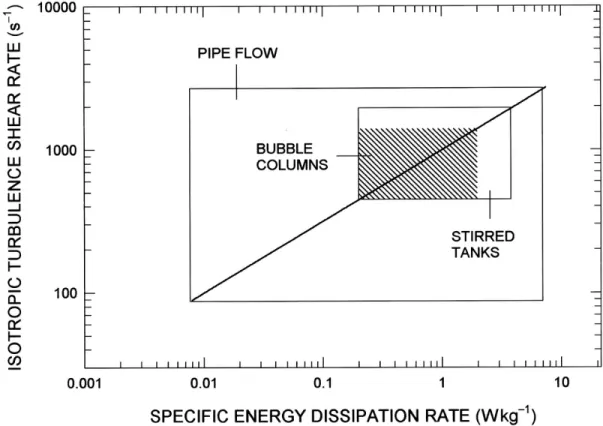
As shown in Figure 9, there is the same relationship between the shear rate of isotropic turbulence and the specific energy dissipation rate in different culture devices; however, since different devices operate in different ranges of specific energy dissipation rates, the shear rates experienced by cells are different under the culture conditions commonly used. The relationship between isotropic turbulence shear rate and specific energy dissipation rate is the same (solid line) in different culture devices, but the devices operate in different specific energy dissipation ranges. In flow systems such as pipes and channels, the shear velocity at the wall is always greater than in bulk flow (Figure 6).
In still other cases, the fluid-eddy shear rate and the maximum shear rate at the fan will have to be taken into account.
Effects of Suspended Particles on Turbulence
Shear Effects on Cells
Other approaches to calculating shear stresses in turbulent flow are discussed by Cherry and Kwon.72. In summary, several possible shear rate values can be calculated for a given situation in a bioprocess device. For pneumatically stirred bioreactors, when the turbulence characteristics in the bulk liquid are relevant, the preferred approach is to use Eq.
There are two reasons for this: (1) due to hydrodynamic forces, cells typically move away from the walls; and (2) a relatively high shear rate in laminar flow of the boundary layer adjacent to walls is less detrimental than a comparable shear rate in turbulent flow away from walls.5 In some cases, the relevant shear rate may be that at the interface of a rising bubble unless the turbulence is so intense that bubbles cannot rise freely. Factors such as the frequency with which a sensitive biocatalyst passes through a high shear region may need to be considered for cyclical flows such as those encountered in stirred tanks, airlift devices, and recycle loops.
Suspended Cells
Hybridomas and Suspension-Adapted Cells
It was assumed that the interfacial tension between the droplet and the medium was equal to the membrane tension of the cell. The viscosity of the drop was taken as the internal viscosity of the hybridoma. However, the precise value of the viscosity was not crucial for predicting the extent of cell damage in a shear field.11.
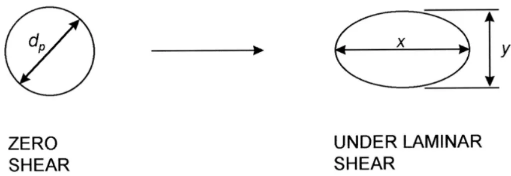
The deformation, dfb, is a weak function of the relative increase in cell surface area, and an average value of dfb can be used11 in Eq. In theory, the model should allow a prediction of cell survival behavior from mechanical property data measured by micromanipulation. Abu-Reesh and Kargi82 varied the rotation speed of the inner cylinder and the viscosity of the suspension fluid to obtain different values of Taylor number.
Shiragami,15 is the mean shear stress acting on cells suspended in a capillary flow, the shear stress on capillary walls if the ratio of cell diameter to capillary diameter.
Blood Cells
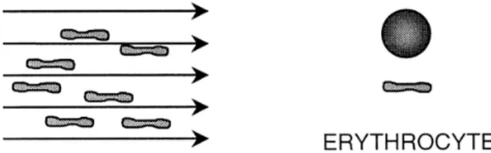
Erythrocytes are highly deformable cells that orient in laminar flow so that the smallest possible disc-shaped cell surface is perpendicular to the flow (Figure 12). Based on measurements on turbulent jets, a critical lytic shear stress level of 4000 N⋅m-2 has been reported for very short exposures (~10-5 s).76 Measurements on erythrocytes of various mammals reveal that the critical stress of shear increases dramatically as cell volume decreases. Some chemical lysines and some antigen-antibody reactions cause perforation of the cell membrane and leakage of intracellular material.76 Cholesterol enrichment or depletion.
In tubular flow, erythrocyte shell wall roughness correlates with hemolysis.76 Bubbles trapped in surface imperfections appear to promote lysis. In tubes, hemolysis correlates with shear rate and surface-to-volume ratio.76 This type of lysis occurs at shear stress thresholds lower than those required to produce lysis in a fluid shear field. Wall contact related lysis has been observed to depend on the chemical nature of the wall material.
Lysis may decrease over time as surfaces become passivated by prolonged contact with plasma proteins.76.
Adherent Cells
Cells on Stationary Surfaces
In capillaries with a diameter of ~1 mm, an upper limit to the average tube velocity of 6 m⋅s-1 has been suggested for capillaries with sharp-edged inlets and blood with a viscosity of 4 × 10⁻³ Pa⋅s. 76 This corresponds to a Reynolds number of 1500 inside the capillary and an average wall shear velocity of about 4800 s-1. The slight decrease in lysis rate at 100 rpm (Figure 14) was associated with improved surface aeration compared to a static flask.103. Leukocytes adhering to the vascular endothelium detach when the shear stress is between 26.5 and 106.0 N⋅m-2.105 According to the work cited by Prokop and Bajpai,86 a shear stress level of 60 N⋅m-2 applied over 10 minutes should about a quarter of the leukocyte population is released.
In another study, sublethal shear stresses of 10 and 20 N⋅m-2 applied over 10 min in a Couette viscometer affected the biochemical response of human T cells relative to unsheared controls.106 Therefore, it appears that that circulating cells in vivo are apparently more resistant to shear than those studied in vitro. In studies with anchorage-dependent cells attached to the flat glass walls of a rectangular flow channel, Shiragami and Unno113 observed increased activity of lactate dehydrogenase (LDH) in cells that had been exposed to a steady state shear stress of 0.5 N⋅m-2 for 12 hours; In vivo hemodynamic forces have been implicated in various physiological and pathophysiological processes.109 Atherosclerotic lesions in humans tend to develop in zones of flow sepa-.
Arteries adapt to chronic changes in blood flow, increasing in circumference under strong flow and narrowing under reduced flow.109 Shear stress signals transmitted from the vascular cell through cytoskeleton and biochemical elements result in changes in structure, metabolism, and gene expression. 109.
Cells on Suspended Microcarriers
Resistance of a cell to impact cracking (i.e., burst resistance) is a potential measure of cell survivability in the culture environment. Sinskey et al.121 advanced the concept of an integrated shear factor (ISF) - a measure of the strength of the shear field between the impeller and spinner vessel walls - to correlate shear damage to mammalian cells. To achieve a more homogeneous shear field in stirred culture vessels (i.e., for the minimum value of the maximum-to-average shear rate ratio), Croughan et al.50 noted an optimal vessel geometry corresponding to ri/rT = 0.74.
Relative extent of growth of human diploid fibroblasts on microcarriers as a function of the integrated shear factor in spinner vessels. Relative extent of growth of human diploid fibroblasts on microcarriers as a function of the Kolmogoroff microscale of turbulence in spinner vessels. Under conditions typical of microcarrier culture in airlift bioreactors, the specific energy dissipation rates are different in different zones of the vessel.
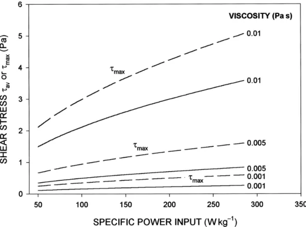
For a given specific input power, the shear stress increases with increasing viscosity of the slurry fluid, as shown in Figure 20.
Shear Effects on the Cell Cycle
Concluding Remarks
Effects of culture viscosity and specific input power on mean and maximum shear stress values on the surface of suspended microcarriers. Turbulent shear stress is generally more damaging than laminar shear stress of the same magnitude. Influence of protein concentration on mechanical cell damage and fluid dynamics in air transport reactors for mammalian cell culture.
Turbulent shear stress - effect on mammalian cell culture and measurement using laser doppler anemometer. A quantitative analysis of shearing effects on cell suspension and cell culture of Perilla frutescens in bioreactors. Protective effect of methyl cellulose and other polymers on insect cells subjected to laminar shear stress.
The role of plasma membrane fluidity on the shear sensitivity of hybridomas grown under hydrodynamic stress.