Divulgence of Bacterial diversity and Corresponding Antibiotic Susceptibility Profile of Coastal Waters in Saint Martin's Island,
Bangladesh
By
Sauti Malabika Nadi ID: 18136015 Marzuq Mohammad Alam ID: 18136043 Tazrian Jannath Protiva ID: 18136064
A thesis submitted to the Department of Mathematics and Natural Sciences in partial fulfillment of the requirements for the degree of Bachelor of Science in Biotechnology
Biotechnology Program,
Department of Mathematics and Natural Sciences BRAC University
© 2022. BRAC University All rights reserved
1 | P a g e Declaration:
It is hereby declared that
1. The thesis titled “Divulgence of Bacterial diversity and Corresponding Antibiotic Susceptibility Profile of Coastal Waters in Saint Martin's Island, Bangladesh.” is our own original work while completing the degree at BRAC University.
2. The thesis does not contain material previously published or written by a third party, except where this is appropriately cited through full and accurate referencing.
3. The thesis does not contain material that has been accepted or submitted, for any other degree or diploma at a university or other institution.
4. We have acknowledged all main sources of help.
Student’s Full Name and Signature:
Sauti Malabika Nadi ID: 18136015 Bachelor of Science, Biotechnology
Department of Mathematics and Natural Sciences
BRAC University, Dhaka, Bangladesh
Marzuq Mohammad Alam ID :18136043
Bachelor of Science, Biotechnology
Department of Mathematics and Natural Sciences
BRAC University, Dhaka, Bangladesh
Tazrian Jannath Protiva ID :18136064
Bachelor of Science, Biotechnology
Department of Mathematics and Natural Sciences
BRAC University, Dhaka, Bangladesh
2 | P a g e
Approval
The thesis “Divulgence of Bacterial diversity and Corresponding Antibiotic Susceptibility Profile of Coastal Waters in Saint Martin's Island, Bangladesh.” submitted by Sauti Malabika Nadi ( ID: 18136015 ) , Marzuq Mohammad Alam ( ID :18136043 ) and Tazrian Jannath Protiva ( ID :18136064 ) of Spring 18 , 2018 has been accepted as satisfactory in partial fulfillment of the requirement for the degree of Biotechnology on July 2022.
Examining Committee:
Supervisor: _______________________________
Fahmina Akhtar Senior Lecturer,
Department of Mathematics and Natural Sciences BRAC University
Co-supervisor: _______________________________
Akash Ahmed Senior Lecturer
Department of Mathematics and Natural Sciences BRAC University
Program Coordinator: _______________________________
Iftekhar Bin Naser Assistant Professor,
Department of Mathematics and Natural Sciences BRAC University
Departmental Head: _______________________________
(Chair) A F M Yusuf Haider Professor and Chairperson
Department of Mathematics and Natural Sciences
3 | P a g e
Acknowledgment:
It is by the grace of The Almighty that we have been able to complete this research, so at first He should be thanked for granting us the strength, good health and opportunity to work on this thesis.
We would like to express our gratitude to the Chairperson of the Department of Mathematics and Natural Sciences, Professor A F M Yusuf Haider for allowing us to complete our undergraduate thesis.
Our sincere obligation and indebtedness goes to our respected supervisor Fahmina Akhtar, Lecturer, Department of Mathematics and Natural Sciences, BRAC University for her constant guidance, constructive criticism and encouragement to give our best effort and pursue our highest potential. We would like to express our appreciation and gratitude toward our respected co- supervisor Akash Ahmed, Lecturer, Department of Mathematics and Natural Sciences, BRAC University for guiding us with his experience and assisting us tirelessly throughout the project. It for their cordial support and subsequent directions for the whole journey, that we have been able to reach the goal.
Moreover, we would like to thank Professor Mahboob Hossain, Microbiology Program, Department of Mathematics and Natural Sciences, BRAC University for his guidance in the laboratory. Our sincere appreciation goes out to Laboratory Officers Mahmudul Hasan, Shamim Akhter Chowdhury and Asma Binte Afzal for their guidance and support during our work.
Finally, we would like to appreciate everyone working beside us in the laboratory and those who assisted us with their services.
4 | P a g e
Title: Divulgence of Bacterial diversity and Corresponding Antibiotic Susceptibility Profile of Coastal Waters in Saint Martin's Island, Bangladesh.
Abstract:
Considered to be the largest reservoir of microorganisms, ocean water holds massive potential for scientific research. The Saint Martin’s Islands of Bangladesh, located in the north-east part of the Bay of Bengal, is one such reservoir with very limited study having been conducted on its biodiversity. Moreover, the island is a popular tourist destination, so it often faces overpopulation.
Hence, it has become a potential home for a diverse culture of bacteria. The purpose of this study was to identify the bacterial diversity of the coastal waters in Saint Martin’s Island and to determine the corresponding antibiotic effectiveness profile. The main concern was to identify whether or not the bacterial presence in the island’s water could become a public health concern. The samples were collected and analyzed through biochemical testing, antibiotic susceptibility testing, PCR, and gel electrophoresis. The results showed that the marine water stores a significant number of bacteria belonging to the Enterobacteriaceae, Bacillaceae, Staphylococcaceae and Vibrionaceae families. Furthermore, at lesser majorities, different species belonging to the Yersiniaceae, Paenibacillaceae, Brevibacteriaceae, Aeromonadaceae, and Morganellaceae families were also identified. The characteristics of the identities pointed to be mostly rod-shaped (70%) and tested type Gram negative (59.4%). Altogether, 100% of the identifications were tested catalase positive which indicated aerobic nature. Subsequent antibiotic susceptibility profiling yielded in 97.8%
effectiveness by Amikacin, 95.7% for Meropenem, 89.4% for Norfloxacin, 85.1% for Amoxicillin + Clavulanic acid, 61.7% for Tetracycline, 57.4% for Azithromycin/Erythromycin, 46.8% for Amoxicillin, 44.6% for Colistin Sulphate, 34% for Vancomycin, and 31.9% for Cefixime.
Following PCR runs resulted in the detection of mcr-1 gene presence in the Colistin Sulphate resistant isolates, justifying the compromised efficacy of the last-resort drug. To this regard, presence of disease causing, antibiotic resistant bacteria in coastal waters indicates declining water quality. This further demonstrates the increasing possibility of severe health risks being linked to interactions with the coastal region if nature is not given sufficient opportunities to heal on its own.
5 | P a g e
Table of Contents
Chapter Pages
Chapter 1 Introduction………... .10
1.1 Ubiquitous nature of diversity and importance……….. .10
1.2. Saint Martin’s Island and its biodiversity………10
1.2.1. Threats to the Island biodiversity………..10
1.3. Overview of antibiotics and antibiotic resistance genes (ARG)……….11
1.4. Overview of Polymyxin resistance……….12
1.5. Novelty of research, established techniques and necessity of identification………..12
1.6. Overview of motive………..13
1.6.1. Objectives of the study………..13
1.7. Research Question………13
Chapter 2 Methodology 2.1. Sample Site and Sample Collection 2.1.1 Description of sample sites………..14
2.2. Sample Collection………...16
2.3. Sample processing………...16
2.3.1. Culture in Nutrient Agar and Differential Media………16
2.3.2. Spread Plating, Colony Selection and Isolation………17
2.4. Biochemical Test………..17
2.4.1.Gram Staining………17
2.4.2. Catalase Test………..18
2.4.3. Oxidase test……….18
2.4.4.Triple Sugar Iron test (TSI)………..18
6 | P a g e
2.4.5. (Motility-Indole-Urease) MIU test………19
2.4.6.Citrate test………...19
2.4.7.MR - VP test………20
2.5. Antibiotic Susceptibility Testing………..20
2.5.1. KirbyBauer Disc Diffusion Method……….20
2.6. Determination of the presence of mcr-1 Polymyxin Antibiotic Resistance Gene……...21
2.6.1. DNA extraction by Boiling Method………..…...21
2.6.2. Performance of the Polymerase Chain Reaction (PCR)………...……….21
2.6.3. Gel electrophoresis………22
Chapter 3 Results 3.1. Bacterial identification per sample zones………...23
3.2. Cultured colony characteristics on selective/differential (SD) media………..23
3.3. Results of biochemical testing………..23
3.4. Antibiotic susceptibility profiling………....26
3.5. Determination of mcr-1 amplicon presence via Gel electrophoresis………27
Chapter 4 Discussion 4.1. Overview of identification determination………...…39
4.2. Growth summary………..39
4.3. Recognized habitats of the identified bacterial families………39
4.4. Facilitation of bacterial spread across the marine environment………..40
4.5. Cross referencing with Gram stain results and known habitation………..40
4.6. Pathogenic capabilities of the identified species……….41
4.7. Poor healthcare situation on the island………...41
4.8. Overview of antibiotic susceptibility testing………..42
7 | P a g e
4.9. Meropenem susceptibility pattern and reasoning……….42
4.10. Amikacin susceptibility pattern and reasoning………42
4.11. Amoxicillin and Vancomycin susceptibility pattern………43
4.12. Use of adjuncts, Clavulanic acid, Cefixime, Azithromycin/Erythromycin, and Tetracycline susceptibility patterns and reasoning...……….43
4.13.Norfloxacin susceptibility patterns and reasoning……….44
4.14. Effect of the mcr-1 gene in facilitating resistance against Colistin Sulphate………...44
4.15.Genetic testing to determine mcr-1 presence in CLR bacteria………...45
Chapter 5 Conclusion……….46
Supplementary Tables……….47
Appendix-I Media Compositions ………...55
Appendix- II Reagents and buffers………60
References……….63
List of Figures: Figure no. Page no. Figure 1: Saint Martin’s Island Sample Sites……….………15
Figure 2: Gram positive bacilli………...24
Figure 2.1: Gram negative cocci………24
Figure 3: Results of the TSI test………...25
Figure 4: MIU negative (All 3 attributes)………..25
Figure 4.1: MIU positive (All 3 attributes)………25
Figure 5: Citrate positive……….26
8 | P a g e
Figure 5.1: Citrate negative………26
Figure 6: MR negative……….26
Figure 6.1: MR positive………...26
Figure 6.2: VP negative………...26
Figure 7: Antibiotic susceptibility profiling on MHA agar………..27
Figure 8: Gel run result Batch 1……….28
Figure 8.1: Gel run result Batch 2………..28
Figure 9: Representation of antibiotic resistance profile against Gram positive bacteria…37 Figure 9.1: Representation of antibiotic resistance profile against Gram negative bacteria………..37
9 | P a g e List of Tables
Table no. Page no.
Table 1: TSI result interpretation………..19 Table 2: PCR conditions utilized for targeted amplification of the mcr-1 gene………22 Table 3: Identified bacteria per site of sampling………..29 Table 4: Representation of biochemical testing results of the identified bacteria………….30 Table 4.1: Representation of biochemical testing results of the identified bacteria………..31 Table 4.2: Representation of biochemical testing results of the identified bacteria………..33 Table 5: Representation of the antibiotic susceptibility status of the identified bacteria….34 Table 5.1: Representation of the antibiotic susceptibility status of the identified bacteria………..35 Table 5.2: Representation of the antibiotic susceptibility status of the identified bacteria………..36
10 | P a g e
1.0. Introduction:
1.1. Ubiquitous nature of diversity and importance:
Microbes, explicitly bacteria, are ubiquitous organisms which can be found in most places of the environment. At particular geographic locations, the bacterial diversity can have profound impacts on the regional ecology, affecting local health, food environment, and subsequently, trade and commerce (Horner-Devine et al., 2004). Among the vast assortment of different species, marine bacteria are the type which resides in marine environments, such as regions directly associated with seas and oceans. They have been indicated as being part of the earliest residents of habitable planet Earth (AIMS, n.d.). These bacteria play key roles as primary producers, guiding oceanic cycling of energy and nutrients (Bienfang et al., 2011). Moreover, they also are a vast source of medicinal components, natural-products, genetic resources, and bioremediation (McCarthy et al., 1998; De et al., 2014). Henceforth, the exploration of this diversity is of paramount importance since it directly pertains to the divulgence of vital resources whilst also contributing towards the sustenance of environmental health (Colwell, 1997).
1.2. Saint Martin’s Island and its biodiversity:
The location of choice for this study is the Saint Martin’s Island, a coral island located at the south- east periphery of Bangladesh. Endowed with a variety of physiographic landscapes, the island boasts a comprehensive distribution of lagoons, marshes, sandy beaches, and coral constellations with a rich co-habitation of species such as corals, seaweeds, algae, and mangroves (Muhibbullah
& Sarwar, 2017; UNDP, 2010). Furthermore, as reported by Hossain and Islam (2006), the coastal shores of this region, linked to the Bay of Bengal, provide habitation to around 234 species of fish (Hossain & Islam, 2006). This biodiversity is further enhanced by the tropical climate of the subcontinent along with direct linkage to the Indian Ocean. It is to be noted that this richness not only applies to visible organisms, but also extends to the microbiological world as it has been studied that the environment acts as a viable habitat to a significant variety of bacterial species. As reported in an investigation by Padmanaban et al. (2019), phylogenetic analysis of deep-sea sediments yielded in the identification of a plethora of gram-positive bacteria along with species of Proteobacteria, Actinobacteria and many more (Padmanaban et al., 2019). Such findings have been further verified by additional studies. Furthermore, with augmentation of next-generation sequencing techniques, scopes to further characterize these relatively unexplored waters holds massive potential (Akter et al., 2020).
1.2.1. Threats to the island biodiversity:
Due to effects of pollution and increasing anthropological activities, the local biodiversity of the coastal waters reaching the island shores faces significant threats. Due to recent infrastructural
11 | P a g e
developments, accessibility to the island during the active season has increased significantly (Hasan, 2009). This was proven by a local report which found that the daily tourist-population intake is of around 4000 individuals, which largely surpasses the capacity of 1250 (Ahamed, 2021).
Considering this, pollution of all forms has increased hand-in-hand. Consequently, the potential degradation in marine water quality has become a major concern. It is projected that whilst the key focus of the local administration was in facilitating tourism, ignorance may have befallen on managing the waste that is generated during the process. As reported in a study by Muhibullah and Sarwar (2017), the island lacks an organizational drainage-system whilst the land-use pattern follows a random mode of arable and/or free land allocation with a bias towards hotels and resorts (Muhibbullah & Sarwar, 2017). As a result, it is anticipated that the coastline, constituting vicinities with human (or animal) presence, has undergone an upsurge in the population of enteric bacteria. This type comprises of a collection of species, such as Staphylococcus and Vibrio, which can be generally found in the human (and animal) gut (Donnenberg, 2000). Of further note is that sediments, which are in abundance at these regions, are rich reservoirs of such species (Hassard et al., 2016; Rehmann et al., 2009). Accordingly, the collective health conditions of the locals who are reliant on this ecosystem and the tourists in contact, both become impacted as enteric pathogens can directly cause gastrointestinal infections (Hossain & Islam, 2006; Santamaría, 2003).
Antimicrobial agents of different classes are regularly prescribed to mitigate the aforementioned health complications caused by such bacteria. Thus, pairing this situation with the upsurge of antibiotic resistance development, both as a cause and an effect, paints a grim picture in both the tourism and health sectors.
1.3. Overview of antibiotics and antibiotic resistance genes (ARG):
Antibiotics, based on chemical structures and mechanism of action, make up around 16 major antibiotic families, including β-lactams, aminoglycosides, macrolides, tetracyclines, glycopeptides, and many more (Zhuang et al., 2021). Based on their capabilities of particularly neutralizing bacterial cells, these compounds act as a source of significant selective pressure to such populations. Henceforth, the development of spontaneous mutations in the genes of such cells is not an unlikely scenario. In this manner, any alterations to the gene products (proteins or cellular components) that were targeted by a certain class of antibiotics can result in the development of resistance. Furthermore, this may also develop based on bacterial efflux enhancements, target-site modification, and enzymatic degradation of the antibiotic compound (Waglechner & Wright, 2017). Subsequent duplication of such cells further forwards the resistive traits among the species.
In this manner, a total of around 3000 antibiotic resistance gene (ARG) subtypes have been elucidated till date (Zhuang et al., 2021). The most significant subtypes are the Polymyxin, Tetracycline, Sulfonamide, and Carbapenem-resistance genes (Zhuang et al., 2021). Thus, with continuing exertion of selective pressure via emerging classes of antibiotics, development of resistance will increase as well, which is further accelerated by a lack of adequate anthropogenic carefulness during usage (Larson, 2007).
12 | P a g e
1.4. Overview of Polymyxin resistance:
Among the Polymyxin subtypes, a prominent example is the mcr-1 gene which encodes for a membrane bound protein, phosphoethanolamine transferase (Li et al., 2020). This enzyme subsequently functions to attach a phosphoethanolamine moiety to lipid A molecules situated on the outer bacterial cell membrane of Gram negative cells (Gao et al., 2016). In this manner, the preliminary barrier towards antibiotic entry becomes modified, causing an alteration in the permeability. This facilitates a spill-over effect on the action of the Polymyxin antibiotics which particularly act to alter bacterial membrane permeability by disruption of Ca2+ and Mg2+ cations (El-Sayed Ahmed et al., 2020). Among these, Colistin Sulphate has been further addressed as a
“last-resort drug” against Gram negative cells (Conly & Johnston, 2006). This drug is particularly prescribed to treat multi-drug resistance due to it possessing proven susceptibility patterns against these certain targets (Conly & Johnston, 2006). It is against this much-hailed agent that the mcr-1 gene poses a significant threat since the gene-product directly alters the target site and entry- pathway of the drug compound, preventing it from reaching a necessary concentration for optimum function. Of greater concern is brought on due to multiple studies having reported that the gene can be transferred among bacterial species via diverse plasmid-mediated pathways (Liu et al., 2016). This was also displayed by the same plasmids having been isolated from the human gut microbiota (Gao et al., 2016). However, an interesting notion has been exhibited in a study by Li et al. (2020) which displayed that upon acquiring plasmid mediated mcr-1 gene, loss of cell membrane integrity is enhanced (Li et al., 2020). Thus, it can be perceived that Polymixin resistance-development brings on a form of evolution which comes with a hand-off and necessitates further research so that enhanced characterization can be conducted.
1.5. Novelty of research, established techniques and necessity of identification:
It is to be noted that marine bacteria have advantages over terrestrial bacteria in that they can survive in hostile environments. Microorganisms that are thermophiles, halophiles, alkalophiles, and polyextremophiles, coupled with their ability to multiply by using adaptation techniques that include a variety of cellular metabolic systems, are likely to get identified from this Saint Martin’s Island (Hossain & Islam, 2006). Thus, based on its geographical location, speculations arise regarding a potentially bacterial-species rich aquatic environment. However, this is still theoretical because such bacterial classification has not been conducted yet. The utilization of biochemical and genetic testing methods to conduct microbiological classification of coastal, beach waters are gaining significant traction in identifying the bacterial biodiversity. This also extends to the associated potential antimicrobial-resistive traits that the organisms might possess. Whilst genetic testing enhances the specificity of detection, biochemical methods also offer a significant degree
13 | P a g e
of accuracy since they directly test for phenotypic characteristics, such as specific enzymatic presence, whilst being more cost-efficient, attainable, and requiring less specialization to interpret (Franco-Duarte et al., 2019). Subsequently, they enable for rapid identification of potential contamination at risk zones where public health concerns might arise. For instance, a study conducted by Oun et al. (2017) in Michigan using biochemical-based identification of E.coli as fecal coliform indicators, discovered that around 50% of the water samples in their study area had surpassed the guidelines provided for standard water quality (Oun et al., 2017). Another study conducted by Pérez et al. (2008) in the Iberian Peninsula, also using fecal coliform (FC) indicators, learned that the majority of the concentrations of FCs were present near river mouths and beach areas (Garrido-Pérez et al., 2008). The utilization of genetic methods to conduct this classification would have been more resource demanding and redundant to a large extent. Moreover, a preliminary investigation in Saint Martin’s Island concerning sponge-associated bacteria, conducted by Paul et al. (2001), also utilized biochemical testing methods to identify around 15 bacterial genera (Paul et al., 2021). As a result, the prospect of analyzing the coastal waters of Saint Martin’s opens windows for high potential research. Such is the case because the divulgence of the bacterial repository will subsequently enable in further understanding of the region.
Consequently, this will facilitate in the innovation of strategies to conserve the natural biodiversity whilst also avoiding a potential public health concern.
1.6. Overview of motive:
To this regard, the following study was conducted with the key motive of identifying the bacterial diversity and potential coliform presence in the coastal waters of the Saint Martin’s Island. For this venture, the use of traditional culture methods, biochemical testing, and genetic analysis was employed. Furthermore, we subsequently aimed to determine the presence of antimicrobial- resistance capabilities of the discovered species, and divulge genetic reasoning behind the traits.
1.6.1. Objectives of the study:
Identification of bacterial diversity.
Determination of the potential presence of antibiotic resistance in the identified bacteria.
Detection of fecal coliform presence in water sources (ocean and community).
Genetic analysis and classification in terms of mcr-1 gene presence.
Determination of the presence of Polymixin resistance in identified bacteria.
1.7. Research Question:
In terms of the bacterial diversity and its corresponding characterization, does the coastal water surrounding the Saint Martin’s Island have the potential of becoming a significant public health concern?
14 | P a g e
2.0. Methodology
2.1. Sample Site and Sample Collection 2.1.1 Description of sample sites
Saint Martin’s Island is situated at the northeast side of the Bay of Bengal and is approximately 9 km south of the Cox’s Bazaar - Teknaf peninsula. The Island is primarily divided into two sections with a narrow piece of land as the connection point at the center. Furthermore, it lies between 92°18' and 92°21'E longitudes and 20°34' and 20°39'N latitudes with reefs being located around 10-15 km to the west-northwest (Khan & Mamunul, 2010). Moreover, the land is mostly of a flat plane and is situated at 3.6m above sea level. Of further note is that the entire island is surrounded by a continuous beach which allows people to have access to almost all sides of the it, but there is a difference in the intensity of anthropogenic activity at different regions (Khan & Mamunul, 2010). To that regard, more people come in contact with the waters near the jetty for multiple reasons. As the island is only a two-and-a-half-hour boat ride away from the mainland, some tourists come in for a day-long trip. Most of these people opt to stay near the jetty since it is more economically feasible and it also allows easy access to the main market area which is composed of eateries, shops, and all other necessary amenities. Hence, human activity near the jetty is greater compared to the other locations around the island.
The sample sites were selected randomly with significant efforts having been placed to ensure inclusivity of all varieties and intensities of anthropogenic activities and exposures to seawater.
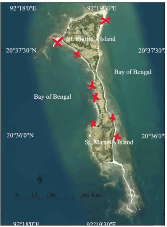
Thus, a total of 8 sample sites were selected around the island. The sites include the jetty, the most visited beach on the northwest part, largely rocky areas, reefs, beaches adjacent to farmlands, beaches near fishing area, and so on. The locations were selected once before arrival and then after inspection of the island in person. The coordinates of the samples sites are :- 20.63377052856086, 92.32811941494049 ( Jetty ) , 20°37'26.4"N 92°19'09.6"E, 20°36'48.6"N 92°19'29.6"E, 20°36'44.3"N 92°19'31.5"E, 20°36'33.3"N 92°19'30.0"E, 20°36'08.4"N 92°19'54.5"E, 20°36'12.8"N 92°19'53.1"E, 20°36'20.6"N 92°19'49.7"E.
15 | P a g e
Figure 1: Saint Martin’s Island Sample Sites, marked with red ink. ( Saha, S., Sehrin, S., Ahsan Habib, K., & Abdul Baki, M. (2018). NEW RECORDS OF TWO LUTJANUS SPECIES (TELEOSTEI: PERCIFORMES: LUTJANIDAE) WITH RE-DESCRIPTION OF SIX LUTJANIDS FROM SAINT MARTIN’S ISLAND OF THE BAY OF BENGAL, BANGLADESH Human health risk assessment of heavy metals in tropical fish and shellfish collected from the river Buriganga, Bangladesh View project DNA barcoding of marine fishes of Bangladesh View project.
https://doi.org/10.3329/bjz.v46i.39056 )
16 | P a g e
2.2. Sample Collection
Water samples were collected during the occurrence of incoming waves, at a height of around 2.5 feet from the depth and at a distance of around 35-40m from dry land. This was done via the use of sterile syringes and the collected samples were stored in sterile falcon tubes. The falcon tubes were then stored in fixed positions within sealed ice boxes to protect the samples from sunlight.
This facilitated the prevention of heat shock and minimized the likelihood of mechanical shearing.
Also, the tubes were kept upright inside the boxes using Styrofoam frames to avoid sample loss during transportation. To minimize skin exposure to the water as well as the inner surfaces of the tubes, gloves were worn along with synthetic clothing and rubber boots throughout sampling. The samples were subsequently brought to the lab within 24 hours of collection. From every site, 2 sets of samples were collected: - Raw-water sample and sample mixed with Nutrient Enrichment broth (Peptone water). Peptone water is an enrichment medium that helps repair injured cells and assists in protecting the organism volume. It was noted earlier that the loss of organism-volume is inevitable during transportation and sample processing, and therefore, samples were collected in peptone water as reinforcement of the raw sample.
2.3. Sample processing:
2.3.1. Culture in Nutrient Agar and Differential Media
Collected water samples were cultured on selective/differential (SD) media as well as Nutrient Agar (NA) media. With regards to selective/differential media, MacConkey agar, m-FC Agar, UTI agar, Mannitol Salt (MSA) agar, XLD agar, and TCBS agar were used to culture and subsequently make preliminary identification notations of different species of bacterial colonies as each medium selectively promotes the growth of specific bacteria whilst inhibiting others. Furthermore, due to the differential capabilities of the media, the different species growing on the same plate could be differentiated between based on characteristic colony attributes. MacConkey Agar is recommended for the selective isolation and differentiation of lactose fermenting and lactose non- fermenting enteric bacteria (MacConkey Agar M081B Composition, n.d.). m m-FC Agar utilizes comparatively higher temperatures (44.5°C)and the membrane filtration technique to conduct the detection and accounting of faecal coliforms (M-FC Agar Base M1122 Composition, n.d.). UTI is a chromogenic differential medium and is used to identify enteric bacteria found in specimens such as urine, which may contain a large amount of protease and other pathogenic gram–positive organisms (UTI Agar, Modified Composition, n.d). MSA agar is used to identify pathogenic Staphylococci spp. from clinical and non-clinical samples (Mannitol Salt Agar Composition, n.d.). XLD agar or Xylose-Lysine Deoxycholate Agar is specifically used to isolate Salmonella species. TCBS is used to grow enteropathogenic Vibrio species. Nutrient Agar (NA) is a general- purpose media.
17 | P a g e
2.3.2. Spread Plating, Colony Selection and Isolation
The collected samples were processed to isolate the significant bacterial colonies. At first, raw samples were subjected to serial dilution to avoid lawn growth on the media plates. For this procedure, 0.9% NaCl solutions were used, and the dilutions were conducted on a trial-error basis where dilutions of 10-2 and 10-3 were determined as being ideal in providing single colonies.
Subsequently, the upper limit of the dilution used was 10-9, which was during Peptone water usage for Site 1.The diluted samples were then spread plated on Nutrient agar and other differential media mentioned above. It is to be noted that strict measures were taken to ensure that the plating was being done in a sterile environment to avoid contamination. Upon plating, the culture media plates were incubated in 37° C for 24 hours, except for the m-FC media. At this media, the culture plates were incubated at 44° C, and for 48 hours. After the incubation and following observation with the naked eye, based on their characteristics on different media, single colonies were chosen and sub-cultured on NA media.
2.4. Biochemical Tests
Several biochemical tests were performed on the selected bacterial isolates to characterize their biochemical and enzymatic activities. The results were subsequently used to identify the unknown samples. The tests performed were Gram staining, Catalase test, Oxidase test, TSI test, MIU test, Citrate test, Methyl Red test, and Vogues Proskauer test. To obtain optimum results for all the assessments, fresh cultures (incubated for 24 hours) were used.
2.4.1. Gram Staining
For Gram staining, previously published (Smith, 2019), standard procedures were followed to identify and differentiate between the Gram-positive and Gram-negative bacteria. Single colonies of bacterial isolates were transferred into autoclaved distilled water. The colonies were subsequently smeared over respective glass slides and then heat fixed. Following this, a few drops of Crystal Violet were added onto the area of the smear on the slides with a dropper and then left for 60 seconds. This is the primary dye, which is positively charged, so it adheres to the cell membrane of both Gram positive and negative cells. After 60 seconds, the Crystal Violet stain was carefully washed with distilled water, and Gram’s iodine was added. Gram’s iodine is a mordant that helps Crystal Violet to adhere to the Gram positive organisms. 60 seconds were again given after the Gram’s iodine addition, following which; the slide was washed with distilled water. Then, the organisms were washed with 95% ethanol. This step differentiates the Gram positive and negative organisms. In Gram positive organism, the Crystal Violet- iodine complex is trapped in the cell due to a thicker peptidoglycan cell wall. This shrinks due to alcohol dehydration, thus becoming more constricted and lowering cell wall permeability. In Gram-negative cells however,
18 | P a g e
the cell boundary contains a thinner peptidoglycan wall and a higher concentration of lipids, which are soluble in alcohol. The ethanol, acting as a decolorizer, dissolves the lipid layer and increases the permeability of the cell membrane of the Gram-negative cells. After the ethanol rinse, a counterstain, Safranin was added to the glass slides which were then washed after 45 seconds.
The Gram-positive bacteria appeared dark purple under the microscope and the Gram-negative bacteria appeared dark pink or red.
2.4.2. Catalase test
For the catalase test, bacterial colonies were smeared onto sectioned filter papers and 3% hydrogen peroxide solution was placed on the respective smears. Following this, observation for bubble formation was conducted. The organisms which contained the catalase enzyme formed bubbles/froth and were classified as catalase positive. On the other hand, the organisms that did not possess the enzyme consequently did not form bubbles/froth. These were classified catalase negative.
2.4.3. Oxidase test
For the oxidase test, a Whatman filter paper was soaked in freshly prepared oxidase reagent (1%
tertramethyl-p-phenylene-diamine dihydrochloride). Then, with the tip of a sterilized needle, a single colony of bacteria was smeared on the filter paper and observed for color change.
Appearance of a blue color indicated the presence of the cytochrome C oxidase enzyme.
Organisms which contain cytochrome C as a part of their respiratory chain are oxidase positive and turn the reagent into dark blue. Organisms devoid of the enzyme do not produce any color.
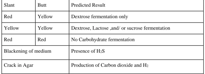
2.4.4. Triple Sugar Iron test (TSI)
The TSI test was performed by preparing triple sugar iron agar, at a slanted manner, within sterile test tubes. The media were then inoculated with the bacterial isolates, incubated for 24 hours, and then observed according to a pre-determined reference (Aryal, 2019). TSI test differentiates organisms according to their carbohydrate fermentation pattern and hydrogen sulfide production.
Carbohydrate fermentation is indicated by the change of color of the pH indicator present in the media which may or may not be associated with gas production.
19 | P a g e
Expected results chart:
Table 1: TSI result interpretation (The Triple Sugar Iron (TSI) Test – Principle, Procedure, Uses and Interpretation. (n.d.).)
Slant Butt Predicted Result
Red Yellow Dextrose fermentation only
Yellow Yellow Dextrose, Lactose ,and/ or sucrose fermentation
Red Red No Carbohydrate fermentation
Blackening of medium Presence of H2S
Crack in Agar Production of Carbon dioxide and H2
2.4.5. (Motility-Indole-Urease) MIU test
MIU test was performed using fresh bacterial culture inoculated in MIU media base for 24 hours at 37° C. Following this, observation was conducted for the presence of bacterial motility and the urease enzyme. If the inoculated colony clouded the stab-line with growth extension, then the organism was motility positive, and if the media base showed color changes to pink/red, then it indicated urease positive activity. The color change would occur due to the presence of Phenol red in the media shifting to an alkaline condition. Upon the addition of Kovac’s reagent, the appearance of a pink-red colored ring at the top indicated indole positive organisms.
2.4.6. Citrate test
As for the Citrate test, the organisms were inoculated in Simmons citrate medium and incubated for at least 48 hours at 37°C, and then observed for color change. A Prussian blue color indicated a positive result whilst unchanged color indicated a negative result. It is to be noted that if growth was observed without the media color having been changed to Prussian blue, the result was taken to be positive. Positive result indicates that the bacterial colony possesses the citrate permease enzyme and uses citrate as its carbon and energy source. The presence of a pH indicator (bromothymol blue) facilitates the visible color change when an alkaline by-product of citrate metabolism is generated.
20 | P a g e
2.4.7. MR - VP test
The Methyl Red (MR) and Vogues Proskauer (VP) tests were performed using Glucose phosphate broth. The samples were incubated for 48 hours, and each sample was incubated in pairs of test tubes, with two test tubes containing broths incubated with the same organism. For the MR test, methyl red reagent was added to the 48 hours-incubated broth, and color change from red to yellow indicated the change of pH. An organism showing positive results can utilize glucose and produce a stable acid that changes the color of methyl red from yellow to red. In the other test tube of the same class, same volume of Barritt’s Reagent A and Barritt’s Reagent B were added and observed.
Positive test was indicated by the appearance of eosin pink color within 30 minutes of periodic shaking. Vogues Proskauer test determines an organism's ability of producing acetylmethyl carbinol from glucose.
2.5. Antibiotic Susceptibility Testing:
2.5.1. Kirby Bauer Disc Diffusion Method:
The Kirby Bauer Disc Diffusion method was utilized to determine the nature of susceptibility or resistance of the isolated aerobic bacterial colonies to a selected collection of antibacterial agents.
For the disc diffusion test, MHA (Mueller–Hilton Agar) media and commercial antibiotic discs were used. Moreover, the procedure was run following the guidelines provided by the CLSI (Hudzicki, 2009). The antibiotics that were used are as follows: - Colistin Sulphate (CT) 10, Cefixime (CFM) 5, Vancomycin (VA) 30, Norfloxacin (NX) 10, Amoxicillin + Clavulanic acid (AMC) 30, Meropenem (MRP) 10, Azithromycin (AZM) or Erythromycin 15, Amikacin (AK) 30, Tetracycline (TE) 30, and Amoxicillin (AML) 10.
At first, freshly cultured organisms were inoculated in sterile 0.9 % saline at 0.5 McFarland standard of turbidity. The cultures were then inoculated on MHA Agar using sterile cotton swabs.
Following this, different antibiotic discs were placed on the Mueller Hilton agar plates and the media were incubated at 37° C for 24 hours. The antimicrobial agent diffuses out from the disk to spread around the surface of the agar in a relatively circular pattern. According to their respective potencies of being effective, they will inhibit the growth of the organism inoculated on that plate.
To this regard, after the incubation time had passed, the zone of inhibition was measured and compared with the CLSI guideline to determine whether the organism developed resistive traits, or whether they were moderately or fully susceptible.
The reference diameters are displayed in Table 1 of the Supplementary section as reference.
21 | P a g e
2.6. Determination of the presence of mcr-1 Polymyxin Antibiotic Resistance Gene:
2.6.1. DNA extraction by Boiling Method
According to the results of the antibiogram, the isolates showing resistance to Colistin Sulphate were selected for DNA isolation and the PCR procedure. The mcr-1 gene is a plasmid mediated Colistin resistance (CLR) conferring gene. The presence of it in bacteria threatens the clinical utility of the antibiotic, as Colistin Sulphate is considered as a last-resort antibiotic in treating multi drug resistant bacterial infections. This agent has activity against most Gram negative bacteria.
Phenotypic Colistin resistance is related to the presence of plasmid mediated mobile Colistin resistance (mcr) gene (Islam, 2020).
The selected organisms for DNA isolation and PCR were as follows :- MSA RS1, MSA RS5 (1), MAC RS1 (2), UTI RS1 (G), MAC RS8 (2), RS7 10^-6, MSA RS5 (2), TCBS RS2 (3), MSA RS5 (1), TCBS RS2 (2), XLD RS1, XLD RS6, XLD RS 8 (2), XLD RS8 (1), MAC RS6, MSA RS3, MSA RS5 (2), MSA RS5 (1), UTI RS4, MFC RS6, TCBS RS1, TCBS RS2, TCBS RS1.
Subsequently, the selected isolates were inoculated in respective test tubes containing sterile LB broth and then subsequently incubated at 37° C for 24 hours in a shaker incubator. The next day, for each isolate, 700µl of the broth was transferred in respective Eppendorf tubes and centrifuged at 13000 rpm for 10 minutes. The supernatants were then carefully discarded and 300µl of PBS (Phosphate Buffer Saline) were added and mixed with slow re-pipetting motions. Following this, the mixtures were again centrifuged at 14000 rpm for 5 minutes. The supernatants were discarded, 200µl of TE (Tris-EDTA) buffer was added, and the mixtures were subjected to boiling at 100°C for 15 minutes inside a water-bath. After this, the mixtures were immediately cooled in a freezer at -20°C for 10 minutes and were then finally centrifuged at 14000 rpm for 5 minutes. The debris were discarded this time and the supernatants were collected in fresh Eppendorf tubes. The extracted DNA collections were securely stored at -20°C until further use.
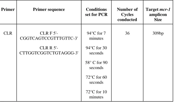
2.6.2. Performance of the Polymerase Chain Reaction (PCR):
To determine the presence of the CLR gene (mcr-1) among the colistin-resistant bacterial colonies, PCR was carried out on the respective samples of DNA which had been previously extracted from the colistin-resistant colonies. A total of 32 pairs of forward and reverse primers were prepared to make up a total volume of 5µl. The sequences of the primers in the pair were as follows: - Forward Primer 5’-CGGTCAGTCCGTTTGTTC-3’ and Reverse Primer 5’-CTTGGTCGGTCTGTAGGG- 3’. Firstly, 5µl of nuclease-free water was added to each PCR Eppendorf to help avoid DNA
22 | P a g e
degradation and then 11µl of master mix was added to each tube. The final volume of the total PCR mixes in each Eppendorf totaled to 21µl. As templates for PCR, the previously extracted DNA isolates of colistin-resistant organisms were added to the PCR tubes at a volume of 4µl. All PCR processes were performed in a thermal cycler following a set among a list of previously published, standardized conditions (Yi-Yun Liu , 2015; Salequl Islam, 2020).
The condition for the PCR is given in the table below:
Table 2: PCR conditions utilized for targeted amplification of the mcr-1 gene.
Primer Primer sequence Conditions set for PCR
Number of Cycles conducted
Target mcr-1 amplicon
Size
CLR CLR F 5'-
CGGTCAGTCCGTTTGTTC-3' CLR R 5'-
CTTGGTCGGTCTGTAGGG-3'
94°C for 7 minutes 94°C for 30
seconds 58° C for 90
seconds 72°C for 60
seconds 72°C for 10
minutes
36 309bp
2.6.3. Gel electrophoresis:
The sizes of the PCR products were resolved by 1.2% agarose gel electrophoresis. A 100 bp ladder was used for the sizing and quantification of the PCR products and the procedure was performed in a Biometra Electrophoresis machine. For visualization under a UV spectrophotometer, 4.5 µl of Ethidium Bromide was added to the loading gel. 1X concentration of TAE buffer was used as the running buffer of the electrophoresis procedure and no extra loading dye was needed as the PCR products already contained green-colored dye from the master mix. The whole process of gel-run was conducted for about 45 minutes and 80 Volts were used. Subsequently, the results were photographed and noted down.
23 | P a g e 3.0. Results:
3.1. Bacterial identification per sample zones:
The conduction of bacterial identification was one of the major phases of this study. It was conducted via the use of data representing the respective colony characteristics and the biochemical analyses. This data was subsequently cross referenced with use of the ABIS software, standard published online resources, and the Bergey’s Manual of Systematic Bacteriology. The results were formulated into Table 3. Here, the first column shows the 8 different zones on the island from which samples were collected whilst the second column demonstrates the divulged identities of the bacteria found with respect to each site. Sites 1 (26%) and 8 (19%) yielded in the largest and second-largest number of bacterial isolates, respectively. On the contrary, site 2 yielded in the least amount. With respect to the identities, bacterial species of the Enterobacteriaceae families were found to be present at the highest amount with bacteria of Staphylococcus spp. and Bacillus spp. occurring most frequently.
Table 2 of the Supplementary section summarizes the identified organisms and their characteristics.
3.2. Cultured colony characteristics on selective/differential (SD) media:
The SD media were utilized in culturing each of the samples upon dilution. For each respective zone, a uniform cohort of the media was allocated so that the conditions and likelihood of growth of all viable organisms were maximized as much as possible. To this regard, the samples were cultured for 24 hours except for the cultures on the m-FC media. That was cultured for 48 hours at 44° C. Upon the completion of culturing, respective tables for each media type were developed with the columns representing the source of the sample plate, the total number of the colonies and the number of visibly different colonies. Along with these, the forms, elevation, color, and surface nature of the colonies were also noted. The results were formulated into tables which are provided in the Supplementary section.
3.3. Results of biochemical testing:
A total of 8 biochemical tests were conducted to check for 15 different attributes of the sampled colony isolates. The results of the tests were formulated into Table 4, 4.1 and 4.2. Here, the columns represent positive or negative test results regarding each respective test while the leftmost and rightmost columns represent the sample names and the associated identified bacteria. The identification was done upon attaining and cross-referencing the respective results between the
24 | P a g e
ABIS software and the Bergey’s Manual of Systematic Bacteriology. It was observed that a majority of the identities were rod shaped (70%) and of the Gram negative type (59.4%).
Furthermore, a 100% of the samples tested were catalase positive, indicating an aerobic nature.
Also, among the sugar sources, glucose utilization was observed to be widespread throughout the samples. This was indicated in the TSI (Triple Sugar Iron test) results, where 68% of the total tested organisms showed glucose utilization. Moreover, as the Methyl Red test results were observed, 93.6% of the total tested organisms had shown positive results which indicated glucose phosphate utilization in the MR broth. In contrast to the higher percentage occupancies, urease activity, gas production, and H2S formation were less frequently observed among the samples.
Positive and negative results were distributed at moderate frequencies with respect to the other tests. Upon the completion of the investigations, the results were further cross-referenced with established databases for each respective test to strengthen the confidence behind the identity determinations.
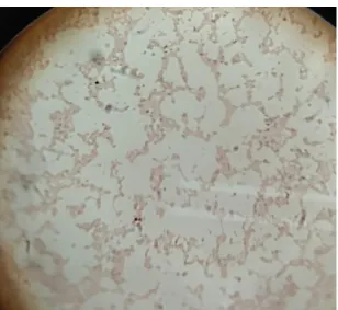
The images, given below (Figures 2-6) were obtained during biochemical testing and are presented here as references. They represent the positive and negative results of each respective test which have been conducted.
Figure 2.1: Gram negative cocci Figure 2: Gram positive bacilli
25 | P a g e
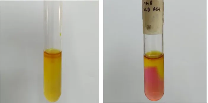
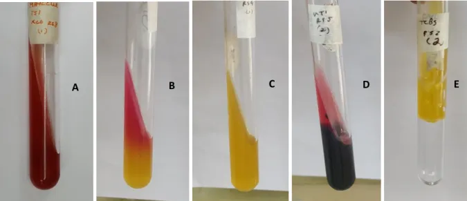
Figure 3: Results of the TSI test (A) No gas (NG), No H2S (N H2S), Red butt and slant. (B) NG, NH2S, Yellow butt and red slant. (C) NG, NH2S, Yellow butt and slant. (D) NG, H2S, Yellow butt and red slant. (E) Gas, NH2S, Yellow butt and slant.
A B C D E
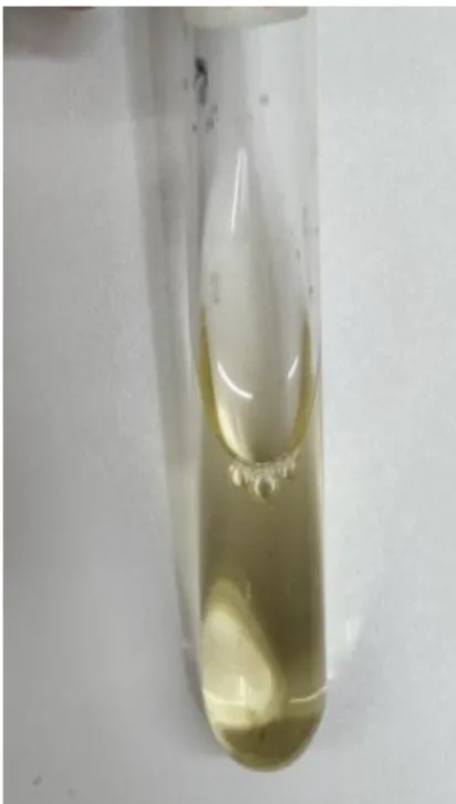
Figure 4: MIU negative (All 3 attributes) Figure 4.1: MIU positive (All 3 attributes)
26 | P a g e
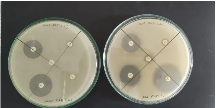
3.4. Antibiotic susceptibility profiling:
The identified samples were subjected to antibiotic susceptibility testing in order to determine the effectiveness of a selected cohort of modern antibiotics. The data obtained with regards to the zones of inhibition were subsequently compared to the established guideline of CLSI (Clinical and Laboratory Standards Institute). Accordingly, the organisms were classified as being susceptible
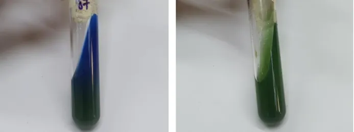
Figure 5: Citrate positive Figure 5.1: Citrate negative
Figure 6: MR negative Figure 6.1: MR positive Figure 6.2: VP negative
27 | P a g e
“S”, intermediate “I”, or resistant “R”. Tables 5, 5.1, and 5.2 represent the antibiotic susceptibility profile of the samples to 10 antibiotics. It was observed that Amikacin, Meropenem, Norfloxacin, and Amoxicillin + Clavulanic acid possessed susceptibility at 97.8%, 95.7%, 89.4%, and 85.1%
respectively. These were at the higher tier of effectiveness. Tetracycline and Azithromycin/Erythromycin were at the middle tier of effectiveness with susceptibility percentages of 61.7% and 57.4% respectively. At the lower tier of effectiveness were the remaining antibiotic agents, namely Amoxicillin, Colistin Sulfate, Vancomycin, and Cefixime with corresponding susceptibility percentages of 46.8%, 44.6%, 34% and 31.9%.
Figure 7 is presented below as a reference.
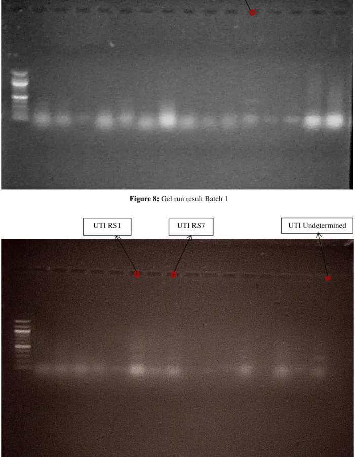
3.5. Determination of mcr-1 amplicon presence via Gel electrophoresis:
The process of gel electrophoresis was subsequently conducted after PCR to confirm the prevalence of the targetted mcr-1 gene among the variety of the samples which possessed resistance to Colistin Sulphate. The figures 8 and 8.1 displayed below represent the results of the gel run. It was observed that a total of 4 isolates possessed the CLR conferring mcr-1 gene.
Figure 7: Antibiotic susceptibility profiling on MHA agar.
28 | P a g e
MAC RS1
UTI RS1 UTI RS7 UTI Undetermined
Figure 8: Gel run result Batch 1
Figure 8.1: Gel run result Batch 2
29 | P a g e
Table 3: Identified bacteria per site of sampling.
Sample zones
Bacterial Identities
Site 01 Cedecea neteri, Staphylococcus aureus, Yersinia mollaretii, Citrobacter gillenii, Bacillus novalis, Bacillus tequilensis, Paenibacillus macerans, Brevibacterium iodinum, Vibrio pacinii, Aliivibrio logei, Photorhabdus asymbiotica (Emerging Human pathogen)
Site 02 Vibrio furnissii, Enterobacter spp., Aeromonas schubertii, Xenorhabdus beddingii Site 03 Staphylococcus epidermidis, Lysinibacillus sphaericus, Escherichia spp.
Site 04 Providencia rustigianii, Lysinibacillus sphaericus, Aneurinibacillus aneurinilyticus Site 05 Staphylococcus aureus, Staphylococcus epidermidis, Bacillus carboniphilus,
Lysinibacillus sphaericus, Salmonella spp.
Site 06 Moellerella wisconsensis, Shimwellia blattae, Serratia fonticola, Viridibacillus neidei, Providencia alcalifaciens
Site 07 Bacillus aminovorans, Serratia fonticola, Staphylococcus spp.
Site 08 Klebsiella pneumoniae, Shimwellia blattae, Buttiauxella brennerae, Citrobacter youngae, Paenibacillus macquariensis, Shigella dysenteriae, Salmonella spp., Shigella boydii
30 | P a g e
Table 4: Representation of biochemical testing results of the identified bacteria
Sample Number XLD RS1 XLD RS6 XLD RS8 (1) XLD RS8 (2) XLD RS4 MAC RS6 MAC RS8 MSA RS1 MSA RS5 (1) MSA RS5 (2) MAC RS1 (1) MAC RS1 (2) MAC RS8 (1) MAC RS8 (2) RS 4 (10^-4) RS 7 (10^-6) RS 8 (2) UTI RS 1 (G) UTI RS 5 (1) MSA RS 3 TCBS RS2 (2) TCBS RS2 (3)
Colony Morphology Bacilli Bacilli Bacilli Bacilli Bacilli Bacilli Bacilli Staphylococci Staphylococci Staphylococci Cocci Cocci Bacilli Bacilli Bacilli Bacilli Bacilli Bacilli Bacilli Cocci Bacilli Bacilli
Gram Stainin g - - - - - - - + + + - - - - + + + + + + - -
Catalase + + + + + + + + + + + + + + + + + + + + + +
H2S - - - - - - - - - - - - - - - + - - - - - -
Gas - - - - - - - - - - - - - - - - - - - - + +
Citrate + + - - - + + + - + - - + + + - - - - + + +
Indole - - - - + - - - - + - - - - - - - - - + - -
MR + + + + - + + + + + + + + + + + + + + + + +
VP - - - - - - - - - - - - - - - - - - - - - -
31 | P a g e
Motility + - - - + - - - - + - + - - - + + + + + + +
Urease - - - - + - + + + + - - + - + + - - - + - -
Glucose Utilization + + + + + - - - - + + + + - - + + + - + + +
Lactose Utilization + + - - - - - - - + + + - - - - + - - + + +
Sucrose Utilization + - - - - - - + + + + + - - - - + - - + + +
Bacteria Cedecea neteri Moellerella wisconsensis Klebsiella pneumoniae Klebsiella pneumoniae Providencia rustigianii Shimwellia blattae Shimwellia blattae Staphylococcus aureus Staphylococcus aureus Staphylococcus epidermidis Yersinia mollaretii Citrobacter gillenii Buttiauxella brennerae Citrobacter youngae Lysinibacillus sphaericus Bacillus aminovorans Paenibacillus macquariensis Bacillus novalis Bacillus carboniphilus Staphylococcus epidermidis Vibrio furnissii Vibrio furnissii
Table 4.1: Representation of biochemical testing results of the identified bacteria
Sample Number XLD RS8 XLD RS8 XLD RS8 XLD RS6 XLD RS7 UTI RS5 UTI RS4 UTI RS3 UTI RS5 UTI RS1 UTI RS1 UTI RS1 UTI RS4 XLD RS8 MFC RS6 MFC RS2 MSA RS7 MFC RS2 TCBS RS1 TCBS RS2 TCBS RS1
Colony Morpholog y Bacilli Cocci Bacilli Bacilli Bacilli Bacilli Bacilli Bacilli Bacilli Bacilli Bacilli Cocci Bacilli Cocci Bacilli Bacilli Cocci Bacilli Bacilli Bacilli Bacilli
Gram Staining - - - - - + + + + + + + + - - - + - - - -
Catalase + + + + + + + + + + + + + + + + + + + + +