i | P a g e
A study on prevalence of gastrointestinal parasites in pig at Rangamati Sadar upzila of Rangamati district
A clinical report submitted in partial satisfaction of the requirement for the Degree of Doctor of Veterinary Medicine (DVM)
Submitted by:
Anika Binte Belal Roll No: 18/53 Reg No: 03015 Intern ID: 49 Session: 2017-18
Faculty of Veterinary Medicine
Chattogram Veterinary and Animal Sciences University Khulshi, Chattogram – 4225, Bangladesh
November 2023
ii | P a g e
A study on prevalence of gastrointestinal parasites in pig at Rangamati Sadar upzila of Rangamati district
Approved by:
( Dr. AMAM Zonaed Siddiki Professor
Department of Pathology and Parasitology)
Faculty of Veterinary Medicine
Chattogram Veterinary and Animal Sciences University Khulshi, Chattogram – 4225, Bangladesh
November 2023
iii | P a g e
TABLE OF CONTENTS
Contents………..……….………Page no
List of contents……….iii
List of tables………..iv
List of figures……….………..………..iv
List of Acronyms Symbols Used……….………..…v
ABSTRACT……….…….vi
CHAPTER I: INTRODUCTION…….………01-03 CHAPTER II: REVIEW OF LITERATURE……….…04-09 2.1 Epidemiology………..04
2.2 Diagnosis of GI parasites……….04-05 2.3Prevalence of GI parasites around the world………...06-09 CHAPTER III: MATERIALS AND METHODS………...10-12 3.1 Period of the study……….10
3.2 The study area………10
3.3 Selection of samples………...11
3.4 Fecal examination………..11
3.5 Experimental Procedure………...12
3.6 Statistical analysis……..………12
CHAPTER IV: RESULTS……….14-18 4.1 Overall prevalence of gastrointestinal parasitic infections………...…14-15 4.2 Age specific prevalence of gastrointestinal parasitic infections………….………15
4.3 Sex-specific prevalence of gastrointestinal parasitic infections………….…..…..16
4.4 Rate of gastrointestinal parasitic infection in pig according to rearing system...17
4.4 Rate of gastrointestinal parasitic infection in pig according to deworming...17-18 CHAPTER V: DISCUSSION………19-20 CHAPTER VI: LIMITATION OF THE PRESENT STUDY……….20
CHAPTER VII: CONCLUSION………...21
ACKNOWLEDGEMENT………...22
REFERENCES………...27
BIOGRAPHY………...………29
iv | P a g e
LIST OF TABLES
Tables Topics Page
no.
Table
1 Overall prevalence of gastrointestinal parasitic infections 14 Table
2
Age-specific occurrence of gastrointestinal parasitic
infections 15
Table 3
Sex wise percentage of gastrointestinal parasitic infection
of pig 16
Table 4
Rate of gastrointestinal parasitic infection in pig according
to rearing system 17
Table 5
Rate of gastrointestinal parasitic infection in pig according
to deworming 17
LIST OF FIGURES
Figure Topics Page
no.
Fig.1
Study area map 10
Fig.2 Experimental Design (at a glance). 11
Fig 3 Microscopic pictures of eggs of gastrointestinal parasites of pig
13
Fig.4 Overall infection rate of gastrointestinal parasitic infections in study area.
14
v | P a g e
List of Acronyms Symbols Used:
Abbreviation Elaboration
% Percentage
et.al And his association
GI Gastrointestinal
Spp. Species
P-value Probability value
N Sample size
CVASU Chattogram Veterinary and Animal
Sciences university
vi | P a g e
ABSTRACT
Gastrointestinal (GI) parasites in pigs pose a significant challenge in the hilly areas of Bangladesh, where pig farming is a common livelihood. The aim of the study is to investigate the prevalence of GI parasites of pigs in Bangladesh. The study was conducted in different areas of Rangamati Sadar Upzila in May 2023. Fecal samples were collected from 13 pig farms (N=38) and examined by using direct smear and Stoll’s ova counting techniques. All animals were found infected with one or more species of parasites. Four species were identified, namely Ascaris suum (47.36%), Balantidium coli (39.48%), Fasciolopsis sp (7.90%), Strongyloides sp (5.26%). Age-specific analysis revealed that pigs aged between 6 and 12 months were more susceptible to parasitic infections. Ascaris suum was highly prevalent in this age group (45.24%). The study also indicated that female pigs exhibited a slightly higher susceptibility to gastrointestinal parasitic infections, with Ascaris suum being the most prevalent (50%). In terms of rearing systems, pigs in extensive rearing systems had a higher prevalence of Ascaris suum (46.67%) and Balantidium coli (36.67%) infections. Pigs with no prior anthelminthic exposure showed a higher prevalence of parasitic infections, particularly Ascaris suum (47.83%) and Balantidium coli (43.46%). Data showed that there was no significant (p>0.05). It can be said that this study represents a novel contribution to the field and can serve as a benchmark for future research in this area. Further research and improved management practices are recommended to decrease the high prevalence of gastrointestinal parasitic infections in indigenous pig populations in this region.
Keywords: Prevalence, parasitism, gastrointestinal parasite, pig,
Bangladesh, Rangamati.
1 | P a g e
CHAPTER 1 INTRODUCTION
Pigs are known for their rapid growth and high reproductive rates, as indicated by several studies (Durranc and Maxson 2008; Prakash et al. 2008; Phookan et al. 2006; Taylor et al. 2006). They are widely regarded as one of the most prolific livestock breeds. Pigs hold a significant place in the livestock industry as they offer a rich source of animal protein, particularly for those who include pork in their diet, and this protein source is available at a relatively low cost.
Pigs play a vital role in the livestock industry because they are primarily raised by individuals from socioeconomically disadvantaged segments of society. Pork stands out as a significant source of animal protein in human diets, and its affordability positions it as one of the most promising ways to enhance animal protein consumption for those with limited financial resources. Pigs are noteworthy for their prolific reproduction and rapid growth, making them a highly valuable livestock breed.
In order to thrive, pigs require certain essential conditions, including warmth, a dry and comfortable resting place, and protection from both winter cold and summer heat.
Additionally, pigs have specific minimal needs when it comes to space, access to fresh air, hygienic surroundings, and easy access to food and water. It's imperative that their living conditions and accommodation do not predispose them to illness or injury (Moore et al. 2002).
The majority of pigs in Bangladesh are found in rural and hilly regions, primarily raised by low-income households with subpar hygiene practices. Nonetheless, the traditional pig farming approach remains appealing to resource-constrained farmers, (Verhulst, 1993 and Phiri et al. 2003.) In Bangladesh, pig production methods predominantly fall into two categories: first, there is the semi-intensive system, which is mainly adopted by tribal communities who rear pigs in their residential areas, and second, the free-range or extensive system, which is followed by nomadic groups that raise pigs in herds by constantly shifting their scavenging locations.
Globally, there are widespread reports of swine infections caused by gastrointestinal parasites, and these infections are heavily influenced by the methods used for managing swine, (Nansen et al. 1999). In rural areas of many developing countries, the practice of raising free-range pigs is common, despite its drawbacks, such as inefficient food conversion, elevated mortality rates, and the production of lower-quality products.(
Kagira et al. 2010 ; Greve et al. 2012)
2 | P a g e .
Additionally, domestic pigs serve as significant reservoirs for parasites that can affect other animals and pose health risks to humans,.(Popiołek et al. 2009). While many parasitic infections in pigs often have no obvious clinical symptoms, there can be instances of symptomatic infections, particularly in younger pigs,.(Weng et al.2005) The most common errors made by pig owners in controlling parasitic infections include the failure to conduct fecal sample tests on animals to identify specific parasite issues on the farm, the improper administration of anti-parasitic medications, and the ineffective disinfection of facilities,(Balicka-Ramisz et al. 2020).
In pigs, gastrointestinal (GI) parasites are widespread. The principal effects of these parasites involve a reduced appetite, diminished daily weight gain, inefficient food utilization, and an increased susceptibility to other pathogens. Gastrointestinal parasites lead to significant reductions in productivity within the swine and broader livestock industry, (Boes et al. 2000 ; Joachim and Daugschies 2000).
Indigenous pigs are the dominant breed in smallholder regions, where they are reared through a free-range system and primarily depend on limited nutritional resources, (Mashatise et al. 2005). The pigs primarily function as scavengers, making use of food scraps discarded by people. The roaming behavior of these pigs exposes them to internal parasite eggs, heightening their susceptibility to internal parasitic infestations, (Roepstorff and Nansen 1994). Additionally, the warm and humid tropical conditions, combined with the insufficient treatment of local pigs against parasitic diseases, consistently result in these pigs carrying significant loads of GI nematodes ( Mashatise et al. 2005)
Indigenous pigs are prevalent among smallholders, where they are raised under a free range system and thrive on meager diets. These pigs mainly scavenge for food scraps discarded by people, and their wandering behavior exposes them to internal parasite eggs, making them highly susceptible to internal parasite infestations. Additionally, the warm and humid tropical conditions, combined with inadequate treatment for parasitic diseases, lead to these pigs carrying substantial loads of gastrointestinal nematodes (Sarker et al.
2016).
Gastrointestinal parasites are common in domestic pigs across various production systems worldwide. Pigs, being omnivores, consume a wide range of foods, including dead insects, worms, tree bark, decaying carcasses, garbage, kitchen waste, and even human excreta. Swine raised in intensive operations are less vulnerable to gastrointestinal infections, although they are often susceptible to large roundworms, such as Ascaris spp (Weng et al. 2005 and Eijck and Borgsteede 2005).
3 | P a g e In temperate pig farming, major helminth species include Ascaris suum, Trichuris suis, and Oesophagostomum sp. Although infections are typically subclinical, pigs with these parasites experience reduced food utilization and growth rates, along with occasional liver damage due to migration of Ascaris suum larval stage.(.Roepstorff et al.2011).
Some pig parasites can be transmitted to humans, particularly farmers, directly or indirectly. Ascaris suum and Trichuris suis are zoonotic parasites closely related to Ascaris lumbricoides and Trichuris trichiura, which infect millions of people worldwide (De Silva et al.2003). Gastrointestinal parasite infections in pigs are prevalent globally and are influenced by the type of pig management practices. Poor husbandry practices, coupled with extensive management, are identified as risk factors for pig infections with GI parasites.
Despite the significant economic losses caused by gastrointestinal parasites, this issue is often overlooked because most infected animals exhibit few noticeable clinical signs throughout their productive lives, (Reza et al. 2010).
Therefore, the present study was conducted with following objectives:
1) To investigate the presence of GI parasites in pigs in Rangamati.
2) To determine the impact of various factors, including age and sex, on the occurrence of these diseases.
4 | P a g e
Chapter II Review of Literature
This chapter serves as a review of various pieces of literature focused on gastrointestinal parasitism in pigs, along with their prevalence. Its primary objective is to offer insights into the research conducted in this field. Below, we have outlined essential information pertinent to the present study under the following headings:
2.1 Epidemiology:
The pig-rearing systems in Bangladesh exhibit significant diversity.
2.1.1 Factor affecting the size of gastrointestinal infection
Gastrointestinal nematode infection size depends on the following factors:
The number of infective larvae/eggs ingested by the host, influenced by climate, vegetation, livestock density, and grazing patterns.
The rate at which host resistance develops, affected by parasite and host species, genetics, nutrition, and physiological stress (e.g., parturition).
The intrinsic multiplication rates of parasite species, controlled by fecundity, pre-patent period, and environmental factors.
Management, especially grazing patterns (Radostits et al. 1994).
Geographical distribution and availability of intermediate hosts.
The use of anthelmintic, including timing and frequency of administration (Radostits et al, 1994 and Hansen and Perry, 1993).
2.2 Diagnosis of gastrointestinal parasitism
To diagnose gastrointestinal parasitic infections in herbivores, it is essential to recover the parasites or their eggs/larvae from the animal's digestive tract or fecal material. The key steps involved in this process include:
Collecting fecal samples.
Separating eggs/larvae from fecal materials and concentrating them.
Microscopically examining the prepared specimens.
fecal cultures preparation.
Isolation and identification of larvae from culture (Baermann apparatus techniques)
5 | P a g e The following Qualitative and Quantitative tests were used for the diagnosis of gastrointestinal parasitism:
Fecal sample
(Urquhart et al., 1996, Hansen and Perry., 1993, Soulsby, 1982 and Benbrook and Sloss, 1962)
Qualitative test Quantitative test
Direct method Indirect method
Direct smear
Floatation
Simple
floatation Test tube floatation
Centrifugal floatation
Simple sedimentatio
n
Centrifugal sedimentation
McMaster technique
Stoll’s ova counting technique
Sedimentation
6 | P a g e 2.3 Prevalence of Gastrointestinal parasites in around the world:
Yadav et al. (1989) investigated the prevalence of gastrointestinal parasites in 496 domestic pigs in sub-tropical and high-rainfall areas of India over 18 months. The study identified 11 different parasite species, including Ascaris suum, Oesophagostomum dentatum, and others. Ascaris suum, the most common species (51.67%), is possibly zoonotic. The overall infection rate was significantly higher (76.42%) in low-altitude regions compared to high-altitude areas (62.50%).
Nsoso et al. (2000) conducted a study in Botswana, it was found that 82% of the pigs were infected with Ascaris suum, with a prevalence rate of 54.6%.
Weng et al. (2005)in Guangdong Province, China, from July 2000 to July 2002, a study was conducted to investigate the prevalence of intestinal parasites in intensive pig farms.
The research examined fecal samples from 3,636 pigs of various sexes and age groups from 38 representative intensive pig farms employing different parasite control strategies.
Out of the sampled pigs, 209 (5.7%) were found to be infected with Trichuris suis, 189 (5.2%) with Ascaris sp, 91 (2.5%) with Oesophagostomum spp., 905 (24.9%) with coccidia (Eimeria spp. and/or Isospora suis), and 1,716 (47.2%) with Balantidium coli.
These infections were primarily observed in pigs from farms without a systematic anti- parasite treatment plan. Co-infections with multiple parasites were common, and T. suis was the most prevalent nematode, affecting breeding, young, and mature pigs.
Dutta et al. (2005) in a study conducted in West Bengal, specifically in Kolkata and Jalpaiguri districts, the prevalence of gastrointestinal parasites in pigs managed under both backyard and scientific management systems was investigated. A total of 1,074 fecal samples and 93 gastrointestinal tracts of pigs were examined to detect both parasite eggs and adult parasites. The study found that parasitic infections were most intense during the rainy season and least prevalent in the summer. Trematode and nematode infections were most common in the semi-intensive management system, while protozoan infections were more frequent in the free-range system. Helminthic infections were most common in adult pigs (older than 2 years), while protozoan infections were more prevalent in piglets (less than 6 months). The identified parasites included Fasciolopsis buski, Gastrodiscoides hominis, Schistosoma suis, Ascaris suum, Trichuris suis, Metastrongylus sp., Ascarops strongylina, Physocephalus sexalatus, Strongyloides ransomi, Oesophagostomum dentatum, Hymenolepis sp., as well as oocysts of Eimeria spp. and Balantidium coli.
7 | P a g e Eijck and Borgsteede, (2005) a study conducted from November 2001 to October 2002 aimed to assess the prevalence of gastrointestinal parasites in sucking piglets, fattening pigs, and sows across different farm types, including 16 free-range farms (FRF), 11 organic farms (OF), and 9 conventional farms (CF). Through fecal examinations of composite samples taken during four visits to each farm over three-month intervals, the study found varying rates of parasitic infections. Coccidian infections were most prevalent on organic farms (90.90%), while Ascaris suum had a higher prevalence on both organic (72.7%) and free-range farms (50%). Oesophagostomum spp. infections were observed in all farm types, with sows having the highest rates. Trichuris suis was found in significant numbers, especially among sows on free-range and organic farms.
Marufu et al. (2008) An investigation was conducted in Zimbabwe, specifically in the Hama-Mavhaire communal area of Chirumhanzu District, to assess the prevalence of gastrointestinal nematodes in indigenous pigs. A total of 143 pigs, encompassing both sexes and various age groups (< 5 months, 5 - 12 months, and > 12 months), were randomly selected from 10 villages. Their rectal fecal samples were collected for the identification and quantification of nematode eggs. Among the 143 pigs, 58.7% tested positive for gastrointestinal nematodes, with 17.5% exhibiting mixed infections. The study identified four parasite species, with Oesophagostomum species being the most prevalent (54.6%), followed by Strongyloides ransomi (14%), Ascaris spp (7%), and Trichuris suis (4.2%).
Morris et al. (2009) A research investigation was conducted in Oklahoma to determine the prevalence of swine gastrointestinal parasites on swine farms. A total of 975 fecal samples were collected from 98 farms. Parasites were recovered from pig feces on these farms as follows: Ascaris spp (53.0%), Strongyles sp (53.1%), Trichuris sp (35.7%), Spirurids sp (6.1%), Strongyloides sp (19.4%), coccidia (57.1%), and Balantidium coli (55.1%). Notably, a higher percentage (16.5%) of hogs kept on cement floors tested positive for Ascaris sp compared to those on dirt lots (11.9%) or slatted floors (9.9%).
However, pigs on dirt lots had a higher percentage of coccidia infections (21.0%) compared to those on cement or slatted floors (8.5% and 6.0%, respectively). The prevalence of Trichuris sp infections was similar (ranging from 6.8% to 11.3%) in hogs from all three management practices.
Uysal et al. (2009) A study in Turkey investigated intestinal parasites in pig feces with potential human pathogens. A total of 238 pig fecal samples were collected from pig farms in Çorlu (Tekirdağ), Ayazma, and Arnavutköy (Istanbul) during the summer. The findings revealed that 8.8% of fecal samples contained Cryptosporidium spp., 3.7% had Giardia spp., 1.6% showed Balantidium coli cysts, and 4.1% had Ascaris suum eggs.
Among pigs younger than 6 months, Giardia lamblia was found in 7.6% of cases, Cryptosporidium spp. in 11.4%, and Balantidium coli cysts in 1.5%. In pigs older than 6
8 | P a g e months, Giardia lamblia was found in 0.7%, Cryptosporidium spp. in 6.7%, Balantidium coli cysts in 1.5%, and Ascaris suum eggs in 6.7%.
Ismail et al. (2010) conducted an investigation in Korea to assess the infection status of intestinal parasites in pigs and beef cattle in rural areas of Chungcheongnam-do. Between November 2009 and April 2010, a total of 241 fecal samples, including 136 from pigs and 105 from beef cattle, were examined using direct smear and centrifugal sedimentation methods. The findings revealed that the overall positive rates of intestinal parasites were 73.5% among pigs and 4.8% among beef cattle. Additionally, the double infection rate was 10.3% in pigs. Among the 136 pig specimens, 64.7% were infected with Balantidium coli, 17.6% with Ascaris suum, and 3.7% with Entamoeba spp. In the case of the 105 beef cattle specimens, 4.8% were found to be infected with Entamoeba spp. These results indicated a high B. coli infection rate and a moderate A. suum infection rate among pigs raised on rural farms in Chungcheongnam-do.
Tomass et al. (2013) A study was conducted in Ethiopia to investigate the prevalence of gastrointestinal parasites and Cryptosporidium species in extensively managed pigs in Mekelle and urban areas of the southern zone of Tigray Region during June to September 2012. A total of 714 pigs of different ages and sexes were chosen for fecal sample collection, and 25 soil samples were also collected. Gastrointestinal tract parasites were examined using flotation and sedimentation techniques, while the Modified Ziehl Neelsen technique was used to examine oocysts of Cryptosporidium species from 276 randomly selected fecal samples. The findings revealed that out of the 714 pigs examined, 27.3% were infected by at least one gastrointestinal parasite. Ascaris suum (25.9%) was the most prevalent parasite, followed by Fasciola hepatica (1.8%), Eimeria spp (1.7%), and Trichuris suis (0.3%). There was no significant association between sex and the prevalence of parasites. However, the age of pigs had an effect on the prevalence of parasites. About 7% of the examined pigs tested positive for oocysts of Cryptosporidium spp. Additionally,72% of the soil samples were found to be contaminated with eggs of Ascaris spp. in the study area. In addition to causing morbidity in infected pigs, the potential for Ascaris sp parasites in pigs to infect humans and vice versa, combined with poor environmental hygiene, could complicate the epidemiology and control of Ascariasis in the study areas. The study also suggested that extensively managed pigs may act as potential reservoirs for zoonotic infections from Cryptosporidium sp.
9 | P a g e Md. Nur-E-Azam et al.(2015) A three-month cross-sectional study was conducted in two upazillas of Dinajpur, Bangladesh in 2015 to investigate gastrointestinal parasitism in pigs. A total of 100 pig fecal samples were collected for analysis. The study revealed that among different gastrointestinal parasitic infections, the prevalence of Ascaris suum infection was the highest at 38% in the study population. The second most common parasitic infection was caused by Macracanthorhynchus hirudinaceus (22%), followed by Strongyloides ransomi (20%). The lowest parasitic infections were recorded for Trichostrongylus axei (1%) and Fasciolopsis buski infection (1%).The study also found a comparatively higher prevalence of Ascaris suum, Macracanthorhynchus hirudinaceus, and Strongyloides ransomi in pigs in relation to their age and sex. The occurrence of gastrointestinal parasitic infection was higher in pigs aged over 6-12 months and in female pigs.
Sarker et al. (2016) conducted a study in various areas of Chittagong district from May to August 2014 to investigate gastrointestinal parasitism in pigs. They collected fecal samples from 86 pigs and reported a 52.3% prevalence of the infection in pigs in Chittagong, Bangladesh. They identified five species of parasites, namely Schistosoma sp (24.41%), Fasciolopsis sp (66.27%), Ascaris sp (70.93%), Strongyloides sp (38.37%), and Balantidium sp (52.32%).
Krishnamoorthy et al. (2022) conducted a study in various regions of India to assess the prevalence of gastrointestinal parasites in pigs. They found that the overall prevalence of GI parasites in pigs was 54%. Interestingly, a higher prevalence of these parasites was observed in samples (84%) collected from slaughterhouses compared to fecal samples (46%) from pigs. Among the various parasite species, coccidia (29%) were the most commonly reported, while Globocephalus urosubulatus (0.7%) was observed at a sporadically low rate. Regarding nematodes and trematodes, a higher prevalence was found in Ascaris spp. (27%) and Amphistomes (12%).
10 | P a g e
C
HAPTER III
MATERIALS AND METHODS
3.1 Period of study: The study lasted 20 days, from May 9th to May 29th.
3.2 The study area: Pigs were typically raised by tribes mostly in the Chittagong Hill tracts. So I chose Rangamati regions as my research area to investigate. The sample is collected from three union oh Rangamati sadar upzila. The unions are: Magban, Asambosti and Sapchori.
Fig. 1: Study area map
11 | P a g e
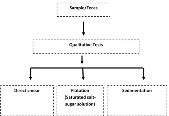
Sample/Feces
Qualitative Tests
Direct smear Flotation
(Saturated salt- sugar solution)
Sedimentation
3.3 Selection of samples: Due to the limited study period, the sample was collected from different unions of Rangamati sadar upzila. Indigenous/crossbred pigs were selected for this study as target animal. To assess the age susceptibility of various parasitic infections, the pigs were divided into three sub-groups: Piglets (≤6 months), Growers (>6-12 months), and Adults (>12 months) based on their age. A total of 38 fecal samples were randomly collected from these 13 pig farms. Additionally, a questionnaire was utilized to record information such as the area, rearing system, housing, age, sex, and other relevant factors.
3.4 Fecal examination
The fecal samples underwent a thorough examination process. All collected fecal samples were subjected to direct smear, sedimentation, and flotation techniques for coproscopy.
Sugar Salt Solution was utilized as the flotation fluid. To identify the developmental stages of parasites, such as eggs, cysts, and oocysts, the study adhered to the appropriate morphological characteristics described by various authors, including Hendrix (2006), Urquhart et al. (1996), Hansen and Perry (1993), Soulsby (1982), Benbrook, and Sloss (1962).
Fig. 2: Experimental Design (at a glance)
12 | P a g e
3.5. Experimental Procedure
3.5.1 Direct smear: A small amount of feces from each sample was placed on a clean microscope slide, and a drop of tap water was added to create a relatively uniform and transparent preparation. The larger particles were gently moved aside, and a cover glass was placed over the clear liquid. The preparation was systematically examined under low magnification as per the guidelines provided by Hendrix (2006), Urquhart et al. (1996), and Soulsby (1982).
3.5.2 Sedimentation technique: For each sample, 3 grams of feces were transferred to the first container and suspended in 40-50 ml of distilled water. The mixture was stirred thoroughly with a stirring device (e.g., tongue blade or fork) and passed through a tea strainer, with the filtrate collected in a second container. The filtrate was then poured into a test tube and allowed to settle for 20-30 minutes. Afterward, the supernatant was carefully discarded, and the sediment was examined under a microscope using magnifications ranging from 10X to 40X, following the procedures outlined by Hendrix (2006), Urquhart et al. (1996), and Soulsby (1982).
3.5.3 Floatation technique (Test tube floatation): Each sample, containing 2 to 5 grams of feces, was placed in a suitable container and suspended in 50 ml of floatation fluid.
The mixture was stirred thoroughly with a stirring device (e.g., tongue blade or fork) and passed through a tea strainer, with the filtrate collected in a second container. The filtrate was then poured into a test tube until a meniscus formed at the top of the tube (a convex meniscus). A glass cover slip was placed over the meniscus and allowed to stand for 15- 30 minutes. Finally, the cover slip was lifted off from the tube, along with any adhering fluid, and immediately placed on a glass slide for observation under a microscope using magnifications from 10X to 40X, following the procedures described by Hendrix (2006), Urquhart et al. (1996), and Soulsby (1982).
3.6 Statistical analysis
The collected data were imported, stored, and coded using Microsoft Excel-2010 and then subjected to analysis using STATA-13.0. The results were expressed as percentages, along with p-values from the Chi-Square Test. Statistical significance was considered when p < 0.05.
13 | P a g e
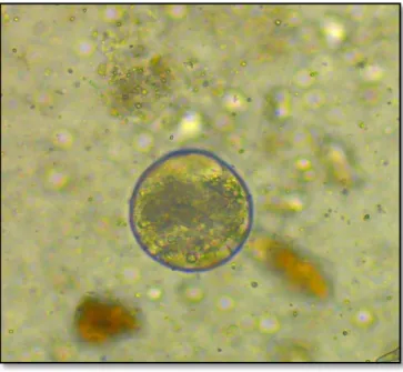
Fig. : Egg of Ascaris suum
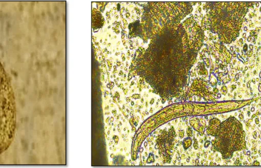
Fig. : Egg of Fasciolopsis sp
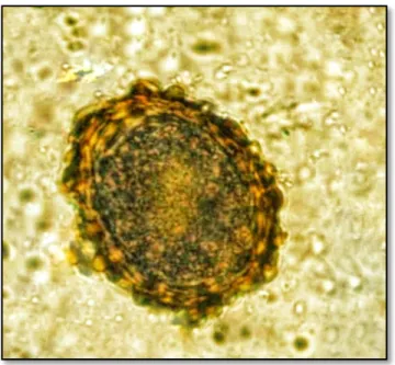
Fig. : Cyst of Balantidium coli
Fig. : Filariform stage of Stongyloides sp
Fig 03: Microscopic pictures of eggs of gastrointestinal parasites of pig
14 | P a g e
0 10 20 30 40 50
Ascaris suum Balantidium coli Fasciolopsis spp Strongyloides spp
No of infected
Ascaris suum Balantidium coli Fasciolopsis spp Strongyloides spp
CHAPTER IV RESULTS
4.1 Overall percentage of gastrointestinal parasites of pig
Throughout the study, 38 fecal samples were analyzed, and it was discovered that all of them (100%) were infected with one or more species of endoparasites. Specifically, four distinct types of GI parasites were identified.
(Table: 1 Overall prevalence’s of GI parasites in pigs in the study areas)
(Figure 4: Overall prevalence’s of GI parasites in pigs in the study areas)
Among different gastrointestinal parasitic infections, prevalence of Ascaris suum infection was the highest which was 47.46%% in study population. The second most common parasitic infection was caused by Balantidium coli (39.48%%) followed by
Parasite identified Number of infected Prevalance (%)
Ascaris suum 18 47.36
Balantidium coli 15 39.48
Fasciolopsis sp 3 7.90
Strongyloides sp 2 5.26
Total N=38 100
15 | P a g e
Fasciolopsis spp (7.9%). The lowest parasitic infections were also recorded for Strongyloides sp (5.26%) (Table 01, Figure: 01)
4.2 Age specific occurrence of GI parasitic infections
*Significant when P<0.05
The occurrence of gastrointestinal parasitic infections was also influenced by the age of the animals. The investigation revealed that pigs aged between >6 months and 12 months were more susceptible to various parasitic infections compared to the other two age groups .Among the different parasitic infections, it was observed that Ascaris suum was highly prevalent in pigs aged >6-12 months, accounting for 45.24% of the cases, followed by pigs aged ≤6 months at 69.23%. Additionally, Balantidium coli infection (52.94%) was more common in pigs aged >6-12 months compared to those aged ≤6 months (15.38%). Strongyloides sp infection was most common in the >12 months age group, while Fasciolopsis sp. infection was more common in pigs aged >12 months.
Parasitic infections Age Category Pearson chi2 value
P Value
≤6 months >6-12 months >12months
Ascaris suum 41.18(7) 69.23(9) 25(2) 4.35 0.11
Balantidium coli 15.38(2) 52.94(9) 50(4) 4.81 0.09
Fasciolopsis sp 5.88(1) 7.69(1) 12.50(1) 0.32 0.84
Strongyloides sp 0(0) 7.69(1) 12.50(1) 1.93 0.37
16 | P a g e
4.3 Sex specific infection rate of GI parasitic infection in pig
*Significant when P<0.05
The age of the animals significantly influenced the occurrence of gastrointestinal parasitic infections. The investigation revealed that pigs aged between 6 to 12 months were more susceptible to various parasitic infections when compared to the other two age groups. Among the different parasitic infections, it was noted that Ascaris suum was highly prevalent in pigs aged between 6 and 12 months, with a prevalence of 45.24%, followed by pigs aged below 6 months, with a prevalence of 69.23%. Furthermore, Balantidium coli infection was more common in pigs aged between >6-12 months, with a prevalence of 52.94%, compared to those ≤ 6 months old, with a prevalence of 15.38%.
Strongyloides sp infection was more common in pigs older than 12 months. Additionally, Fasciolopsis sp infection was prevalent in pigs > 12 months of age.
Parasitic infections Sex catagory Pearson chi2 value
P Value
Male Female
Ascaris suum 45(9) (50)9 0.09 0.7
Balantidium coli 45(9) 33.33(6) 0.51 0.4
Fasciolopsis spp 5(1) 11.11(2) 0.48 0.4
Strongyloides ransomni 5(1) 5.56(1) 0.00 0.9
17 | P a g e
4.4 Rate of GI parasitic infection in pig according to rearing system.
*Significant when P<0.05
The occurrences of gastrointestinal parasitic infections were also influenced by rearing system animal. During this investigation, it was observed that prevalence of Ascaris suum infection was the highest in extensive rearing (46.67%). Occurrence of Fasciolopsis sp infection in extensive system was 10% and Strongyloides sp infection was also was 6.67%. The occurrence of protozoan infection such as Balantidium coli infection was also higher in extensive rearing in compare to semi-intensive.
4.5 Rate of GI parasitic infection in pig according to deworming
*Significant when P<0.05
Parasitic infections Rearing system Pearson chi2 value
P Value Semi-intensive Extensive
Ascaris suum 7.45 (4) 46.67(14) 0.02 0.8
Balantidium coli 8.25(4) 36.67(11) 0.47 0.49
Fasciolopsis sp 0(0) 10(3) 0.8 0.35
Strongyloides sp 0(0) 6.67(2) 0.5 0.45
Parasitic infections Deworming Pearson
chi2 value
P Value
Yes No
Ascaris suum 38.67(7) 47.83(11) 0.00 0.9
Balantidium coli 33.33(5) 43.46(10) 0.30 0.5
Fasciolopsis sp 6.67(1) 8.70(2) 0.05 0.8
Strongyloides sp 0(0) 13.33(2) 1.30 0.2
18 | P a g e
Occurrences of gastrointestinal parasitic infections were also influenced by deworming animal. In non-dewormed chicken, the prevalence of Ascaris suum, Balantidium coli Fasciolopsis sp, Strongyloides sp was 47.83%, 43.46%, 8.70, and 13.33(respectively, whereas in dewormed chicken, the prevalence of Ascaris suum, Balantidium coli , Fasciolopsis sp, Strongyloides sp was 38.67%, 33.33%, 6.67% and 0% respectively.
19 | P a g e
CHAPTER V DISCUSSION
During the study period, total of 38 fecal samples were examined. All the samples (100%) were found to be infected with one or more species of gastrointestinal parasite.
The present study aimed to assess the prevalence and diversity of gastrointestinal (GI) parasites in indigenous pigs from Rangamati district, Bangladesh. Our findings indicated an overall prevalence of GI parasites in the pigs, with a rate of 100%. This prevalence aligns with similar studies conducted in various regions, such as Indonesia (100%;
Widisuputri et al., 2020), another study in Bangladesh (100%; Sarker et al., 2016), Burkina Faso and Uganda (91%; Nissen et al., 2010; Tamboura et al., 2006), a different study in Bangladesh (96.4%; Dey et al., 2014), and Brazil (93.1%; Barbosa et al., 2015).
However, our results showed a lower prevalence compared to reports from Bangladesh (65%; Md. Nur-E-Azam et al., 2015), Kenya (83%–84.2%; Kagira et al., 2012; Obonyo et al., 2013), Tanzania (83%; Nonga & Paulo, 2015), South Africa (79.2%; Nwafor et al., 2019), and Korea (73.5%; Ismail et al., 2010).
The variation in parasite prevalence among these studies can be attributed to several factors, including the age and sex of the pigs, their rearing conditions, pig breeds, and immune responses, as well as climate, sampling seasons, geographical landscapes, farming practices, sample sizes, and laboratory techniques for fecal analysis.
One significant factor contributing to the elevated prevalence of GI parasites in our study may be the suboptimal rearing conditions of the pigs. Many farmers in the study region lacked knowledge about effective pig-rearing and farm management practices.
Another potential factor contributing to the elevated prevalence of gastrointestinal parasites in our study could be the methodological variation. We employed a range of fecal analysis techniques, including direct wet mount, floatation, sedimentation, acid-fast staining, and sporulation. The cumulative use of these methods might have led to higher detection rates of enteric parasites. Additionally, it's worth noting that pigs themselves serve as natural reservoirs for many of the GI parasites found in this study (Ji et al., 2019;
Schuster & Ramirez‐Avila, 2008). Indigenous pig breeds are also known to naturally harbor a high prevalence of GI parasites (Murthy et al., 2016), which could have contributed to the high prevalence observed in the fecal samples examined.
In terms of parasite diversity, Ascaris suum exhibited the highest prevalence rate (47.36%) in this study, which was slightly higher than the findings of Tamboura et al.
20 | P a g e
(2006) in Burkina Faso, Nigeria, who reported 40%. However, our results were lower than those reported by Yadav et al. (1989), Morris et al. (2009), and Nwoha and Danie (2011), with prevalence rates of 51.67%, 53%, and 50%, respectively, in various locations, which were greater than the results of our present study. The elevated prevalence of Ascaris suum in our study could be attributed to its thick eggshell, which provides resistance to adverse environmental conditions, enabling prolonged survival in the soil (Soulsby, 1982). Additionally, habitat contamination and the scavenging behavior of pigs may have contributed to the higher prevalence of such parasitic infections in our study population (Johnstone, 2000).
It is noteworthy that Balantidium coli, a commonly occurring parasite in pigs, was detected in 39.48% of the samples. This prevalence was lower than what was reported in Indonesia (79% in Widisuputri et al., 2020), Bangladesh (40% in Dey et al., 2014), and India (29.48% in Patra et al., 2019). However, it was higher than the prevalence reported in China (22.79% in Lai et al., 2011).
The occurrence of Fasciola sp. infection in this study was 7.90%, which is significantly lower than the findings from the Tigray region of Ethiopia (1.8% in Tomass et al., 2013).
The prevalence of Strongyloides spp. was 5.26%, which was also lower than the reported rates in Bangladesh (29.1% in Dey et al., 2014), Ghana (11% in Atawalna et al., 2016), and India (11.10% in Patra et al., 2019).
CHAPTER VI:
LIMITATION OF THE PRESENT STUDY
There are some limitations in this study. They are-
Breed and age differences were not considered in the current study.
Seasonal variations in disease prevalence were not addressed.
Worm load, an important factor in parasitic infections, was not examined.
Additional extensive research is needed to overcome these study limitations.
Investigate the potential impact of pig parasitic infestations on public health.
Aim to identify crucial predictors related to these parasitic diseases.
21 | P a g e
CHAPTER VII CONCLUSION
The primary objective of this research was to determine the prevalence of GI parasites in pigs. The investigation discovered a notably higher occurrence of Ascaris suum and Balantidium coli in pigs concerning their age and gender. Gastrointestinal parasitic infections were most prevalent in female pigs aged over 6 to 12 months. The study suggested that the likelihood of gastrointestinal parasitism is elevated in regions with hot and humid climates, which are ideal for the proliferation of these parasites. However, suboptimal pig management practices, inadequate nutrition, and a lack of awareness about deworming also contribute to the elevated infection rates. It's important to note that this study had certain limitations, including time constraints, variations in topography, seasonal disease patterns, and the inclusion of both local and crossbred pigs. As a result, it is advisable to conduct more extensive research on gastrointestinal parasitism to address these limitations and uncover key factors associated with these diseases.
22 | P a g e
ACKNOWLEDGEMENTS
The author extends heartfelt gratitude to the divine presence of Almighty "ALLAH," the supreme authority and ruler of the universe, for granting the strength and guidance to successfully complete this endeavor.
A profound debt of appreciation is owed to the internship supervisor Professor Dr.
AMAM Zonaed Siddiki, Department of Pathology and Parasitology, Faculty of Veterinary Medicine, Chattogram Veterinary and Animal Sciences University. His expert guidance and scholarly oversight have been invaluable in shaping this report.
The author is deeply grateful to Dr. Lenin Dey, Senior Assistant Director of Pig Development Farm, Rangamati and all the staff members of the Pig Development Farm for their scholarly guidance, invaluable suggestions, and inspiring contributions that have greatly enriched this study report.
The author also wishes to thank Dr, Prima Mohajan, Former Vs of Rangamati Sadar Upzila and Dr. Debaraj Chakma,VS of District livestock office for their cooperation and amazing guidelines.
The author extends sincere gratitude to Professor Dr. A. S. M. Lutful Ahsan, the esteemed Vice Chancellor of Chattogram Veterinary and Animal Sciences University, for his invaluable support. Special thanks are also owed to Professor Dr. Mohammad Lutfur Rahman, Dean of FVM, and Professor Dr. AKM Saifuddin, Director of External Affairs, for providing an exceptional internship program and research experience.
The author is equally appreciative of the unwavering assistance from her family, friends, and colleagues during the questionnaire survey and samples collection, which significantly contributed to the completion of this work.
23 | P a g e
REFERENCES
Abdu, S. and Gashaw, A. 2010. Production system dynamism and parasitic interaction of swine in and around Holetta, Ethiopia, Ethiop. Vet. J., 14 (1): 71-81.
Nansen, P.; Roepstorff, A. Parasitic helminths of the pig: Factors influencing transmission and infection levels. Int. J. Parasitol. 1999, 29, 877–891
Kagira, J.M.; Githigia, S.M.; Nganga, J.C.; Kanyari, P.W.N.; Maingi, N.; Gachohi, J.M.
Prevalence of gastrointestinal protozoa and association with risk factors in free- range pigs in Kenya. J. Protozool. Res. 2010, 20, 1–9
Greve, J.H. Internal parasites: Helminths. In Diseases of Swine, 10th ed.; Zimmerman, J.J., Karriker, L.A., Ramirez, A., Schwartz, K.J., Stevenson, G.W., Eds.; John Wiley & Sons: Chichester, UK, 2012; pp. 912–913.
Popiołek, M.; Knecht, D.; Boruta, O.; Kot, M. Effect of breeding conditions, phenology, and age on the occurrence of helminths in pigs. A preliminary study. Bull. Vet.
Inst. Pulawy 2009, 53, 213–220.
Weng, Y.B.; Hu, Y.J.; Li, Y.; Li, B.S.; Lin, R.Q.; Xie, D.H.; Gasser, R.B.; Zhu, X.Q.
Survey of intestinal parasites in pigs from intensive farms in Guangdong Province, People’s Republic of China. Vet. Parasitol. 2005, 127, 333–336.
Balicka-Ramisz, A.; Wi´sniewski, J.; Stadnytska, O. Extensity and intensity of intestinal parasite infections in pigs in different types of farm organization. Acta Sci. Pol.
Zootech. 2020, 18, 47–50.
Soulsby, E. J. L. (2012). Helminths, arthropods and protozoa of domesticated animals (7th ed.). Affiliated East‐West Press Private Limited 1
Phiri IK, Ngowi H, Afonso S, Matenga E, Boa M, Mukaratirwa S, Githigia S, Saimo M, Sikasunge C, Maingi N, Lubega GW, Kassuku A, Michael L, Siziya S, Krecek RC, Noormahomed E, Vilhena M, Dorny P, Willingham AL . Noormahomed E, Vilhena M, Dorny P, Willingham AL The emergence of Taenia solium
cysticercosis in Eastern and Southern Africa as a serious agricultural problem and public health risk (2003).
Verhulst A (1993) Lessons from field experiences in the development of monogastric animal production. In: Mack S (ed) Strategies for sustainable animal agriculture in developing countries: proceedings of the FAO expert consultation held in Rome, Italy 10–14 December 1990
Hale OM and Stewart TB (1979). Influence of an experimental infection of Trichuris suis on performance of pigs. Journal of Animal Sciences, 49: 1000–1010.
Hale OM, Stewart TB and Marti OG (1985). Influence of an experimental infection of Ascaris suum on performance of pigs. Journal of Animal Sciences, 60: 220–225.
24 | P a g e
Hale OM, Stewart TB, Marti OG, Wheat BE and Mc Cormick WC (1981). Influence of an experimental infection of nodular worms (Oesophagostomum spp.) on performance in pigs. Journal of Animal Sciences, 52: 316-322
Hossain ME, Chakma S, Khatun MM, Hasanuzzaman M, Miah MY and Biswas MAA (2011). Production systems of swine in the rural areas of Rangamati and Khagrachari Districts of Bangladesh. Bangladesh Journal of Animal Sciences, 40(1-2): 28-33.
De Silva NR, Brooker S, Hotez PJ, Montresor A, Engels and Savioli L (2003). Soil transmitted helminth infections: Updating the global picture. Trends Parasitology, 19: 547–551.
Krishnamoorthy, P., Lakshmi, H. K., Siju, S. J., Suresh, K. P., & Shome, B. R. (2022). A scientometric study on prevalence of gastrointestinal parasites in pigs (Sus scrofa) of India. Indian J Anim Sci, 92(11), 1264-1273.
Adesiyun, A.A., Kaminjolo, J.S., Ngeleka, M., Mutani, A., Borde, G., Harewood, W and Harper, W. 2001. A longitudinal study on enteropathogenic infections in Trinidad.
Revista da Sociedade Brasileira de Medicina Tropical, 34 (1): 29-35.
Alam, J. 1993. Livestock sector for more investment in Bangladesh. Asian Livestock, 18:
77-78.
Alim, M.A., Das, S., Roy, k., Sikder, S., Mohiuddin., Masuduzzaman., M and Hossain, M.A.(2011). Prevalence of haemoprotozoan and gastrointestinal parasitic diseases in cattle of Chittagong, Bangladesh. Masters Thesis, Submitted to Department of Pathology and Parasitology, Faculty of Veterinary Medicine, Chittagong Veterinary and Animal Sciences University.
Anonymous. 2013 .Helminth-Infections-Of-Wild-Boars-Sus-Scrofa-In-The-Bursa- Province http://www.bioportfolio.com.Helminth-Of.html.6, June, 2013.
Bachal, B., Phullan, M.S., Rind, R. and Soomro, A.H. 2002. Prevalence of Gastrointestinal Helminths in Buffalo calves. Online J. Bio. Sci., 2(1): 43-45.
Bowen, L.2005. Pig industry in Grenada and the Caribbean. In: proceedings of the St.
George university seminar series on concepts in porcine farming, P. 4-5.
Bugg, R.J., Robertson, I.D, Elliot, A.D. and Thompson, R.C.1999. Gastrointestinal parasites of urban dogs in Perth, Western Australia. Vet. J., 157(3): 295-301.
Cole, H.H. 1996. Introduction to Livestock production. 2nd edn. Freeman and co, San Francisco and London.
D.L.S. 2008. Livestock and Poultry profile Bangladesh, Sunil Chandra Ghosh, D.G., DLS.
de-la. Muela, N., S Hermandez de.L, L and Ferre, I. 2001. Helminths of Wild Boar in Spain, J. Wild life Dis., 37(4): 840-843.
25 | P a g e
Ejick, I.A.I.M. and Borgsteede, F.H.M. 2005. A survey of gastrointestinal parasites on free-range, organic and conventional pig farms in Netherland, Vet. Res. Comm., 29: 407-414.
Gibbens, J.C., Gibbens, N.P. and Fielding, W.J. 1989. An abattoir survey of the prevalence of gastrointestinal helminths and Stephanurus dentatus in pigs in Belize. Trop. Anim. Hlth Prod., 21: 197-204.
Gracia, J.A., Rodriguez-Diego, J.G., Torres-Hernandez, G., Mahuieu, M., Garcia, E.G.
and Gonzal-Garduno, R. 2007. The epizootiology of ovine gastrointestinal strongyles in province of Matanzas. Small Rumin. Res., 72: 119-126.
Hansen, J. and Perry, B. 1993. The Epidemiology, Diagnosis and Control of Helminth Parasites of Ruminants. 2nd edn, Nairobi,Kenya; ILRAD.pp. 20-22.
Hindsbo, O., Nielsen, C.V., Andressen, J., Willingham, A.L., Bendixen, M., Nielsen, M.
and Nielsen, N.O. 2000. Age-dependent occurrence of the intestinal ciliate Balantidium coli in pigs at a Danish research farm, Acta. Vet. Scand., 41:79–83.
Hossain, M.E., Chakma, S., Khatun M.M., Hasanuzzaman, M., Miah, M.Y. and Biswas, M.A.A. 2011. Production systems of swine in the rural areas of Rangamati and Khagrachari districts of Bangladesh, Bang. J. Anim. Sci., 40 (1-2): 28-33.
Intervet. (2011). www, intervetusa.com.
Ismail, H.A., Jeon, H.K., Yu, Y.M., Do, C. and Lee, Y.H. 2010. Intestinal Parasite Infections in Pigs and Beef Cattle in Rural Areas of Chungcheongnam-do, Korea, Korean J. Parasitol., 48 (4): 347-349.
Jang, D.H. 1975. Survey for internal parasites of swine in Korea, Korean J. Vet. Res., 15:309–314.
Kagira, J.M., Githigia, S.M., Ng’ang’a, J.C., Kanyari, P.W.N., Maingi, N. and Gachohi, J.M. 2010. Prevalence of gastrointestinal protozoa and association with risk factors in free-range pigs in Kenya, J. Protozool. Res., 20:1-9.
Kakar, M.N. and Kakarsulemankhel, J.K. 2008. Prevalnce of endo (trematodes) and ecto- parasites in cows and buffaloes of Quetta, Pakistan. Pak. Vet. J., 28(1): 34, 34-36.
kyadav, A. and Tandonn, V. 1989. Nematode Parasite Infections of Domestic Pigs in a Sub-tropical and High-rainfall Area of India, Vet. Parasitol., 31: 133-139.
Lay, K. K., Hoerchner, H.C.F., Morakote, N. and Kreausukon, K. 2008. Prevalence of Cryptosporidium, Giardia and Oher Gastrointestinal Parasites in Dairy Claves in Mandalay, Myanmar. Proc of the 15th Congress of FAVA 27-30 October FAVA- OIE Joint Symposium on Emerging Diseases, Bangkok, Thailand.pp. 273-274.
Long, T.F., Johnson. R.K. and Keele, J.W. 1990.Intensive production system of swine. J.
Anim. Sci., 68: 4069-4078.
Martin, L.J., Gibbs, H.C. and Pullin, J.W. 1974. Gastrointestinal parasites of swine in Québec, Can. Vet. J., 15(3): 72–76.
26 | P a g e
Marufu, M.C., Chanayiwa, P., Chimonyo, M. and Bhebhe, E. 2008. Prevalence of gastrointestinal nematodes in Mukota pigs in a communal area of Zimbabwe, Afr.
J. Agric. Res., 3(2): 91-95.
Mercy, A.R., de Chaneet, G. and Emms, Y. 1989. Survey of internal parasites in Western Australian pig herds, Aust. Vet. J., 66(1):4-6.
Morris, R.G., Jordan, H.E., Luce, W.G., Coburn, T.C. and Maxwell, C.V. 1984.
Prevalence of gastrointestinal parasitism in Oklahoma swine, Am. J. Vet. Res., 45(11):2421-2423.
Nahar, N., Uddin, M., Gurley, ES., Khan, MS., Hossain, MJ., Sultana, R and Luby, SP.
2012. Pig illnesses and epidemics: a qualitative study on perceptions and practices of pig raisers in Bangladesh- International Centre for Diarrhoeal Disease Research, Bangladesh (ICDDR, B).
Napravnik, J. and Berka, T. 2002. Cause and effect of ascariosis in pig herds.
Helminthologia, 39:171-183.
Nsoso, S.J., Mosala, K.P., Ndebele, R.T. and Ramabu, S.S. 2000.The prevalence of internal and external parasites in pigs of different ages and sexes in Southeast District, Bostwana, Onderstepoort J.Vet. Res., 67: 217-220.
Nwoha, R.I.O. and Daniel, G. 2011. Department of Veterinary Medicine and Department of Pathology, Michael Okpara University of Agriculture, Prevalance of Gastrointestinal Nematodes Parasites In Intensively Managed Pigs Of Different Ages And Sexes In Umuahia City Of Abia State, Continental J. Vet. Sci., 5 (1): 11 – 17.
Permin, A., Yelifari, L., Bloch, P., Steenhard, N., Hansen, N.P. and Nansen, P. 1999.
Parasites in crossbred pigs in upper East region of Ghana, Vet. Parasitol., 87(1):
63-71.
Reza, A.M., Murtaza, S., Bachaya H.A., Qayyum, A. AND Zaman, M.A. 2010. Point Prevalence of Toxocara vitulorum in Large Riminants Slaughtered at Multan Abattior, Pak. Vet. J., 30(4): 242-244.
Roepstorff, A., Mejer, H., Nejsum, P. and Thamsborg, S.M. 2011. Helminth parasites in pigs: new challenges in pig production and current research highlights. Vet.
Parasitol., 180(1-2):72-81.
Solaymani, M.S., Mobedi, I., Rezaian, M., Massoud J, Mohebali M, Hooshyar ,H., Ashrafi, K.and Rokni, M.B.2003. Helminth parasites of the wild boar, Sus scrofa, in Luristan province, western Iran and their public health significance. J.
Helminthol., 77(3):263-267.
Soulsby, E.J.L. 1982. Helminthes, Arthropods and Protozoa of Domesticated Animals, 7th edn. Baillere Tindall, London.pp:707-717, 729-735.
Tamboura, H.H., Banga-Mboko, H., Maes, D., Youssao, I., Traore, A., Bayala, B. and Dembele, M.A. 2006. Prevalence of common gastrointestinal nematode parasites in scavenging pigs of different ages sexes in eastern centre province, Burkina Faso, Ondersteport J. Vet. Res., 73:53-60.
Taylor G and Roese G (2006). Basic pig husbandry. NSW, New South Wales.
27 | P a g e
Tomass, Z., Imam, E., Kifleyohannes, T., Tekle, Y. and Weldu, K. 2013. Prevalence of gastrointestinal parasites and Cryptosporidium species in extensively managed pigs in Mekelle and urban areas of southern zone of Tigray region, Northern Ethiopia, vetworld., 6(7): 433-439.
Urquhart, G.M., Armour, J., Duncan, J.L. and Jennings, F.W. 1996. Vet. Parasitol., Black well Science Ltd., 2nd edn.pp. 242-251.
Uysal, H.K., Borali, O., Metiner, K and Ilgaz, A. 2009. Investigation of Intestinal Parasites in Pig Feces, Türkiye Parazitoloji Dergisi., 33 (2): 218 – 221.
Visco, R.J, Corwin, R.M. and Selby, L.A. 1977.Effect of age and sex on the prevalence of intestinal parasitism in dogs, J. Am. Vet. Med. Assoc., 170(8): 837-935.
Weng, Y.B., Hu, Y.J., Li, Y., Li, B.S., Lin, R.Q., Xie, D.H., Gasser, R.B., and Zhu, X.Q.
2005. Survey of intestinal parasites in pigs from intensive farms in Guangdong Province, People’s Republic of China, Vet. Parasitol., 127 (3-4): 333–336.
Adhikari, R. B., Adhikari Dhakal, M., Thapa, S., & Ghimire, T. R. (2021).
Gastrointestinal parasites of indigenous pigs (Sus domesticus) in south‐central Nepal. Veterinary Medicine and Science, 7(5), 1820-1
Dey, T. R., Dey, A. R., Begum, N., Akther, S., & Barmon, B. C. (2014). Prevalence of endo parasites of pig at Mymensingh, Bangladesh. Journal of Agriculture and Veterinary Science, 7, 31–38. 10.6084/M9.FIGSHARE.1226224
Apt, W., Aguilera, X., Vega, F., Alcaíno, H., Zulantay, I., Apt, P., González, V., Retamal, C., Rodríguez, J., & Sandoval, J. (1993).
Prevalence of fascioliasis in humans, horses, pigs, and wild rabbits in 3 Chilean provinces. Boletin De La Oficina Sanitaria Panamericana Pan American Sanitary Bureau, 115, 405–414. Eysker, M., Boerdam, G., Hollanders, W., &
Verheijden, J. (1994). The prevalence of Isospora suis and Strongyloides ransomi in suckling piglets in the Netherlands. Veterinary Quarterly, 16, 203–205. 10.1080/01652176.1994.9694449
Argenzio, R. A., Armstrong, M., & Rhoads, J. M. (1997). Role of the enteric nervous system in piglet cryptosporidiosis. Journal of Pharmacology and Experimental Therapeutics, 279, 1109–1115.
Atawalna, J., Attoh‐Kotoku, V., Folitse, R., & Amenakpor, C. (2016). Prevalence of gastrointestinal parasites among pigs in the Ejisu Municipality of Ghana.
Scholars Journal of Agriculture and Veterinary Sciences, 3, 33–36.
Bernard, A. N., Daminabo, V., Ekam, E., Okonkwo, E., Nwuzo, A., Afiukwa, F., & Agah, M. (2015). Prevalence of intestinal parasites in faecal droppings of swine in Pankshin urban, Pankshin local government area, Plateau state, Nigeria. American Journal of Life Sciences, 3, 119–122.
10.11648/j.ajls.20150302.19
Eijck, I., & Borgsteede, F. (2005). A survey of gastrointestinal pig parasites on free‐
range, organic and conventional pig farms in the Netherlands. Veterinary Research Communications, 29, 407–414. 10.1007/s11259-005-1201-z
Ejinaka, O. R. A., & Onyali, I. O. (2020). Parasitic gastrointestinal helminths and protozoa in pigs at Enugu, Nigeria. The Biomedical Diagnostics, 4, 67–74.
Tamboura, H., Banga‐Mboko, H., Maes, D., Youssao, I., Traore, A., Bayala, B., &
Dembele, M. (2006). Prevalence of common gastrointestinal nematode parasites in scavenging pigs of different ages and sexes in eastern centre
28 | P a g e
province, Burkina Faso. Onderstepoort Journal of Veterinary Research, 73, 53–
60. 10.4102/ojvr.v73i1.169
Zhang, S.‐X., Zhou, Y.‐M., Xu, W., Tian, L.‐G., Chen, J.‐X., Chen, S.‐H., Dang, Z.‐S., Gu, W.‐
P., Yin, J.‐W., & Serrano, E. (2016). Impact of co‐infections with enteric pathogens on children suffering from acute diarrhea in southwest China.
29 | P a g e
Biography
I am Anika Binte Belal, daughter of MD Belal Hossain and Nasima Akther. I have completed my Secondary School Certificate examination from Lakers’ Public School and College , Rangamati in 2015 and Higher Secondary School Certificate from Chattogram Cantonment Public School and College, Chattogram in 2017. I am an intern veterinarian at Chattogram Veterinary and Animal Sciences University, Bangladesh under the Faculty of Veterinary Medicine.I hold a profound passion for veterinary medical research, and I am eager to leverage my skills and creativity to make a positive impact on our country's challenges in this field. My goal is to contribute significantly to overcoming the current difficulties we encounter in veterinary medicine through innovative research and solutions.