i
Role of Mesenchymal Stem Cells & MicroRNA in the Treatment of Osteoporosis
By
Mohammad Rafi Hassan Chowdhury 18146053
A thesis submitted to the School of Pharmacy in partial fulfillment of the requirements for the degree of
Bachelor of Pharmacy (Hons.)
School of Pharmacy Brac University
April, 2022
© 2022. Brac University All rights reserved.
ii
Declaration
It is hereby declared that
1. The thesis submitted is my/our own original work while completing degree at Brac University.
2. The thesis does not contain material previously published or written by a third party, except where this is appropriately cited through full and accurate referencing.
3. The thesis does not contain material which has been accepted, or submitted, for any other degree or diploma at a university or other institution.
4. I/We have acknowledged all main sources of help.
Student’s Full Name & Signature:
Mohammad Rafi Hassan Chowdhury ID: 18146053
iii
Approval
The thesis/project titled “Role of Mesenchymal Stem Cells & MicroRNA in the Treatment of Osteoporosis” submitted by Mohammad Rafi Hassan Chowdhury (18146053) of Spring, 2018 has been accepted as satisfactory in partial fulfillment of the requirement for the degree of Bachelor of Pharmacy (Hons.) on 12th April, 2022.
Examining Committee:
Supervisor:
(Member) _______________________________
Dr. Hasina Yasmin Professor, School of Pharmacy
Brac University
Program Coordinator:
(Member)
_______________________________
Namara Mariam Chowdhury Lecturer, School of Pharmacy
Brac University
Deputy Chairperson:
(Member)
Departmental Head:
(Dean)
_______________________________
Dr. Hasina Yasmin Professor, School of Pharmacy
Brac University
_______________________________
Dr. Eva Rahman Kabir
Professor and Dean, School of Pharmacy Brac University
iv
Ethics Statement
Since this project is a review article, therefore it didn’t perform any animal or human trials of any sort.
v
Abstract
Osteoporosis is currently the most difficult and painful disease endangering the lifestyle of the geriatric population. Through the gradual degeneration of osteoblastic cell in various parts of the bones, osteoporosis has caused a massive healthcare problem and cost for a significant part of the world populace. Despite having conventional therapies, like bisphosphonates, the disease still has no sustainable treatment option because the conventional drugs only emphasize on preventing bone degradation, not bone regeneration. As such, increasing researches on regenerative treatment options are getting huge attention especially in mesenchymal stem cells and it’s relationship with microRNAs. This novel treatment techniques introduces a sustainable, cost efficient and long- lasting options for the osteoporotic patients. As such, this review mainly focuses on the tonsil derived mesenchymal stem cell therapy along with the therapeutic effects of microRNA in order to examine whether their therapeutic and regenerative potential are truly an amazing alternative or not.
Keywords: Osteogenesis; osteoblast; mesenchymal stem cell; microRNA; ovariectomy; hydrogel.
vi
Table of Contents
Declaration... ii
Approval ... iii
Ethics Statement... iv
Abstract ... v
List of Tables ... ix
List of Figures ... x
List of Acronyms ... xii
Chapter 1 Introduction... 1
1.1 Osteoporosis ... 1
1.2 Current Existing treatment of Osteoporosis ... 2
1.3 Stem Cell Therapy ... 4
1.4 Mesenchymal Stem Cell Therapy ... 4
1.5 Advantages & Disadvantages of MSC therapy ... 5
1.6 Aim & Objectives of the study ... 7
Chapter 2 Methodology ... 8
Chapter 3 Pathophysiology of Osteoporosis ... 9
3.1 Osteoporosis ... 9
3.2 Osteoimmunology ... 10
3.3 Cellular Senescence... 11
vii
3.4 Microbiomes of Gastrointestinal Tract ... 12
Chapter 4 Tonsil derived Mesenchymal Stem Cell Therapy ... 15
4.1 Background ... 15
4.2 Advantages of the TMSC therapy ... 15
4.3 Mechanism of Action of TMSC therapy ... 16
4.4 Delivery method of the TMSC ... 16
4.5 Preparation of Preclinical mouse model... 18
4.6 Therapeutic & Biological analysis of the TMSC-GHH ... 20
4.6.1 Serum Osteocalcin (OCN) level ... 20
4.6.2 The stability & efficiency of the GHH ... 21
4.6.3 Therapeutic efficacy of TMSC ... 22
4.6.4 Serum OCN, ALP & Total Calcium levels ... 23
4.6.5 Biocompatibility of the GHH scaffold ... 26
4.6.6 Potential for Visceral fat reduction ... 28
Chapter 5 Mesenchymal Stem Cell & microRNA Combination Therapy ... 30
5.1 Introduction ... 30
5.2 Potential advantage of incorporating miRNA in MSC ... 31
5.3 Examples of MiRNA in Osteogenic differentiation of MSC ... 32
5.3.1 miR-23a... 32
5.3.2 miR-29a... 33
viii
5.3.3 miR-138 ... 33
5.3.4 miR-346 ... 34
5.4 Delivery method ... 36
5.4.1 The use of HA/TCP scaffold... 36
5.4.2 Photoactivation by UV light ... 37
Chapter 6 Conclusion & Future Aspects of MSC Therapy ... 38
References ... 40
ix
List of Tables
Table 01: A summary of different microRNAs for osteoporosis treatment………. 35
x
List of Figures
Figure 01: Different sources of Mesenchymal Stem Cells (MSCs)………..05
Figure 02: Pathophysiological effect of Cellular Senescence in Osteoporosis……….12 Figure 03: A schematic diagram showing how the GIT microbiome can possibly influence Osteoporosis………..14 Figure 04: GHPA polymers backbones are being enzymatically crosslinked to form GHH scaffold, where TMSC are then fixed in the hydrogel meshwork………...18 Figure 05: Comparison of serum OCN levels & bone condition between OVX and Non-OVX mice………..20 Figure 06: The stability & longevity of GHH while incorporating TMSC for 03 months…...21 Figure 07: Micro CT images and bone mineral density of femoral head trabeculae at the experimental
endpoint………...22 Figure 08: Serum osteocalcin (OCN) level in OVX mice after 03 months……….24 Figure 09: Serum Alkaline Phosphatase (ALP) level in OVX mice after 03 months……….25 Figure 10: Serum total calcium level in OVX mice after 03 months………...26 Figure 11: Macroscopic morphology of postmortem kidneys and liver obtained from each OVX group after euthanization………...26 Figure 12: Mean body weight, adjusted kidney mass & adjusted liver mass of each OVX mice group three months after first treatment………..27
xi
Figure 13: The Mean visceral fat mass and mean body weight adjusted visceral fat mass of each OVX mice groups after 03 months……….28
xii
List of Acronyms
MSC Mesenchymal stem cell
RANKL Receptor Activator of Nuclear factor-kB ligand SERMs Selective Estrogen Receptor Modulators
CAF Carcinoma-associated fibroblast TAF Tumor-associated fibroblast miRNA MicroRNA
TMSC Tonsil derived mesenchymal stem cell BM-MSC Bone marrow mesenchymal stem cell OVX Ovariectomized
BMD Bone Mineral Density
HepG2 Human hepatocellular carcinoma cell line Skov-3 Human ovarian cancer cell line
MVs Micro vesicles
SASP Senescence-associated secretory phenotype SCFA Short chain fatty acid
PTH Parathyroid hormone IBM Intra-bone marrow
xiii HRP Horseradish peroxidase
GHPA Gelatin-hydroxyphenyl propionic acid GHH Gelatin-hydroxyphenyl hydrogel OCN Osteocalcin
ALP Alkaline phosphatase
hBMSC Human bone marrow mesenchymal stem cells HA/TCP Hydroxyapatite/Tricalcium phosphate
hADSC Human adipocyte derived stem cell
1
Chapter 1 Introduction 1.1 Osteoporosis
Osteoporosis is a chronic, long-term disorder of the skeletal system, which is seen more pronounced in geriatric patients, typically in men after the age of 65 and women after 55 years. In a review by Noh et. al., (2020), a report about osteoporosis conducted in 1993 by the WHO, osteoporosis is recognized as “progressive skeletal disease identified by the incidence of low bone mass and decay of the microarchitecture of the bone tissue, which leads to an eventual increase in bone fragility and vulnerability to fracture”. To this day, the incidence of osteoporosis has steadily increased with each passing year. Studies support this fact by displaying that around one among two women and on the other hand one among four men aging 50 or above it endure osteoporosis.
As a result, it has become an enormous public health crisis for governments all over the world.
According to the International Osteoporosis Foundation (2022), the disease is seen globally in around 6.3% of men above the age of 50 and 21.2% of women in the same range of age. The predominant reason for women to have a high risk of getting osteoporosis is because during their menopause, their estrogen level is very low and causes osteoporosis. Estrogen is a potent hormone that controls and manages calcium metabolism in the body and thus the lack of it causes the risk factor of contracting osteoporosis. Furthermore, if we consider the earth’s population as whole, the International Osteoporosis Foundation (2022) also gives a figure of 500 million men and women suffering from osteoporosis worldwide with an annual number of more than 8.9 million fractures, which equates to an osteoporosis fracture occurring every three seconds. The principal bones affected and more susceptible to osteoporotic fractures are the nearest ends of the humerus
2
and femur to their respective joints, the remote end of the radius from it’s joint and the spine.
According to Sarafrazi et. al., (2021), in a NCHS data brief in 2017–2018, the prevalence of osteoporosis at the end of the femur or on the lumbar region of the spine or both of these bones among adults aged 50 and above was 12.6% and among women it was 19.6%; which is 4.4%
higher if we compare with men. The impact of osteoporosis on the economy is also high as the average burden of fractures on annual medical cost was $8,600, which totals with a cost of $14 billion in the USA. On the other hand, nearly half of the non-fracture osteoporosis patients receive conventional drug treatment, which averages $500 per patient or $2 billion nationwide in the USA (Blume & Curtis, 2011). Given that the majority of patients suffering from this disease are old and in their retirement age, this massive cost lowers their pension fund to a great degree and thus becomes a big crisis for their end stage of life.
1.2 Current Existing treatment of Osteoporosis
Since the Bone Mineral Density (BMD) of the patient is gradually reduced in osteoporosis, the principal aims of osteoporosis therapy are to decrease the incidence of bone fracture along with loss of bone tissues and thus in the meantime prevent disability due to it and manage pain. So based on these goals, besides some lifestyle changes (ex. quitting smoking and alcohol), there are therapeutic ways of treating it. First of all, the most common treatment is to give calcium and vitamin D supplements. But supplements aren't always effective to treat osteoporosis. So, there are drugs, especially anti-catabolic and in some cases catabolic drug classes are used.
Bisphosphonates, like risedronate and zoledronic acids, are the most common drugs for osteoporosis, which works by hampering bone resorption or deletion by connecting to the hydroxyapatite binding sites on top of the bones when resorption phase of it is active. Such mechanism of action resists osteoclasts from developing a border and clinging to the surface of
3
bones, which ultimately inhibits the synthesis of protons necessary for osteoclasts to perform it’s action. The drug also resists the osteoclast progenitor cell to develop properly, while in the same time promotes osteoclast apoptosis. However, the major limitation of these drugs are that they are useful in decreasing any risk of future fractures in patients who have already sustained a fracture due to osteoporosis (Samwald et al., 2020). Furthermore, oral dosage of this drug is necessary to be ingested as soon as possible in the morning, preferably before breakfast. The patient in order to prevent the possible side effect of esophageal ulcer, should remain in a supine position, keeping an empty stomach for roughly from half an hour to one hour maximum after taking the drug (Gupta and March, 2016). Such a strict dosage system along with adverse effects, like bisphosphonate- related osteonecrosis of the jaw makes bisphosphonate not a popular medicine for patient adherence.
Another form of treatment is by using monoclonal antibodies like denosumab. Denosumab inhibits RANKL (receptor activator of nuclear factor-kB ligand), a key receptor for osteoclast regulation and thus maintains a balanced bone mass (Theill et al., 2002). Denosumab is appropriate for administration in postmenopausal women and in case men, it is used for those who are at advanced risk for fractures. The monoclonal antibody has proved its effectiveness in reducing the risk for hip and vertebral fractures. However, it is not appropriate for all ages, as in patients under the age of 18 years. Furthermore, the antibody also has side effects like skin infections and hypocalcemia, with an idiosyncratic effect of jaw necrosis. Worse still, is that there is an increased chance of vertebral fractures if denosumab is discontinued, thus limiting its use (Cummings et al., 2018).
Since osteoporosis is very common in women, a specific treatment option is available for them.
It’s called Selective Estrogen Receptor Modulators (SERMs), which are artificial ligands for the estrogen receptors. They give a different response compared to female estrogen hormones; like
4
estradiols and give their therapeutic effects by enhancing osteoclast apoptosis. Some prominent examples of SERMs are raloxifene, lasofoxifene and bazedoxifene. Like all therapies, this one is not without it’s side-effects. According to Cummings et al., (2009), despite lasofoxifene showing therapeutic efficacy in reducing risks of bone and spinal fractures observed among postmenopausal women suffering from osteoporosis, in the meantime it also made the risk of contracting venous thromboembolic disorders highly plausible. As a result, SERMs usage are limited and are only applicable as a valid treatment option for women who doesn’t have thromboembolic disease occurred previously in her life (D'Amelio and Isaia, 2013).
1.3 Stem Cell Therapy
To write about stem cell therapy, it’s important to know what a stem cell is. Stem cells are unique cellular apparatus in multicellular organisms. It is a type of immature and undifferentiated cells, which under specific conditions can be induced to be differentiated into other types of cells, such as heart, kidney, blood or in our case, bone cells. As a result, these cells have opened a new door for regenerative medicines because their differentiating ability can be a potential new treatment for degenerative diseases, like osteoporosis (Strauer & Kornowski, 2003).
1.4 Mesenchymal Stem Cell Therapy
Mesenchymal stem cells (MSCs) are multipotent cells, meaning they have the ability to replicate and self-renew by dividing in order to develop into multiple specialized cell types situated in a specific tissue or organ. As a result, this stem cell is researched extensively for being a potential new regenerative therapeutic for treating a variety of degenerative and immune-mediated diseases.
In recent studies, it has been found that this therapy has the added benefit of modulating endogenous tissue and immune cells (Parekkadan & Milwid, 2010). It means that when the stem
5
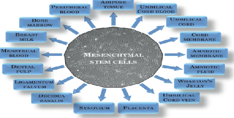
cell is implemented in the body, the complications of hazardous immune response can be reduced or avoided since it bypasses or adjusts in accordance with the donor's cellular ecology. Majority of this therapy is based on the use of bone marrow–derived mesenchymal stem cells (BM-MSC) with other sources include embryonic sources such as: synovium, umbilical cord, amniotic fluid and adult cells such as: dental pulp, periodontal ligament, adipose tissue, tendon and menstrual blood (Musial-Wysocka et al., 2019).
Figure 01: Different sources of Mesenchymal Stem Cells (MSCs) (Musial-Wysocka et al., 2019).
1.5 Advantages & Disadvantages of MSC therapy
One advantage of this therapy is that they don’t induce immunogenic reactions. As a result, no immunosuppressive drugs are needed during transplantation. MSC therapy induces this effect based on their immunomodulatory & immunosuppressive activity. They are able to sustain such effects either by suppressing development or initiation of various types of cells present in the immune system, which in turn are based on direct interaction with immune cells or by other indirect soluble factors (Musial-Wysocka et al., 2019).
However, it’s not without certain disadvantages. There are some risks that comes in the shape of carcinogenesis after stem cell has undergone transplantation. It’s because of their action of
6
proliferation for a long period of time and resistance to apoptosis (Bellagamba et al., 2016). Some factors for risk of carcinogenesis after mesenchymal stem cell transplantation are: the age of donor, the receiver’s tissue morphology, growth factor regulators expressed by receiver’s tissue and receptor control mechanisms involved at the target site. Furthermore, mesenchymal stem cells and its connection to tumorigenesis is also solid due to researches showing the incidence and formation of carcinoma-associated fibroblast (CAF) cells, tumor-associated fibroblast (TAF) cells and endothelial pericyte-like cells during MSC therapy under appropriate conditions. Also, genetic instability, along with chromosomal aberrations are observed when manipulations are simulated and applied for a long time in vitro cultures of MSCs (Barkholt et al., 2013).
Despite the serious side effects, in a wildly converse way, we can also observe a double nature in relation to their tumorigenicity. It’s because researches regarding MSCs has also shown that some factors secreted by these cells have established anti-carcinogenic properties. According to Clarke et al., (2015), when cancer cells derived from the breasts are cultured in MSC-conditioned medium, they have shown to exhibit a significant inhibition of tumor or cancerous cell migrations compared to cells primed in a standardized media. This antitumor action of MSCs may be the result of secretion by proteins, namely TIMP-1 and TIMP-2, which functions by blocking the action of the MMPs that are involved in this relocation processes of cancerous cells.
Similar blocking of cancer cell differentiation was also observed by Bruno et al., (2014), when a human hepatocellular carcinoma cell line (HepG2), a human ovarian cancer cell line (Skov-3) and Kaposi’s sarcoma cell lines were cultured together in the presence of BM-MSCs, which demonstrated a reduction of in vitro growth. Furthermore, micro vesicles separated from MSCs induced considerable reduction in tumor cell growth by retarding cell cycle advancement, facilitating apoptosis and necrosis of the tumor cells. All of these observed effects were validated
7
by in vivo studies in which cancer cell growth was retarded down significantly by the use of BM- MSC-derived MVs.
So, MSC therapy can be a good and efficacious form of treatment for osteoporosis because of it’s low immunogenicity, lack of side effects, easy availability from human sources and low incidence of loss of potency from preservation.
1.6 Aim & Objectives of the study
The main aim of this study is to review two specific methods of MSC therapy for osteoporosis.
First one is the tonsil derived mesenchymal stem cell therapy and the second one is the combination between MSC therapy and microRNAs. In doing so, we will briefly discuss these two methods of therapy and how they are conducted, statistical and biological evaluation of their potency and how they can be an effective alternative in osteoporosis treatment
8
Chapter 2 Methodology
This review paper on the application of mesenchymal stem cell therapy and miRNA for the treatment of osteoporosis has been performed based on recent and relevant research papers and articles from journals having high-impact factor. A comprehensive search has been performed through peer-reviewed journals, official reports, and articles. To enrich the review paper, basic and additional information have been collected from different books. Following search engines have been used to collect data for this paper- Research Gate, Google Scholar, Science Direct, PubMed, MDPI, Elsevier, etc. in which the major publications include- Nature, ACS (American Chemistry Society), AACR (American Association for Cancer Research), Molecular Cell, Cancer Cell, Journal of Molecular Biology, EXCLI, Springer, Journal of Medicine, Science, etc. In-depth screening of the journals followed by narrowing down to the most recent (within the last 5 years) and relevant ones was done to create an ideal quality review on the role of Mesenchymal Stem Cell (MSC) and miRNA therapy for the treatment of Osteoporosis.
9
Chapter 3
Pathophysiology of Osteoporosis 3.1 Osteoporosis
When talking about osteoporosis, it’s important to know how it occurs at first. According to numerous researches, it is well established within scientific consensus that osteoporosis mainly occurs when an imbalance or discrepancy occurs between the formation of bone generating osteoblast cells and bone resorbing osteoclast cells. In such cases, the formation and number of osteoblast cells are much lower compared to osteoclast, which results in brittle bones (Feng and McDonald, 2011). Furthermore, there are also reports about the influence of mesenchymal stem cells on osteoporosis. Because reports stated that since MSCs differentiates and in turn helps to synthesize osteoblasts, there is a positive complementary relationship between these two cell types.
So, the decrease in osteoblastic cells in osteoporosis means that there is decreased bone marrow MSC formation or there are problems associated with it. The BM-MSC mainly produces adipocytes instead of osteoblasts during osteoporosis (Lin et al., 2011).
Now, if we look at the pathophysiologic models of osteoporosis, the mostly researched and acceptable model throughout the years was concerned with that of endocrine mechanisms, for example: the effect of estrogen deficiency on secondary hyperthyroidism in senile populations, the effect of reduced dietary intake, for example the deficiency of vitamin D as a principal determinant of postmenopausal osteoporosis (Clarke and Khosla, 2010). Despite these pathophysiology, studies and researches performed throughout the years show that there are far more new and complex pathophysiological mechanisms available which shows a clearer picture to the onset of osteoporosis. Some newfound models of pathophysiology of osteoporosis are discussed below:
10
3.2 Osteoimmunology
As this name suggests, this model looks into the relationship between bone morphology and the immune system in the pathophysiology of osteoporosis. When we observe the cell responsible for bone resorption i.e osteoclast, we can see that it also acts as a source of immune cells, namely macrophages, dendritic cells and monocytes. Besides a common precursor, the immune and bone cells also have some transcription & signaling factors, chemokines in common between them (Tsukasaki and Takayanagi, 2019). So, it is not hard to imagine the pathophysiological effect that immune cells may have on osteoclasts. For example, a type of T cell, called CD4+ with the help of it’s subdivision called Th17, can start bone resorption performed by osteoclast by initiating the secretion of IL-17, which in turn that stimulates the expression of RANKL (Receptor Activator of Nuclear factor-kB ligand). RANKL is a potent signaling factor for osteoclast synthesis and thus helps to enhance the number of osteoclasts and thus makes osteoporosis a possible reality for patients.
Furthermore, if we observe the effect of regulatory T-cells, the effect of immune systems on bone homeostasis will be clear. Regulatory T-cells aside from regulating excessive immune response or reactions, also prevents excessive tissue damage by preventing inflammation by expressing a transcription factor called FOXP3. So, since no inflammation occurs, cytokines and interleukin expression are controlled and thus osteoclast generation by RANKL expression is also prevented (Bozec and Zaiss, 2017). This phenomenon has also been proven by experiment, where Zaiss et al., (2010), performed an experiment by transferring Treg cells into T-cell deficient mice. After the insertion, the mice showed enhanced bone mass with diminished number of osteoclasts. In another experiment, Foxp3 integrated into mice of transgenic nature were saved from ovariectomy (OVX) induced osteoporosis, thus supporting the idea that a Treg cell can induce the development of bone
11
mass just like estrogen (Zaiss et al., 2010). This result has also proved to be consistent with a report where it is stated that estrogen has been shown in vitalizing the growth and division of Treg cells (Tai et al., 2008).
3.3 Cellular Senescence
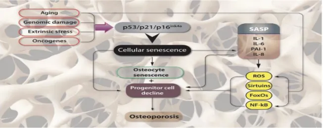
In biology, cellular senescence is a procedure where a cell’s longevity is activated by different types of stress, which in turn creates non-reversible cell cycle restriction and causes opposition to apoptosis (Samwald et al., 2020). Besides this typical characteristic, senescent cells also undergoes senescence-associated secretory phenotype (SASP) state where such cells excessively produce pro-inflammatory cytokines, chemokines and extracellular matrix-degrading proteins (Tchkonia et al., 2013). Coincidentally, in research by the same authors, it was revealed that the number of senescent cell’s number goes up during the natural aging timeframe, which has been shown to play one of the prominent part in age-associated tissue deterioration and the development of age- associated disease such as osteoporosis.
Furthermore, in another study by Farr et al., (2016), the researchers have found and observed that cells within the bone microenvironment becoming senescent with aging are B cells, T cells, myeloid cells, osteoprogenitors, osteoblasts, and osteocytes. Moreover, there were also the observed effect of escalating synthesis of key SASP factors. This confirms the effect of cellular senescence on osteoporosis as the osteoblast are rendered dysfunctional and non-differential due to it.
12
Figure 02: Pathophysiological effect of Cellular Senescence in Osteoporosis (Giovos, G., Yavropoulou, M.P. &
Yovos, J.G, 2019).
3.4 Microbiomes of Gastrointestinal Tract
This new model about the pathophysiology of osteoporosis discusses the possible role of GIT microbiomes on a patient’s bone health and it’s homeostasis. According to Behera et al., (2020), microbiomes not only influence the homeostasis of GIT tissues, but also tissues of tissues related to the synthesis and absorption of nutrients, bodily growth and homeostasis of the immune system.
Likewise, bone related diseases like rheumatoid arthritis have also been found to predispose changes in the composition and environment of GIT microbiomes (Behera et al., 2020).
We can understand this pathophysiological model of the “microbiome-bone” axis if we observe the effects of the GIT microbiome metabolism occurring in the human body. According to Rodrigues et al., (2012), the GIT microbiomes have shown to have the capability to impact the absorption of nutrients necessary for bone expansion such as calcium, phosphates etc. Thus the mineral density of the bones are influenced by these microbiomes. Sometimes intestinal pH controls the absorption of nutrients in our body, which again depends on the structure of the GIT microbiome. Also, the short chain fatty acids (SCFAs) produced as a product of microbial
13
fermentation of foods containing fibers might be able to play a crucial role in bone homeostasis.
In adults, it was found that various prebiotic diets which can be fermented to SCFAs were associated with an enhanced resorption of calcium (Whisner et al., 2014, 2016). Besides, studies have also found that SCFAs efficiently regulate osteoclast differentiation (Zaiss et al., 2019). For example, improvements in bone mass were observed in mice fed with SCFA enriched diet.
Moreover, in some experiments, it was found that in postmenopausal women, bone loss due to the occurrence of inflammation was prevented who had taken SCFA diet, which was possible in particular to the lower incidence of osteoclast growth and bone resorption (Lucas et al., 2018). In light of these findings, it can be elucidated that SCFAs as a GIT derived microbial metabolite has the ability to diffuse into the blood circulation and in turn can control and influence the composition body organs such as the organs of the skeletal system (Zaiss et al., 2019). Since GIT microbiomes can control immune functions to a certain extent, the effects of this microbiome on immune reaction, which in turn regulates the bone homeostasis, provide an insightful and essential correlation between the GIT and the skeletal system.
Cytokines that are vital on the presence of bones, released by immune cells in the gut or immune cells activated in the gut and then passing to the bone are a potential mechanism of action thought to be mediating this GIT microbiome-immune system -bone axis (Pacifici, 2018). As we discussed before in osteoimmunology, the immune cells of Th17 and Treg are suspected to render a principal part in this pathophysiological model. Notably equilibrium between Th17 and Treg cells was shown to be managed by GIT microbiomes (Dar et al., 2018). Also in this case, the role of promotion of growth and division of Treg cells is accredited to SCFAs (Zaiss et al., 2019). In another research, it was shown when parathyroid hormone (PTH) treatment is used to treat any disease of the bone, the osteoblast stimulating effects of PTH therapy depends on a form of SCFA
14
called butyrate, which is synthesized by the microbes living in the gut (Li et al., 2020). The butyrate, along with PTH, induces CD4+ T cells to change into Treg cells, which again vitalizes CD8+ T cells to synthesize Wnt10b (Li et al., 2020). Wnt10b acts as key to initiate the Wnt signaling in stromal or mesenchymal cells and osteoblasts. As a result, due to this initiating signal by Wnt10b, the bone forming osteoblast cells differentiate and thus promote bone formation (Monroe et al., 2012).
Lastly, when bone loss is sometimes initiated by PTH, it mainly happens due to the initiation of the formation of intestinal TNF+ and Th17 T cells. Such phenomenon occurs in reaction to the GIT microbiome and the joining of these cells to the bone marrow (Yu et. al., 2020).
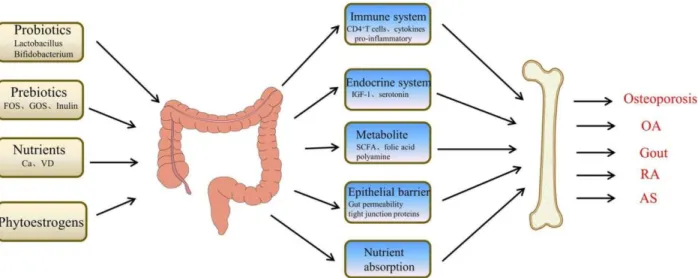
In light of the above discussion, it is clear that GIT microbiome and their metabolites, namely SCFAs, performs an important managing function in bone homeostasis and incidence of osteoporosis.
Figure 03: A schematic diagram showing how the GIT microbiome can possibly influence Osteoporosis (Peng et al., June 2021).
15
Chapter 4
Tonsil derived Mesenchymal Stem Cell Therapy 4.1 Background
In the field of osteoporosis treatment, this particular MSC therapy can be a great addition in treating the bone of older population, especially the women who have just come to their menopause state. It’s because this therapy is especially tested in this regard i.e, it’s pre-clinical testing is done by performing experiments on ovariectomized (OVX) mice. Ovariectomy is the surgery where the ovaries of the subjects are cut down. So, the female subject is deprived of estrogen and thus it becomes easier for the researchers to observe how tonsil derived MSCs (TMSC) can impart its therapeutic effects on mice suffering from hormone deprived osteoporosis.
4.2 Advantages of the TMSC therapy
Like most adult mesenchymal stem cells (MSC), the tonsil derived MSCs do not have adverse effects like cancerous growth or immune rejection. As a result, it has shown serious potential to be used for osteoporosis treatment. According to Kim et al., (2018), discarded or surgically removed tissues of tonsils can be a novel source of MSCs, which can release surface antigens of MSCs and can perform mesodermal proliferation as well as immunosuppression, a characteristic advantage of MSCs. Another added advantage of TMSC is that it’s expressions of MSC-specific surface markers have the ability to not undergo serious morphological changes for a considerable amount of time, thus they live long to give their therapeutic effects uninterrupted in the patient's body (Yu et al., 2014). Moreover, the high proliferative rate of TMSC also gives it an added benefit over other types of MSCs, specially observed in a study where it is demonstrated that the doubling
16
time of TMSC is 37.1 ± 3.4 hours, which is much higher than the bone marrow doubling time of 58.2 ± 2.3 hours (Janjanin et al., 2008).
4.3 Mechanism of Action of TMSC therapy
According to Kim et al., (2018) during their experimentation with TMSCs, they speculated that the therapeutic or bone regenerative effects of the TMSCs are due to the paracrine effect. Paracrine effect can be explained as a phenomenon where specialized donor cells, in this case TMSCs, stimulate the patient's cells to repair the bone tissue without interfering in the formation of the new bone tissue. Due to the donor cell’s stimulation and secretion of special growth factors, the patient's cells change their behavior and the signals are passed from one cell to another (Rogers, November 2012). Furthermore, another mechanism by which TMSC imparts its therapeutic action is through the release of substances like: micro vesicles, secretory factors and exosomes (Spees et al., 2016).
It tends to form a good microenvironment for the tissues because its transcriptome has the ability to produce more protein-binding proteins originated from outside the cell and molecules that can modulate immune system if we compare it to other MSCs (Cho et. al., 2017). Therefore, it can be said that the regenerative efficacy of TMSC is generated through its secretomes, which in turn enforces the differentiation of important bone cells like osteoblasts to develop bone mass.
4.4 Delivery method of the TMSC
During the TMSC therapy, the TMSCs are delivered or in surgical terms, “engrafted” in the site of damaged organs or tissue, in this case the bone tissues. But this procedure faces some challenges for the delivery. According to Kim et al., (2018), intra-bone marrow (IBM) or intra-tail venous injections that they have employed during their experimentation with osteoporotic mice, the TMSC had failed to give their desired therapeutic effects. It’s because systemically delivered MSCs face
17
interference from both blood circulation and the much-dreaded immune reaction. The intra-bone marrow (IBM) route is not exempt from this effect because of the presence of rich blood vessels in this site. The researchers also stated that administering a subcutaneous injection wouldn’t make much of an effect because there is an association of cell loss with this route due to the survival and retention time of stem cells being very low (Kim et al., 2018). So, in order to strengthen the stability of TMSCs in both systemic circulation and the subcutaneous route, a scaffold carrier can be used.
Scaffolds are generally formed of polymeric biomaterials, which provide the structural support for cell attachment, and thus ensure subsequent tissue development (Chang and Leong, 2008). These polymeric biomaterials can solve the problem of apoptosis and promote cellular functions by supporting and ensuring the proper transport of nutrients, waste products and small molecules, like secretory factors needed for healing of the bone (Yousefi et al., 2016). These beneficial effects of scaffolds are validated by numerous researches. For example, in a study by Park et al., (2018), it was shown that the if TMSC was delivered with Matrigel, a type of basement membrane matrix scaffold, then the formation of bone cell increased in an osteoradionecrosis induced rat model.
Furthermore, like Matrigel, TMSC prepared in poly (ethylene glycol)-poly(L-alanine-co-L- phenylalanine) thermogel also provided a better chondrogenic development (Park et al., 2014).
Another research by Moon et al., (2016), showed that TMSC can give well developed osteogenesis in mesocrystals (4–8 μm) of thermogels formed of calcium phosphate and polypeptide as well.
For our review, we looked at the TMSC-immersed gelatin hydrogel (TMSC-GHH) delivery system administered by Kim et al., (2018) on their research about osteoporosis induced by ovariectomized (OVX) mice. The reason the researchers sought gelatin-based hydrogel is due to its extraordinary biocompatibility, biodegradability and non-immunogenicity. Hydrogels have a semblance of a gelatin-like bio-material, which composes of three-dimensional networks of polymers of
18
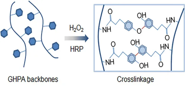
hydrophilic nature. This hydrogel is mainly administered in the site of fixation by horseradish peroxidase (HRP)-catalyzed cross linking for it’s renown of being an efficient medium for cell delivery (Lee et al., 2014). Another reason why this hydrogel is most suitable is because it’s physicochemical properties can be modified, such as: gelation time, matrix strength and degradation rate. GHH also has the advantage of not starting any inflammatory activity when it is degraded on the presence of proteolytic enzymes.
In conclusion, a TMSC engulfed or fixed in a biological scaffold of gelatin-hydroxyphenyl propionic acid (GHPA) hydrogel (GHH) is suitable for subcutaneous administration because of greater biocompatibility, non-immunogenicity and controllability.
Figure 04: GHPA polymers backbones are being enzymatically crosslinked to form GHH scaffold, where TMSC are then fixed in the hydrogel meshwork (Kim et al., 2018).
4.5 Preparation of Preclinical mouse model
In this review, in order to focus on the therapeutic efficacy of TMSC treatment for osteoporosis, we have reviewed the research article, “Tonsil-derived mesenchymal stem cell- embedded in situ cross linkable gelatin hydrogel therapy recovers postmenopausal osteoporosis through bone
19
regeneration” by Kim et al., 2018. At the beginning of the experiment, Kim et al., had procured 50 female OVX mice from the Institute of Cancer Research (ICR), each weighing about an average of 32.4 ± 2.44g. These mice had undergone ovariectomy at 8 weeks of age and were prepared for a 03-month long experiment. After that, the 50 mice were randomly divided into six groups:
Untreated (number= 10), Estrogen (number = 10), TMSC injection applied once (TMSC/1×; n = 5), TMSC injection applied twice (TMSC/2×; n = 5), TMSC-GHH injection applied once (TMSC- GHH/1×; n = 10), and TMSC-GHH injection applied twice (TMSC-GHH/2×; n = 10). Besides these six experimental groups, there were five female mice of ICR that didn’t undergo OVX (non- OVX), was used as controls. The ovariectomized mice were given a diet free of calcium (80 mg calcium/kg diet) in order to make the mice more prone to osteoporosis. The mice which were not ovariectomized, were given a standardized chow diet and all mice were given free rein to food and water. The reason TMSC and TMSC-GHH groups were given injection twice was to determine an effective dosage regimen. At the 3 months long experiment, the blood of OVX induced mice were taken from the vein of the neck of each mouse. The collected blood sample or more specifically the serum were taken to determine serum osteocalcin (OCN) measurement. The reason for measuring serum OCN level is because it is a valid marker of the bone turnover when bone resorption and formation are working cooperatively. Besides OCN, serum alkaline phosphatase (ALP) was also measured as this compound is a known biomarker of bone formation (Kim et al., 2018).
Now, for the experiment to commence, each group was treated differently based on their roles or the type of group they were in. For example, the Estrogen group received five times per week via intraperitoneal injection 17β-estradiol (1.0 mg/mouse), with a volume of 300 μL/injection. For the TMSC and TMSC-GHH groups, mice were first anesthetized by intraperitoneal injection with a
20
mixture of Zoletil (0.25 mL/mouse) and Rompun (0.25 mL/mouse). After anesthesia, TMSC, in the form of adherent mononuclear cells, consisting of 1×10⁵ cells/300 μL/mouse were injected in their subcutaneous region of the back. The groups that were injected once (TMSC/1× and TMSC- GHH/1×) were given injection only at the early stage of treatment, while the groups that were injected twice (TMSC/ 2× and TMSC-GHH/2×) were given injection a second time at 1.5 months after the initial administration of injection. Lastly, among the untreated ovariectomized mice group, none were given treatment (Kim et al., 2018).
4.6 Therapeutic & Biological analysis of the TMSC-GHH 4.6.1 Serum Osteocalcin (OCN) level
Figure 05: Comparison of serum OCN levels & bone condition between OVX and Non-OVX mice (Kim et al., 2018).
The sole reason for calculating the level of serum OCN was due to the fact that it is an essential biomarker for osteoporosis, meaning that it can rise in osteoporotic patients. Such rise in OCN level is also seen in the experiment of Kim et al., where the average initial OCN level of 14.8 ± 3.63 ng/mL rose to 48.9 ± 18.46 ng/mL (Fig 5) after 3 months in the experimental mice. Such significant rise of OCN level difference between baseline and non-ovariectomized controls shows us that osteoporosis occurred as intended. The serum osteocalcin level of ovariectomized mice was
21
seen 1.75 times higher than that of non-ovariectomized mice after three months of surgery.
Furthermore, by using microCT analysis of the hindlimb at necropsy stage, it confirmed the induction of osteoporosis as seen from the figure where femoral head trabeculae of ovariectomized mice had more porosity and cavity in their bone microarchitecture compared to other groups of mice (Fig 5).
4.6.2 The stability & efficiency of the GHH
Figure 06: The stability & longevity of GHH while incorporating TMSC for 03 months (Kim et al., 2018).
With the help of LIVE/DEAD1 assays, Kim et al., has proved in terms of quality and quantity that nearly every TMSC were exceedingly stable and flourishing within each GHH respectively (Fig 6b). The stern preservation of the spindle shape of the TMSCs and their increasing cell number proves that GHH had facilitated a suitable environment of proliferation for TMSCs. It all can be tied down to the fact that as a component of extracellular matrix (ECM), gelatin has proved to have the ability to support cellular adhesion, proliferation and dispersion by its direct contact with the mesenchymal cells (Hoang et al., 2016). Furthermore, the porosity and mesh like structure of the
22
GHH has given substantial support for transporting nutrients to the cells for it’s vitality. Also, in (Fig 6c) Kim et al., found that at the site of injection i.e the dorsum where TMSC-GHH was implanted, the gel successfully retained its shape and presence for the whole three months long experimental period (Fig 6d). In the (Figure 6d), we can observe that there was no sign or any incidence of inflammation or necrosis. This observation proves that TMSC-GHH is perfectly biocompatible and may not cause dismissal by the immune system.
4.6.3 Therapeutic efficacy of TMSC
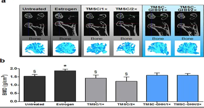
Figure 07: Micro CT images and bone mineral density of femoral head trabeculae at the experimental endpoint (Kim et al., 2018).
In order to measure the magnitude of beneficiary effects of TMSC-GHH on osteoporosis, Kim et al., separated the hind limbs from both the treated and untreated mice and they underwent micro CT analysis. Fig 7a displays the hind limb cross-sectional image and three dimensional images of femoral head trabeculae was also shown. The results were satisfactory, because the femoral heads
23
of osteoporotic mice that were not treated with TMSC-GHH had a porous structure and with the help of estrogen treatment used as positive control, the bone recovery was achieved. Furthermore, the treatment with TMSC-GHH instead of TMSC alone appears to be more efficacious therapeutically as the bone improvement by TMSC-GHH is quite higher than TMSC alone. The reason TMSC alone had such a low therapeutic effect maybe due to disintegration of TMSCs in the systemic circulation. Moreover, compared to the estrogen group, the untreated control group also didn’t show a notable growth in bone mass density (Fig 7b). This observation was not statistically much different if we observe the TMSC-GHH groups, but on the other hand was considerably higher than that of untreated and TMSC-only treated mice. In conclusion, on the basis of figures of bone development samples shown by Kim et al., it can be stated that these results put forward that the BMD growth surge observed in TMSC-GHH-treated mice can be regarded significant in terms of clinical property. Lastly, it is also noteworthy that the bone mineral density after 3 months of treatment didn’t vary between the TMSC-GHH/1× and TMSC-GHH/2× groups, despite the difference of dosage. So, it suggests that the magnitude of dose is not so an important factor here.
4.6.4 Serum OCN, ALP & Total Calcium levels
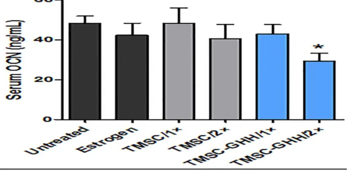
In this segment, we will discuss various improvements that have been observed in the OVX test subjects’ body after 03 months. Kim et al., had collected serum samples, particularly serum OCN, alkaline phosphatase and total calcium from the OVX mice three months after initial treatment.
Firstly, if we observe at the OCN levels, it’s levels were reduced very well in the TMSC-GHH and estrogen groups compared to the untreated OVX mice (Fig 08). This event helped to understand the bone turnover imbalance in postmenopausal osteoporosis because the concentration of serum OCN is always much higher in osteoporotic patient than in non-osteoporotic bones. In the initial
24
stage of the experimental treatment, in accordance with the known increase of OCN, OCN levels in the serum had been higher in the ovariectomized mice compared to non-ovariectomized ones (Figure 05). Its because rise in osteocalcin are known to be balanced when therapeutic recovery of bone metabolism occurs by the TMSC-GHH, the findings suggested by Kim et al., stays true that bone density has returned to normal condition in the estrogen and TMSC-GHH groups. Another interesting finding this research paper showed is that two times injection of TMSC-GHH was effective in diminishing the ovariectomy-related rise of osteocalcin.
Figure 08: Serum osteocalcin (OCN) level in OVX mice after 03 months (Kim et al., 2018).
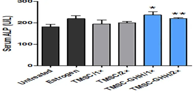
The next important biomarker measurement was that of the level of alkaline phosphatase (ALP) because it is a well known marker for bone formation in living organisms. In the results that Kim et al., got shown that after 3 months of treatment, the estrogen group showed a not very important rise in ALP levels compared to untreated mice. But quite the opposite was found in the case of TMSC-GHH/1× and TMSC-GHH/2×, where both types presented with significantly elevated
25
concentrations of alkaline phosphatase levels (Figure 09). So, it can be stated that TMSC-GHH based treatment can kick start bone foundation regardless of dose frequency and its therapeutic results were more strong than that of estrogen treatment.
Figure 09: Serum Alkaline Phosphatase (ALP) level in OVX mice after 03 months (Kim et al., 2018).
It is well known that every treatment or drug has some side effects, be it mild or severe, associated with it. Mesenchymal stem cells are no exception. Since TMSC induces bone formation by differentiating osteoblast and other mineral absorption like calcium, the incidence of hypercalcemia is a likely event. So, in order to identify the appearance of unfavorable interference in blood calcium levels, Kim et al., had performed total calcium level measurement of the serum at the very end of the experiment with great results. Because it was observed that none of the OVX mice displayed any significant fluctuation from the standard calcium level (Figure 10).
26
Figure 10: Serum total calcium level in OVX mice after 03 months (Kim et al., 2018).
4.6.5 Biocompatibility of the GHH scaffold
As with any other drugs or therapy, the effect of these treatments on the body, especially in the kidney and liver is always present to some extent. Because these organs can metabolize and excrete the drugs that enter our body. Basically, the safety parameter of a drug depends on how well the kidney and liver adjusts with the drug. For this reason, Kim et al., had also observed how the OVX mice reacted to TMSC-GHH. In their study, at the closure of the experiment, all mice
Figure 11: Macroscopic morphology of postmortem kidneys and liver obtained from each OVX group after euthanization (Kim et al., 2018).
were euthanized and their internal organs were harvested for examination. In their observation, Kim et al., found that the livers and kidneys did not lay out any morphological defects that could
27
be characteristic sign of any pathological conditions. Say for example, there was no appearance of hepatic/nephrotoxicity or hepatitis/nephritis, such as: nodules, enlargement, swelling, discoloration or any other variations from regular condition (Figure 11). Besides macroscopic observation, a quantitative observation was also performed by Kim et al., which includes taking the weight of kidneys and liver from each mouse and then adjusting by body weight. After that, each value was measured for any alteration that could indicate toxicity present on these organs stated above (Michael et al., 2007).
Figure 12: Mean body weight, adjusted kidney mass & adjusted liver mass of each OVX mice group three months after first treatment (Kim et al., 2018).
In the end it can be seen that, at the end of the experiments, the ovariectomized mice of all experimental and control groups were more or less had familiar or same features to each other regarding body weight, kidney and liver masses (Fig 12).
Since the kidney and liver masses, as well as their outer morphology remained persistent in groups treated by TMSC-GHH, it can be stated that TMSC and GHH are tolerated and biocompatible with normal body functions of metabolism and excretion (Kim et al., 2018).
28
4.6.6 Potential for Visceral fat reduction
Among bone regeneration abilities, TMSC has also shown potential to diminish visceral fat.
Visceral fat makes 1/10th of all bodily fats of the human body, which is situated and stored deep inside the belly and covers around the organs, namely liver and intestines (Health Direct, May 2021). This type of fats, if uncontrolled, releases toxic substances. Such toxic secretion can also
Figure 13: The Mean visceral fat mass and mean body weight adjusted visceral fat mass of each OVX mice groups after 03 months (Kim et al., 2018).
cause osteoporosis because there are indications that visceral fats can secrete proinflammatory cytokines that can worsen the osteoporosis of suffering patients (Caffarelli et al., 2014). For this reason, Kim et al., in their research they also wanted to know whether TMSC can elucidate visceral fat reduction or not. Surprisingly, TMSC indeed shows this effect. In their research, after euthanization of OVX mice, Kim et al., obtained visceral fats from each mouse and took their weights. It was found that TMSC-GHH/2× mice had lower visceral fat in comparison to TMSC only treatment. After that, Kim et al., adjusted the values of visceral fat mass by the average body weights to adjust the yield of visceral fat mass values (Figure 13). After that, both TMSC-GHH
29
treatment groups, one time and two times, still had significantly lower visceral fat than TMSC only treatment.
30
Chapter 5
Mesenchymal Stem Cell & microRNA Combination Therapy 5.1 Introduction
Before we start discussing this new form of osteoporosis treatment, it is necessary to know what a microRNA or miRNA is. According to Bartel (2018), microRNA can be defined as this minute single-stranded non-coding RNA molecule found in living organisms whose main functions is to perform post-transcriptional regulation of gene expression and RNA splicing. A miRNA is composed of 22 nucleotides and imparts it’s function by base-pairing with complementary sequences within messenger RNA or mRNA molecules (Bartel, January 2009). The way mRNAs are silenced or inactivated by miRNA is either by cleaving of the mRNA strand into two pieces or by destabilizing the mRNA through shortening of its poly(A) tail (Fabian et. al., 2010).
In recent studies, it has been shown that mesenchymal stem cells have been responsive to be controlled and modulated to some extent by microRNAs (miRNAs). MiRNA regulates genes associated with differentiation of bones after the transcription process. But where does this miRNA come from ? MiRNAs are synthesized from previously long primary transcripts which then enter the nucleus of a cell for processing. From there it goes further processing in the cytoplasm of particular cells to build small non-coding RNA (Pasquinelli et al., 2005). The regulation of tissue or more specifically protein synthesis by miRNA is done when it’s sequence becomes complementarity to a mRNA and based on the nature of that complement, miRNA influences either inhibition or degradation of the mRNA after the transcription process. As a result, a mesenchymal stem cell differentiates into an osteoblastic cell if the mRNA gives cell progenitor
31
transcription or else doesn’t induce osteogenesis if the transcription is of inhibitory signal (Budd et al., October 2017).
5.2 Potential advantage of incorporating miRNA in MSC
Since miRNAs have the potential ability to regulate translation, it can thus be used to control cellular processes, such as differentiation. In fact, numerous miRNAs have been identified in recent research which influences the incidence of chondrogenesis and osteogenesis (Fang et al., 2015). In simple words, stem cells could be utilized in the revival of skeletal tissues in combination with miRNAs to build up the growth of transplanted mesenchymal stem cells in the direction of osteogenic origins. Usage of miRNAs could prepare transplanted stem cells for direction toward a desired cell fate, such as: enhancing stem cell differentiation, adhesion and longer residence time in the implanted site. Not only could this novel therapy prime the revival of bone tissues, but if applied in the early stage of treatment, it could prevent the incidence and advancement of osteoporosis (Budd et al., October 2017).
A complete and thorough understanding of miRNA expression and the part that these molecules play in control of gene expression during formation of stem cells gives us a clear comprehension of molecular mechanisms regulating stem cell growth (Budd et al., October 2017). So, if we identify the correct miRNAs which controls the fate of stem cells, then it could be applied to start and increase growth of stem cells. Thus, this system will provide a novel method of cell-based therapy. MiRNAs could strengthen the condition of transplanted stem cells at defected sites of the bone to revive osteoblasts. It can be done by the application of miRNA mimics or miRNA inhibitors, where these molecules then drive mesenchymal stem cell differentiation and towards the desired lineage of osteoblasts.
32
5.3 Examples of MiRNA in Osteogenic differentiation of MSC 5.3.1 miR-23a
This particular miRNA has shown to be down-regulated during osteogenic differentiation of human bone marrow mesenchymal stem cells (hBMSCs). In biology, downregulation is the process by which a cell reduces its quantity of a cellular component, such as RNA, protein or growth factors in response to an external stimulus.
According to Li et al., (2016), miR-23a was shown to directly target the 3’UTR (Untranslated Region) of LRP5 (Low-density lipoprotein receptor-related protein 5). LRP5 is one of the most important part of the Wnt signaling pathway, because Wnt signaling is in charge of bone formation by signaling the proliferation of osteoblasts. During normal circumstances of osteogenesis, miR- 23a is likely to be downregulated and thus enables derepression of LRP5 expression. Derepression is the activation of an operator gene, in this case LRP5 by the deactivation of a repressor gene.
After this derepression, Wnt signaling occurs subsequently to direct osteogenesis (Li et al., 2016).
Furthermore, in order to understand the effect of miR23a clearly, it was found that by the use of miR-23a mimic, the overexpression of miR-23a occurred, which downregulated osteogenic differentiation. This was proved by taking the serum samples of alkaline phosphatase (ALP), osteopontin (OPN), RUNX2 and IBSP mRNA, which showed each of these components' compositions were decreased a lot. ALN and OPN are the most important elements for bone formation and remodeling respectively. So, the opposite effects is seen when with the help of anti- miR-23a resistance to the production of endogenous miR-23a were done and resulted in up regulation of ALP, RUNX2, OPN and IBSP messenger RNAs, which in turn improved osteoblast
33
synthesis. In conclusion, during osteoporosis treatment, the level of endogenous miR-23a should be reduced or limited with a miR-23a inhibitor (Li et al., 2016).
5.3.2 miR-29a
This miR-29a exactly shows the opposite effect of miR-23a, meaning it up-regulates osteogenic differentiation. This phenomenon was observed in an experiment upon human fetal osteoblast cell line (hFOB1.19) (Kessler et al., 2010).
The miR-29a also similarly targets the 3’UTRs, but this time it is the negative modulators of Wnt signaling: Dkk1, Kremen2 and sFRP2. While osteogenesis, miR-29a mostly up-regulates and inhibits the negative regulators of Wnt signaling. So, it kind of indirectly promotes osteogenic differentiation (Kessler et al., 2010).
Now in order to be sure about how well the miR-29a gives osteogenic potential, it’s expression was inhibited by a miR-29a inhibitor inside hFOB1.19 cells. The results were the down-regulation of osteogenesis, which was observed by low levels of biomarkers like that of OCN and ALP mRNA inside the hFOB1.19 cells. Also, further overexpression using miR-29a mimic resulted in the up-regulation of OCN and ALP mRNA. So, enhancement in bone MSC can be achieved by increasing the levels of miR-29a by miR-29a mimic.
5.3.3 miR-138
According to Eskildsen et al., (2011), this particular miRNA’s down-regulation was observed during osteogenesis of human MSCs (hMSCs). Furthermore, MiR-138 elucidated the 3’UTR of PTK2 directly, which encodes focal adhesion kinase (FAK) (Eskildsen et al., 2011). When
34
osteoblast differentiation occurs, it is inhibited by the action of focal adhesion kinase (FAK). The FAK activates Grb2-Sos-Ras pathway and then this pathway induces ERK1/2, resulting in down streaming of genes affecting osteogenesis (Salasznyk et al., 2007).
The anti-osteogenic potential of innermost miR-138 in hMSCs is tested by using anti-miR-138.
When anti-miR-138 was used in hMSC, an increase in biomarkers of osteogenesis like: OCN and ALP mRNA levels occurred. On the other hand, excessive expression of miR-138, using pre-miR- 138, was observed to greatly diminish bone tissue proliferation. Moreover, hMSCs without a miR- 138 when loaded onto a bio scaffold and implanted subcutaneously in mice, showed increased bone growth with up-regulation of ALP and OCN mRNA. So, the recommended technique is to reduce miR-138 levels inside the cell with the help of a miR-138 inhibitor when this miRNA is used for osteoporosis treatment (Eskildsen et al., 2011).
5.3.4 miR-346
Like any pro-osteogenic miRNA, miR-346 also had shown to be up-regulated at the time of osteogenic formation of human bone marrow mesenchymal stem cells (hBMSCs). Here, miR-346 targets the 3’UTR of GSK-3β, a type of negative regulator of Wnt signaling, a pathway for osteogenic differentiation. The method to determine whether Wnt signaling activates is seen when increased gathering of β-catenin occurs inside nucleus of the cell. This typically occurs during the overexpression of miR-346. The negative regulators of Wnt signals are impeded by the upregulation of miR-346, occurring specifically during osteogenesis. Thus, this miRNA indirectly promoting osteogenic differentiation (Wang et al., 2013).
As usual, in order to test the osteogenic potency, miR-346 was overexpressed using a miR-346 mimic, ensued in increased osteogenic division backed by the evidence of increased OPN, ALP
35
and RUNX2 mRNA in the blood serum sample. Conversely, the inhibition of the anti-miR-346 usage gave lowering of osteogenic proliferation as an outcome, shown by decreased levels of osteogenic bio markers, like: mRNA expression, matrix mineralisation and ALP activity. For therapeutic purposes, increased miR-346 levels should be achieved by using and utilizing a miR- 346 mimic (Wang et al., 2013).
Table 01: A summary of different microRNAs for osteoporosis treatment (Budd et al., October 2017).
Name of microRNA (miRNA) Mechanism of Action Potential of use in stimulating osteogenesis
1. miR-23a Induces osteogenesis during
downregulation, which causes derepression of LRP5 expression and then activating Wnt
signaling.
By decreasing miR-23a level by miR-23a inhibitor.
2. miR-29a Directly impacts the negative
controls of Wnt signaling, thus freeing the Wnt pathway to lead osteogenesis.
With the help of miR-29a mimic, increasing the content of miR-29a.
3. miR-138 Negatively impacts osteogenesis
by inducing Grb2-Sos-Ras pathway to downregulate genes helping with osteogenesis.
By decreasing miR-138 level by miR-138 inhibitor.
4. miR-346 Helps in osteoporosis by
inhibiting negative modulators of Wnt signaling.
By enhancing miR-346 level by miR-346 mimic.
36
5.4 Delivery method
5.4.1 The use of HA/TCP scaffold
As we have discussed before, simply injecting the miRNA in the MSC site would not give the sufficient or desired therapeutic effect, because the microRNA would be dissolved in the systemic circulation, interact with undesirable mRNA and lastly may undergo enzymatic digestion. For this reason, a biological scaffold, be it hydrogel, is badly needed for optimum delivery. This system was supported by Budd et al., (2017) In their review, where two miRNAs, miR-138 and miR34a, was enclosed in a hydroxyapatite/tricalcium phosphate (HA/TCP) scaffold and was then applied to the localize stem cells by subcutaneous administration. Furthermore, according to Chen et al., (2014), a similar approach was used to study miR-34a. In their research, Chen et al., got hMSCs transfected with pre-miR34a, anti-miR-34a and control miRNA and then delivered it to a HA/TCP scaffold. After that, the scaffold was implanted into an immunocompromised mouse by subcutaneous administration. Installment of the scaffold of hMSCs transfected with anti-miR-34a resulted in greater than 3.5-fold increase in osteogenesis, because miR-34a was a negative regulator of osteogenesis. Moreover, Eskildsen et al., (2011) have used lipofectamine, a transfecting agent, to transfect pre-miR-138, anti-miR-138 and control miR into hMSCs. A transfecting agent helps to artificially introduce nucleic acids, DNA or RNA, into the cells in question. Moving on, in their research it was found that installation of the scaffold comprising hMSCs transfected with anti-miR 138 resulted in a 2.2- fold increase in bone formation. While, for hMSCs transfected with miR-138 mimic resulted in a 6.7-fold reduction in bone tissue synthesis, reinforcing the observation that miR-138 is not a positive controller of osteogenic formation (Eskildsen et al., 2011).
37
5.4.2 Photoactivation by UV light
In order to successfully deliver miRNA to the desired site in the mesenchymal stem cells, it is important to localize or home the miRNA to the desired cell site. This helps to minimize any miRNA deviating from their destined target site. For solving this problem, Qureshi et al., (2013), had developed a unique delivery system, where photoactivation of nanoparticle conjugated miR- 148b can release the miRNA in the desired site. The reason miR-148b has been selected for this research is because of its previously reported property of up-regulation of osteogenic proliferation in human mesenchymal stem cells (hMSCs) (Eklund et al, 2009). The conjugate is non-toxic in nature and can remain inert until provocations of photoactivation by UV light occurs. The reason UV light can impart this effect was confirmed by a surge in ALP and OCN concentration in photoactivated human adipocyte derived stem cells (hADSCs) compared to cells not treated by UV rays. Moreover, the particular usage of nanoparticle conjugated miR-148b resulted in the precise fixing or homing of miR-148b to inside the cellular compartments of hADSCs without potentially damaging chemical-based methods of transfection stem cells (Budd et al., October 2017).