R E S E A R C H A R T I C L E
Histomorphometric Evaluation of Allogeneic Transplantation of Bone Marrow
Mesenchymal Stem Cells in Azoospermic Mice Model
Ahmad Mozafar
1,2, Davood Mehrabani
3,, Akbar Vahdati
1,2, Ebrahim Hosseini
1,2,
Mohsen Forouzanfar
41Department of Biology, Fars Science and Research Branch, Islamic Azad University, Fars, Iran 2Department of Biology, Shiraz Branch, Islamic Azad University, Shiraz, Iran 3Stem Cell Technology Research Center, Shiraz University of Medical Sciences, Shiraz, Iran
4Department of Biology, Marvdasht Branch, Islamic Azad University, Marvdasht, Iran Corresponding author. E-mail: [email protected]
Received date: Oct 31, 2017; Revised date: Apr 27, 2018; Accepted date: May 15, 2018
B
ACKGROUND: Stem cell-based therapy is one of the newest and evolving techniques in reproductive medicine. The aim of this study was to investigate the effect of allogeneic bone marrow mesenchymal stem cells (BM-MSCs) transplantation on the testis of busulfan induced azoospermia in Balb/C mice.METHODS: Eighteen adult Balb/C mice were divided into three equal groups including control, busulfan and busulfan+cell therapy (busul+CT). For induction of azoospermia, busulfan and busul+CT groups received two injections of 10 mg/Kg of busulfan intraperitoneally with 21 days interval. In the cell therapy group 35 days after the last injection of busulfan, cluster of differentiation (CD)90+/ CD34-/CD45- BM-MSCs were injected into the efferent duct of testis. Eight weeks after the BM-MSCs therapy,
mice were sacrificed and tissues were taken for histological
and histomorphometric evaluations.
RESULTS: In busul+CT group, cellular and total diameters
and cellular and cross-sectional areas significantly increased
in comparison to busulfan group (p˂0.001), but there were
no significant differences between busul+CT and control
group (p˃0.05). Numerical density and tubular count per area
unit in busul+CT and control groups were significantly less
than busulfan group (p˂0.001), but there were no significant
difference between busul+CT and control group (p˃0.05).
The luminal diameter and area showed no significant change
in all groups (p˃0.05). In busul+CT group, spermatogenesis
index significantly increased when compared to busulfan
and control groups (p˂0.001 and p˂0.05, respectively).
CONCLUSION: Histomorphometric findings showed
CD90+/CD34-/CD45- BM-MSCs transplantation on the
testis of busulfan-induced azoospermic in Balb/C mice recovered spermatogenesis.
KEywORDS: mesenchymal stem cell, cell therapy, azoospermia, busulfan, mouse
Indones Biomed J. 2018; 10(2): 171-8
Abstract
Introduction
Nowadays one of the problems of human society is the
infertility problem. Approximately 10 to 15 percent of infertility in men is due to obstructive or non-obstructive azoospermia.(1) Many efforts are being made today to treat azoospermia, especially its non-obstructive type.(2) Cancer is one of the most common diseases around the world, and
in many cases chemotherapy is used to treat it with drugs such as busulfan.(3) Infertility resulting from the use of drugs such as busulfan in the chemotherapy of people with
cancer is considered to be a significant concern in medical
circles.(4) Busulfan (1,4-Butanediol dimethanesulfonate)
is an anti-neoplasia and DNA alkylated drug used to treat
After treatment with busulfan, the most important factor in the degeneration of germ cells is the elimination of the balance between reproduction and apoptosis and germinal epithelium. Animal experiments indicate the ability of stem cells to restore fertility after chemotherapy in animals.(6) Various sources of use for stem cells are used, including embryonic stem cells or pluripotent stem cells, but the use of mesenchymal stem cells (MSCs) is of great interest because of its ease of access and working with it. MSCs can be obtained from various sources such as bone marrow, adipose tissue, dental pulp, and menstrual blood, all of which have a high ability to differentiate into different tissues.(7,8)
MSCs have been shown to secrete cytokines and
growth factors that have anti-inflammatory,
immune-modulating, anti-apoptotic, and proliferative effects.
Some reports confirm the positive effects of MSCs on the
process of repairing body tissues.(9) Meanwhile, bone marrow MSCs (BM-MSCs) have been considered by many researchers because of their high ability to differentiate into a variety of cell lines.(10)
The use of BM-MSCs, due to the possibility of proper extraction and proliferation, as well as the ability of the differentiation of this group of cells into different tissues, has made them an appropriate candidate for cell therapy. (2,5) In this study, the effect of transplantation of cluster of differentiation (CD)90+/CD34-/CD45- BM-MSCs on the treatment of busulfan induced azoospermia in Balb/C mice was investigated.
Methods
Animals
Eighteen male adult Balb/C mice (30±5 g), were provided from Laboratory Animal Center of Shiraz University of Medical Sciences. The animals were kept at controlled temperature (22±2°C), humidity (55±5%) and lightening (12 hours light/dark; from 07:00 a.m. to 19 p.m.) They had free access to food and water. The study was undertaken based on rules and regulations approved by the Ethics Committee for Animal Research at Islamic Azad University of Shiraz, Iran (Ethical code no: IR.Miau 1396.602).
Experimental Design
The mice were randomly assigned into three equal groups of control, busulfan and busul+CT (n=6 in each group). In control group, no busulfan was administered. In busulfan and busul+CT groups, two doses of 10 mg/Kg
Busilvex® busulfan (Pierre Fabre Medicament, Boulogne-Billancourt, France) with 21 days interval were injected intraperitoneally for induction of azoospermia. Based on previous studies, the dose of busulfan was designated in
order to induce azoospermia mice model.(11,12) In the first of experiment, the first dose of busulfan (10 mg/Kg) was
injected. In busulfan and busul+CT therapy groups and 21 days later the second dose of busulfan (10 mg/Kg) was also done, intraperitoneally. In busul+CT group after 35 days the second injection, CD90+/CD34-/CD45- BM-MSCs were injected into the efferent duct of both testes and 8 weeks
after the BM-MSCs therapy all groups were sacrificed for testes removal. On the day of sampling, the sacrifice
was undertaken using ether and cervical dislocation. For histomorphometric and histological evaluations, the
removed testes were fixed in 10% formalin buffer.
Stem Cells Extraction and Culture
Stem cells were isolated from femurs of one donor euthanized Balb/C mouse. In sterile condition the two ends of the bone were cut and injected into the Dulbecco’s
modified eagle medium (DMEM) without fetal bovine
serum (FBS) and penicillin-streptomycin antibiotics. Bone marrow was collected and then, the falcon contains bone marrow cells centrifuged at speeds of 1200 rpm for 5 minutes, after that, the supernatant phase was removed by pipette and the DMEM medium was added to the cells in the falcon. Then cell suspension was transferred to 25 cm2 flask containing DMEM medium with 10% FBS and
1% penicillin-streptomycin antibiotic. And flask was
incubated at 37°C and standard humidity and 5% carbon dioxide concentration. The culture medium was changed
after the first 24 hours and then every 72 hours under
sterile conditions. After increasing the density of the cells
adhering to the flask to 80%, the cells were passaged using
trypsin enzyme, adding the trypsin enzyme for 3 minutes;
the cells sticking to the flask floor were separated. Then,
to neutralize the effect of trypsin enzyme, 2 mL of culture
medium containing 10% FBS was added to the flask. Cells isolated from the medium were called the first passage, and
these actions continued until the third passage.(7)
MSC Characterization: Real Time-Polymerase Chain Reaction (RT-PCR) Evaluation
To confirm the presence of bone marrow mesenchymal
stem cells and the absence of hematopoietic stem cells in this study, RT-PCR was used. In this regard, the presence
of CD90, which is a specific protein of mesenchymal stem
specific proteins of the hematopoietic stem cells were
investigated.
Cells from the third passage were used. In the first step, RNA was extracted by column method using kit. In the second step, cDNA was constructed in reverse transcription
reaction. Then PCR in thermal cycler machine using
200μM dNTPs, 2.5μM cDNA, 0.5 μM MgCl2, 25 μLTaq DNA polymerase, PCR buffer and forward and reverse
primers CD34,CD45 and CD90 was performed (Table 1). The microcentrifuge tubes containing the resultant mixing were transferred into thermocycle. Thirty thermal cycles were performed as follow: 5 minutes at 95°C for the initial denaturation and 30 seconds at 95°C for denaturation were performed.
Osteogenic and Adipogenic Differentiation
After achieving 80% confluence, the CD90+/CD34-/
CD45- BM-MSCs were cultivated in the osteogenic and
adipogenic differentiation medium. For osteogenic medium,
DMEM with 15% of FBS, 200 μM L-ascorbic acid (Wako
Chemicals, Richmond, VA, USA), 100 nM dexamethasone (Sigma-Aldrich, St Louis, USA), 10 mM glycerolphosphate (Sigma-Aldrich) and for adipogenic medium DMEM with
15% of FBS, 200 μM indomethacin (Sigma-Aldrich), and 100 nM dexamethasone, 100 μM L-ascorbic acid
and 0.2 mM L-glutamine (Sigma-Aldrich) were used. Differentiation medium was changed twice a week for 21
days. Before staining, the cells were fixed for 10 minutes
by 10% formalin solution. Alizarin Red staining was used
to indicate the calcified extracellular matrix and osteogenic
differentiation and for adipogenic differentiation Oil Red
O staining was applied that specifically stains lipid
droplets.
Surgical and Transplantation Procedures
For CD90+/CD34-/CD45- BM-MSCs Transplantation, 35 days after last busulfan injection, the mice were anesthetized
using 5 mg/Kg ketamine (Alfazyne, Woerden, Netherlands)
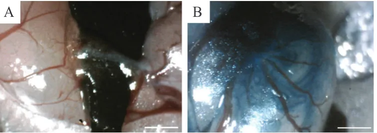
and 20 mg/Kg xylasein (Alfazyne). Then, by providing a 2 cm incision in the posterior abdominal region, access to the peritoneum was provided. Then gently without damaging to the epididymis, the testicles were immobilized by a triangular plastic piece (Figure 1A). To determine the injected, BM-MSCs were mixed with sterile trypan blue
(1:1, v/v). Also, with using a pipette, 100 μL of suspension
(106 BM-MSCs) was injected into the efferent duct of the
both testes till most of seminiferous tubules were filled.
Consequently, the testicle was returned to the abdominal cavity and the abdominal wall and skin were stitched (Figure 1B).(13)
A
B
Figure 1. Transplantation of BM-MSCs in seminiferous tubules of azoospermic mouse. A: To distinguish the translucent efferent duct from the fat tissue and the membrane around it, a hard black plastic triangle was placed under the efferent duct; B: After Injection of CD90+/CD34-/CD45- BM-MSCs suspension into efferent duct of mouse testis seminiferous tubules were partially filled. White bars, 1 mm.
Gene Name-Direction Primer Sequence
CD34-Forward AATGAGTCTGTTGAGGAA CD34-Reverse CTGTCTGAAGTAGTAGGC CD45-Forward AAGTGGATGTCTATGGTTA CD45-Reverse GAAGGAAGTCTCTGGTAT CD90-Forward GAAGACAAGGAGCCAGAAC CD90-Reverse GCAAGGGAAAGAAGAATAAAGG
Table 1. Sequences of RT-PCR primers used to quantify
the expression of BM-MSCs specific marker (CD90) and hematopoietic stem cells’ specific marker (CD45 and CD34) in
Mouse.
At next step, annealing was performed at 64°C, 62°C, and 61°C for 30 seconds, respectively. Finally, extension
was accomplished at 72°C for 30 seconds and the final
Histomorphometric Assessment
After fixation and alcohol dehydration of testicular tissue, it was embedded in paraffin and for tissue section, five
horizontal cross-sections were conducted as described by Panahi, et al. The sections were 5-µm in thickness and staining was conducted using hematoxylin-eosin (H&E) and was visualized using a light microscope Model CX21 (Olympus, Tokyo, Japan). The presence of spermatogonia, spermatocytes and spermatids were evaluated in all tubules. The inner, outer and total diameters were determined in all tubules conducting 10 circular transverse sections from different regions of the tubules. The total and lumen diameters were assessed in transverse sections by Dinocapture software version 2.0 (Dino-Eye, San-Chung, Taiwan). By taking the average of two diameters at right angles including D1+D2/2 and L1+L2/2, the averages of total and Lumen diameters of seminiferous tubules (D and L)
were clarified, respectively.(12)
By use of the diameter data, the luminal, cellular (germinal epithelium) and cross sectional areas were determined. In the seminiferous tubules, the cross-sectional
area (Ac) was evaluated using the equation of Ac=πD2/4; where D was considered as the mean diameter of tubules
and π was regarded as equivalent to 3.142. The seminiferous
tubulecount per unit area was also determined. The sections
of seminiferous tubules per unit area were defined by use
of an unbiased counting frame. In seminiferous tubules,
numerical density (NV) was considered as a number of tubules per unit volume and it was assessed by the following
equation: NV=NA/D+T, where, NA denotes to the number
of profiles per unit area, the tubule mean diameter was
represented by D, and T revealed the mean thickness of the section.(12)
The spermatogenesis index included the presence of spermatogenic cells throughout all testicular tissue compromising the types of cells, the number of cell layers, and the presence of late spermatids in the tubules. Therefore, the number of late spermatids if available were
counted and then seminiferous tubules were classified
based on the scale of 0-7 regarding spermatogenic potential,
while the spermatogenesis index was modified. The scale of 0 to 7 was defined as follows: 0=no germinal cells;
1=only spermatogonia cells; 2=both spermatogonia and spermatocytes cells; 3=spermatogonia, spermatocytes, and less than 50 late spermatids cells per tubule; 4=spermatogonia, spermatocytes, and 50-74 late spermatids cells per tubule; 5=spermatogonia, spermatocytes and 75-99 late spermatids cells per tubule; 6=spermatogonia, spermatocytes and 100-149 late spermatids cells per tubule;
Results
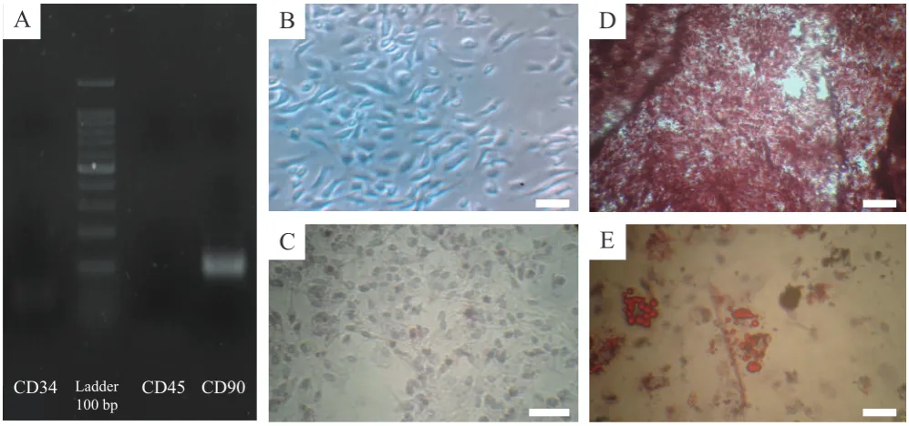
Morphologically, cells in the third passage have a
fibroblastic appearance that is one of the main characteristics
of MSCs (Figure 2A). Also, The results showed that after examination of BM-MSCs by RT-PCR the presence of surface marker for BM-MSCs (CD90) and the absence of surface factors of hematopoietic stem cells (CD34 and
CD45) were identified (Figure 2B). The presence of calcium
deposits in osteogenic differentiation medium after 21 days with alizarin red staining indicated the characteristics of osteoblasts in comparison with control (Figure 2C and 2D). In the adipogenic differentiation medium, the presence of lipid droplets after Oil Red O staining in the same
period confirmed by the differentiation of these cells into
adipoblasts as well (Figure 2E).
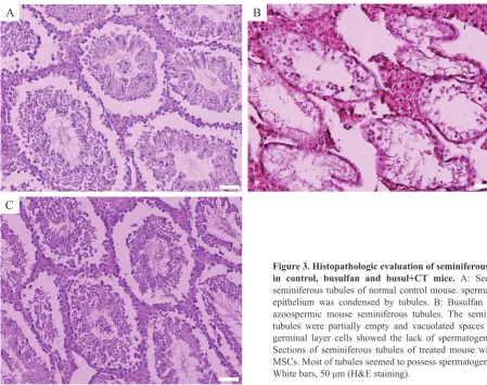
Histologic findings showed that in the control
group, Spermatogenesis was performed completely and spermatogenic cells with Sertoli cells were observed in seminiferous tubules (Figure 3A). In the busulfan group, the
spermatogenic cells were significantly reduced, and the large
vacuoles space were observed in the germinal epithelium of seminiferous tubules. A small number of spermatogonial cells were located on the basement membrane, and Sertoli cells were visible and spermatogenesis was completely degraded (Figure 3B). In the busul+CT group, the vacuolate space was eliminated, and in most seminiferous tubules, there were a variety of cell lines including spermatogonia, spermatocyte, spermatid and sperm (Figure 3C).
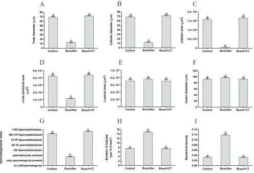
Histomorphometric findings showed that Total and
cellular diameters and cellular and cross-sectional areas were
significantly increased in busul+CT group in comparison to
the busulfan group (p<0.001) and there was no significant
difference in busul+CT and control groups (p>0.05) (Figure 7= all germinal cell types and more than 150 late spermatids cells per tubule.(11,12)
Statistical Analysis
All data were presented as means and standard error (SE). Kolmogorov-Smirnov test was performed to normalization
of data. Statistical analysis was done with one-way ANOVA
and post-hoc Tukey test by SPSS software ver. 20 (SPSS
Inc., Chicago, USA). The Mann-Whitney U test was used
to assess spermatogenesis index. A p-value of less than
0.05 was regarded statistically significant. The graphs are
A
B
C
D
E
CD34 Ladder CD45 CD90
100 bp
Figure 2. Characterization and differentiation of BM-MSCs. A: Agarose gel electrophoresis of products of reverse transcriptase polymerase chain reaction (RT-PCR) demonstrated that specific mesenchymal marker exists (CD90) compared with absence of specific hematopoietic marker (CD45and CD34) of mouse BM-MSCs; B: Spindle shape morphology of mouse BM-MSCs in passages 1 (×40); C: Control group of BM-MSCs in passage 3 (×100); D: BM-MSCs cultivated in osteogenic medium and stained with alizarin red (×100); and E: In adipogenic medium and were stained with Oil Red O at day 21 after induction. White bars, 100 μm.
4A, 4B, 4C and 4D, respectively). There was no significant
difference in the lumen diameter and area in seminiferous tubules in all three groups (p>0.05) (Figure 4E and 4F,
respectively). Spermatogenesis index was significantly
increased in busul+CT group in comparison to the busulfan and control groups (p<0.001 and p˂0.05, respectively)
(Figure 4G). In the busul+CT and control groups the number of tubules per unit area and numerical density of the tubules were less than the busulfan group (p<0.001) and there was
no significant difference in busul+CT and control groups
(p>0.05) (Figure 4H and 4I, respectively).
Discussion
Busulfan is a chemotherapeutic agent which is often used for the treatment of chronic myeloid leukemia (14) and is also used before bone marrow and stem cells transplantation (15,16). Busulfan can inhibit cell division by sticking on
one of DNA strands.(17) Gutierrez, et al., reported that compared to Swiss mice, Balb/C mice were more sensitive to the toxic effects of busulfan and were not fertile after
90 days of treatment.(18) Confirming it, Payehdar, et al., showed that two doses (10 mg/Kg) of busulfan with 21 days interval could induce irreversible azoospermia for more than 90 days.(11) Busulfan induces apoptosis and
destroys mitotic cells including germ cells leading to azoospermia and infertility. (12) Cell transplantation was shown as a new approach for treatment of disorders such as azoospermia.(13) Hormonal therapies or surgical
interventions were demonstrated with low efficacy in
treatment of non-obstructive azoospermia.(19) In our study, BM-SCs were used in treatment of azoospermic Balb/C mice.
Cell therapy with MSCs is a new way for treatment of some disorders. BM-MSCs are able to differentiate between several mesodermal cell lineages including muscle, bone, fat, cartilage and other connective tissues cells.(20) MSCs secrete growth factors and cytokines that have
anti-inflammatory, immunosuppressive, anti-apoptosis, and
proliferative effects that called ‘trophic’ activity.(21) Kuo,
et al., reports that stem cell use improves complications of diabetes on the liver.(22) Also, stem cell use can reduce the complications of heart attack in pigs.(23) Sadraie, et al., showed that BM-MSCs can reduce the side effects of knee rubbing in the guinea pig.(24)
In this study it was shown that transplantation of
CD90+/CD34-/CD45- BM-MSCs in azoospermic Balb/C
mice could successfully recover induce spermatogenesis.
Similar findings demonstrated that Ad-SCs and BM-SCs
cells as immune-privileged for successful cell transplantation without the need for chronic immunosuppression therapy. The immune protective nature of Sertoli cells has been presented in another study.(32) Sertoli cells provide the microenvironment suitable for differentiation and proliferation of spermatogonial stem cells. Our
findings, confirmed that BM-MSCs could reconstitute
tubular microenvironment providing the chance for remained inactivated germinal cells to proliferate in the host seminiferous tubules.(25) There is evidence of the hypoimmunogenic nature of MSCs and so there is implication for allogeneic therapy.(31,33) Intravenous injection of BMSCs was shown to have immunosuppressive effects on production of anti-sperm antibody in allogeneic transfusion in mice after testis rupture.(25)
Mital, et al., reported that survival and protection of the cells after transplantation can be due to the immunological tolerance of Sertoli cells.(32) In seminiferous tubules, some points can be remarked after transplantation of stem cells: 1) blood-testis barrier as the immune privilege character plays an important role after stem cells transplantation in seminiferous tubules (34), 2) Also was shown that BM-MSCs (26). Cakici, et al., showed that Ad-SCs transplantation
after 12 weeks could recover busulfan-treated infertile male rats and induce spermatogenesis.(6) Lue, et al., reported that transplanted BM-SCs in busulfan-treated infertile mouse model could differentiate into germ, Sertoli, and Leydig cells.(27)
Several studies evaluated the differentiation potential of MSCs into spermatozoa in mice and rats regarding intra-seminiferous tubule (2,5) and intra-testicular injection
of BM-MSCs (28). Monsefi, et al., showed that testis in infertile rats receiving BM-SCs could differentiate between germ cells in seminiferous tubules.(28) The differentiation potential of MSCs to spermatozoa was previously reported in rat (2,9,29) and hamster (25). But differentiation did not
happen in all tubules.(6,9) Also, Nayernia, et al., reported that BM-MSCs could differentiation between germ cells in vivo. (30) Chen, et al., reported that human umbilical cord MSCs
transplanted into seminiferous tubule of immunodeficient
mouse could differentiate between sperm.(31) It was shown that cell transplantation in azoospermic animals may be species dependent.(25) Seminiferous tubules are responsible for regulation of spermatogenesis, and Sertoli
A
B
C
Figure 3. Histopathologic evaluation of seminiferous tubule
A
B
C
D
E
F
G
H
I
Figure 4. Mean and standard error of histomorphometric indices of seminiferous tubules in control, busulfan and busul+CT
groups. A: Total diameter (μm), B: Cellular diameter (μm), C: Cellular area (μm2), D: Cross sectional area of the tubule (μm2), E: Luminal
area (μm2), F: Lumen diameter (μm), G: Spermatogenesis index, H: Number of seminiferous tubules per unit area of testicular tissue, I:
Numerical density of the seminiferous tubules. Different superscripts (a, b and c) indicate statistically differences between groups (p<0.05).
to have both hypoimmunogenic and immunosuppressive features after transplantation.(35) Therefore, transplantation of MSCs was considered as a method of choice for azoospermia cell therapy.
Conclusion
In this current study, histological and histomorphometric
findings revealed that intra testicular transplantation
of CD90+/CD34-/CD45- BM-MSCs in seminiferous tubules of busulfan-induced azoospermic Balb/C mice could differentiate between all types of germinal cells and recovered the spermatogenesis. BM-MSCs transplantation could be an effective method to treat non-obstructive azoospermia in patients undergoing chemotherapy such as busulfan and restore fertility in these patients. But more research must be done to verify this issue.
Acknowledgment
The authors wish to thank the Islamic Azad University and Shiraz University of Medical Sciences for their kind support.
References
1. Gudeloglu A, Parekattil SJ. Update in the evaluation of the azoospermic male. Clinics (Sao Paulo). 2013; 68: 27-34.
2. Zhang D, Liu X, Peng J, He D, Lin T, Zhu J, et al. Potential spermatogenesis recovery with bone marrow mesenchymal stem cells in an azoospermic rat model. Int J Mol Sci. 2014; 15: 13151-65.
4. Meistrich ML. Effects of chemotherapy and radiotherapy on spermatogenesis in humans. Fertil Steril. 2013; 100: 1180-6. 5. Zahkook SAM, Atwa A, Shahat MM, Mansour AM, Bakry S.
Mesenchymal stem cells restore fertility in induced azoospermic rats following chemotherapy administration. J Reprod Infertil. 2014; 5: 50-7.
6. Cakici C, Buyrukcu B, Duruksu G, Haliloglu AH, AksoyA, Isık A, et al. Recovery of fertility in azoospermia rats after injection of adipose-tissue derived mesenchymal stem cells: The sperm generation. BioMed Res Int. 2013; 2013: 1-18.
7. Asadi-Yousefabad S-L, Khodakaram-Tafti A, Dianatpour M, Mehrabani D, Zare S, Tamadon A, et al. Genetic evaluation of bone
marrow-derived mesenchymal stem cells by a modified karyotyping method.
Comp Clin Pathol. 2015; 24: 1361-6.
8. Mahdiyar P, Zare S, Robati R, Dianatpour M, Torabi K, Tamadon A,
et al. Isolation, culture, and characterization of human dental pulp mesenchymal stem cells. Int J Pediatr. 2014; 2: 44.
9. Mehrabani D, Hassanshahi MA, Tamadon A, Zare S, Keshavarz S, Rahmanifar F, et al. Adipose tissue-derived mesenchymal stem cells repair germinal cells of seminiferous tubules of busulfan-induced azoospermic rats. J Hum Reprod Sci. 2015; 8: 103-10.
10. Ehninger A, Trumpp A. The bone marrow stem cell niche grows up: mesenchymal stem cells and macrophages move in. J Exp Med. 2011; 208: 421-28.
11. Payehdar A, Hosseini E, Mehrabani D, Forouzanfar M. Busulfan treatment effects on testicular tissue and serum levels of anti-mullerian hormone and testosterone in adult mice. Indones Biomed J. 2017; 9: 106-12.
12. Panahi M, Karimaghai N, Rahmanifar F, Tamadon A, Vahdati A,
Mehrabani D, et al. Stereological evaluation of testes in busulfan-induced infertility of hamster. Comp Clin Pathol. 2014; 24: 1051-6.
13. Rahmanifar F, Tamadon A, Mehrabani D, Zare Sh, Abasi S, Keshavarz S, et al. Histomorphometric evaluation of treatment of rat azoospermic seminiferous tubules by allotransplantation of bone marrow-derived mesenchymal stem cells. Iran J Basic Med Sci. 2016; 19: 653-61.
14. Suttorp M, Millot F. Treatment of pediatric chronic myeloid leukemia in the year 2010: use of tyrosine kinase inhibitors and stem-cell transplantation. Hematology Am Soc Hematol Educ Program. 2010; 2010: 368-76.
15. Le Bourgeois A, Lestang E, Guillaume T, Delaunay J, Ayari S, Blin N, et al. Prognostic impact of immune status and hematopoietic recovery
before and after fludarabine, IV busulfan, and antithymocyte
globulins (FB2 regimen) reduced-intensity conditioning regimen (RIC) allogeneic stem cell transplantation (allo-SCT). Eur J Haematol. 2013; 90: 177-86.
16. Nieto Y, Thall P, Valdez B, Andersson B, Popat U, Anderlini P, et al.
High-dose infusional gemcitabine combined with busulfan and melphalan with autologous stem-cell transplantation in patients with refractory lymphoid malignancies. Biol Blood Marrow Transplant. 2012; 18: 1677-86.
17. Iwamoto T, Hiraku Y, Oikawa S, Mizutani H, Kojima M, Kawanishi
S. DNA intrastrand cross-link at the 5′-GA-3′ sequence formed by
busulfan and its role in the cytotoxic effect. Cancer Sci. 2004; 95: 454-58.
18. Gutierrez K, Glanzner WG, Chemeris RO, Rigo ML, Comim FV,
Bordignon V, et al. Gonadotoxic effects of busulfan in two strains of mice. Reprod Toxicol. 2016; 59: 31-9.
19. Berookhim B.M., Schlegel. Azoospermia due to spermatogenic failure.
Urol Clin. North Am. 2014; 41: 97-113.
20. Caplan AI. Adult mesenchymal stem cells for tissue engineering versus regenerative medicine. J Cell Physiol. 2007; 213: 341-7.
21. Caplan AI, Dennis JE. Mesenchymal stem cells as trophic mediators. J Cell Biochem. 2006; 98: 1076-84.
22. Kuo TK, Hung SP, Chuang CH, Chen CT, Shih YR, Fang SC, et al.
Stem cell therapy for liver disease: parameters governing the success of using bone marrow mesenchymal stem cells. Gastroenterology. 2008; 134: 2111-21.
23. Amado LC, Saliaris AP, Schuleri KH, St John M, Xie JS, Cattaneo S,
et al. Cardiac repair with intramyocardial injection of allogeneic
mesenchymal stem cells after myocardial infarction. Proc Natl
Acad Sci USA. 2005; 102: 11474-9.
24. Sadraie MR, Mehrabani D, Vahdari A. Comparison of therapeutic effects of bone marrow mesenchymal stem cells and liquid culture environment (secreta) in the treatment of induced knee abrasion created in Guinea Pigs. Armaghane danesh. 2016; 20: 651-65.
25. Tamadon A, Mehrabani D, Rahmanifar F, Raayat Jahromi A, Panahi M, Zare S, et al. Induction of spermatogenesis by bone marrow-derived mesenchy-mal stem cells in busulfan-induced azoospermia in hamster. Int J Stem Cells. 2015; 8: 134-45.
26. Hajihoseini H, Vahdati A, Hosseini E, Mehrabani D, Tamadon A. Induction of spermatogenesis after stem cell therapy of azoospermic guinea pigs. Vet Arhiv. 2017; 87: 333-50.
27. Lue Y, Erkkila K, Liu PY, Ma K, Wang C, Hikim AS, et al. Fate of bone marrow stem cells transplanted into the testis : potential implication for men with testicular failure. Am J Pathol. 2007; 170: 899-908.
28. Monsefi M, Fereydouni B, Rohani L, Talaei T. Mesenchymal stem cells
repair germinal cells of seminiferous tubules of sterile rats. Iran J Reprod Med. 2013; 11: 537-44.
29. Sabbaghi MA, Bahrami AR, Feizzade B, Kalantar SM, Matin MM, Kalantari M, et al. Trial evaluation of bone marrow derived mesenchymal stem cells (MSCs) transplantation in revival of spermatogenesisin testicular torsion. Middle East Fertil Soc J. 2012; 17: 243-9.
30. Nayernia K, Lee JH, Drusenheimer N, Nolte J, Wulf G, Dressel R, et al. Derivation of male germ cells from bone marrow stem cells. Lab Invest. 2006; 86: 654-63.
31. Chen H, Tang QL, Wu XY, Xie LC, Lin LM, Ho GY, et al. Differentiation of human umbilical cord mesenchymal stem cells into germ-like cells in mouse seminiferous tubules. Mol Med Rep. 2015; 12: 819-28.
32. Mital P, Kaur G, Dufour JM. Immunoprotective sertoli cells: making allogeneic and xenogeneic transplantation feasible. Reproduction 2010; 139: 495-504.
33. Barry FP, Murphy JM. Mesenchymal stem cells: clinical applications and biological characterization. Int J Biochem Cell Biol. 2004; 36: 568-84.
34. Meinhardt A, Hedger MP. Immunological, paracrine and endocrine aspects of testicular immune privilege. Mol. Cell Endocrinol. 2011; 335: 60-8.