Targeted Treatment in Patients with Non-Small Cell Lung Cancer:
Use of Bevacizumab and its Biosimilars
By Umme Tania ID- 17346054
A thesis submitted to the Department of Pharmacy in partial fulfillment of the requirements for the degree of
Bachelor of Pharmacy (Hons.)
Department of Pharmacy Brac University
April 2021
©2021. Brac University All rights reserved
i
Declaration
It is hereby declared that
1. The thesis submitted is my own original work while completing degree at Brac University.
2. The thesis does not contain material previously published or written by a third party, except where this is appropriately cited through full and accurate referencing.
3. The thesis does not contain material which has been accepted, or submitted, for any other degree or diploma at a university or other institution.
4. I have acknowledged all main sources of help.
Student’s Full Name & Signature:
Umme Tania 17346054
ii
Approval
The thesis/project titled “Targeted Treatment in Patients with Non-Small Cell Lung Cancer:
Use of Bevacizumab and its Biosimilars” submitted by Umme Tania (17346054) of Spring, 2017 has been accepted as satisfactory in partial fulfillment of the requirement for the degree of Bachelor of Pharmacy (Hons.) on April, 2021
Examining Committee:
Supervisor:
Tanisha Khan
Lecturer, Department of Pharmacy Brac University
Supervisor:
Dr. Eva Rahman Kabir Professor, Department of Pharmacy
Brac University
Program Coordinator and
Deputy Chair: Dr. Hasina Yasmin
Professor, Department of Pharmacy Brac University
Departmental Head:
Dr. Eva Rahman Kabir Professor, Department of Pharmacy
Brac University
iii
Ethics Statement
The study does not involve any kind of animal and human trial.
iv
Abstract
Angiogenesis is one of the pivotal factors contributing to the development and metastasis of non- small cell lung cancer which is one of the most commonly diagnosed types of lung cancer.
Bevacizumab, a monoclonal antibody that acts by restricting angiogenesis, is therefore a major choice of medication for advanced and metastatic non-small cell lung cancer treatment.
Bevacizumab is essentially a biologic medicine which is a distinct class of biopharmaceuticals.
Biologics are considerably expensive and this has resulted in the advent of biosimilars which are highly similar to their reference biologics in terms of quality, efficacy, and safety. As biosimilars are adopted as affordable copies of biologics, they possess great potential to influence the healthcare system. In this literature review, the importance of biosimilars with respect to its cost effectiveness and accessibility to the patients were discussed. The study also focused on the availability of biosimilars of bevacizumab as a potential targeted treatment option for NSCLC. In the meantime, regulatory frameworks established in different countries concerning the introduction of biosimilars in the market were reviewed.
Keywords: Biologics, Biosimilars, Bevacizumab, Non-small cell lung cancer, Regulatory frameworks, Targeted therapy
v
Dedication
Dedicated to my parents
vi
Acknowledgement
I am grateful to Almighty Allah for giving me the strength and patience to complete the project and Department of Pharmacy for allowing me to conduct such study for my undergraduation.
At first, I would like to sincerely acknowledge my indebtedness to my respected supervisor, Professor Dr. Eva Rahman Kabir, (Chairperson, Department of Pharmacy, Brac University) for her significant guidance, encouragement and constructive suggestions while conducting the study. Throughout the whole process, I have received her complete support whenever needed for which I convey my gratitude. It has been entirely an honor for me to work under her supervision.
I also owe immense gratefulness to my supervisor, Tanisha Tabassum Sayka Khan (Lecturer, Department of Pharmacy, Brac University) as she has been like a shield throughout completing the whole project. She has shown me every single path to follow in order to perfect the project from the very beginning. Her constant support and sincerity kept me motivated while executing the study.
Finally, I would like to thank my parents and all the loved ones who mentally supported me, motivated me throughout the whole journey.
vii
Table of Contents
Declaration ……….... i
Approval ………... ii
Ethics Statement ………. iii
Abstract ……… iv
Dedication ………. v
Acknowledgement ………... vi
Table of Contents ………... vii
List of Tables ………... ix
List of Figures ………... x
List of Acronyms ………. xi
Chapter 1 Introduction ……… 1-4 Background ………... 1-2 Objectives of the Study ……… 3
Rationale of the Study ………...3-4 Chapter 2 Methodology ………... 5
Chapter 3 Cancer ……….… 6
Cancer: What Is It? ………...…6
Types of Cancer ………... 6
Prevalence of Cancer ………... 9
Chapter 4 Lung Cancer ………... 11
Introduction to Lung Cancer ………... 11
Non-Small Cell Lung Cancer ………..……... 12
Molecular Alterations in NSCLC ……….………. 13
Treatments Available for NSCLC……….. 18
Chapter 5 Biologics and Biosimilars ………... 25
Introduction to Biologics and Biosimilars ……….… 25
Manufacturing Process of Biologics and Biosimilars ……….... 29
Regulatory Framework of Biologics and Biosimilars ………... 33
Chapter 6 Biologics in Non-Small Cell Lung Cancer ……….… 39
Bevacizumab ………..… 40
viii
Biosimilars in Non-Small Cell Lung Cancer ……….… 44 Chapter 7 Conclusion ………... 52 Future Directions……………… 52 References………………... 53-61
ix
List of Tables
Table 1: Definitions of biosimilar according to different country’s regulatory bodies ………... 27 Table 2: The US FDA approved biologics in the treatment of NSCLC ……….. 39 Table 3: Approved and candidate biosimilars of bevacizumab (Avastin) ………... 44
x
List of Figures
Figure 1: The manufacturing process of biologics and biosimilars ………. 32 Figure 2: Mechanism of action of bevacizumab ……….. 43
xi
List of Acronyms
ADA Anti-drug antibody AE Adverse events ADR Adverse drug reaction
AMCP Academy of Managed Care Pharmacy ALK Anaplastic lymphoma kinase
BLA Biologics License Application
BPCIA Biologics Price Competition and Innovation Act CI Confidence interval
CHO Chinese hamster ovary
CTLA-4 Cytotoxic T-lymphocyte-associated protein 4 CMC Chemistry, Manufacturing and Control DOR Duration of response
EGFR Epidermal growth factor receptor EMA European Medicines Agency EU European Union
EEA European Economic Area
FISH Fluorescence in situ hybridization FDA Food and Drug Administration FOB Follow-on biologic
GMR Geometric mean ratio
HER2 Human epidermal growth factor receptor 2 HER4 Human epidermal growth factor receptor 4 HGF Hepatocyte growth factor
ITT Intention to treat
IHC Immunohistochemistry
xii IND Investigational New Drug
KRAS Kirsten rat sarcoma viral oncogene homolog MAP2K1 Mitogen-activated protein kinase 1
MHLW Ministry of Health, Labor, and Welfare NSCLC Non-small cell lung cancer
NTRK1 Neurotrophic tyrosine receptor kinase 1 NGF Nerve growth factor
NDA New drug application
NRAS Neuroblastoma ras viral (v-ras) oncogene homolog NAbs Neutralizing antibodies
ORR Overall response rate OS Overall survival
PFS Progression free survival PFS Progression free survival
PD-1 Programmed cell death protein 1 PD-L1 PD-ligand 1
PIK3CA Phosphatidylinositol-4,5-bisphosphate 3-kinase, catalytic subunit alpha PP Per protocol
PPACA Patient Protection and Affordable Care Act rDNA Recombinant DNA
RT-CPR Reverse transcription polymerase chain reaction RBP Reference biotherapeutic product
RP Reference product
SBP Similar biotherapeutic products SEB Subsequent-entry biologic
TrkA Tropomycin receptor kinase A
xiii TRK Tropomyosin related kinases TKI Tyrosine kinase inhibitor TTP Time to progress
TEAE Treatment-emergent adverse events VEGF Vascular endothelial growth factor
VEGFR Vascular endothelial growth factor receptor WHO World Health Organization
1
Chapter 1 Introduction 1.1 Background
Lung cancer irrefutably can be indexed in the major fights to be won within the oncology sector.
Also, more than 85% of the reported lung cancer cases consist of non-small cell lung cancer (NSCLC). Global Cancer Observatory regulated by the World Health Organization claimed that lung cancer at present comprises the third place in the list of the most commonly developing malignance globally. Additionally, regardless of the declining fatalities since the 90s, this disease happens to be the utmost vicious type of cancer at the moment (Mello, 2020). Different therapeutic strategies utilized for treating NSCLC are surgery, radiation therapy, chemotherapy, targeted therapy and immunotherapy (Non–Small Cell Lung Cancer: Epidemiology, Risk Factors, Treatment, and Survivorship, n.d.). In spite of the expansion of treatment strategies to the newest chemotherapy regimens, such as chemotherapeutic agents based on platinum, the prognosis concerning NSCLC that is in advanced stage, metastatic and not operable stays to be insignificant. A clinical study performed by Eastern Cooperative Oncology group established an extended comparison between a conventional dosage regimen of cisplatin/paclitaxel and three other platinum based chemotherapeutic agents. The ORR (overall response rate) of the study was found to be 19% with a survival rate of 33% one year and 11% two years. Hence, there is an extreme necessity for novel and better therapies in the treatment of NSCLC, as improvement via existing cytotoxic therapy is proven to be inadequate (Herbst & Sandler, 2004).
A better understanding of the mechanisms of tumor growth at the molecular level has discovered several conceivable targeted therapeutic strategies, including angiogenesis pathways to restrain tumor growth more effectively (Herbst & Sandler, 2004; Lauro et al., 2014; Molina et al., 2008).
Angiogenesis has been studied for an extended period of time and is presently reckoned as one of the 10 hallmarks of cancer (Greillier et al., 2016). Vascular endothelial growth factor (VEGF) can be identified as a primary moderator of angiogenesis in tumors. Increased manifestation of vascular endothelial growth factor has been associated with extremely low disease recovery, incorporating an aggravated possibility of recurrence and spread of cancer resulting in fatality
2
(Reinmuth et al., 2019). The functions of VEGF in governing tumor angiogenesis, retaining existing vascularization, causing the tumor to be resistant towards conventional therapeutic strategies accompanying unfavorable clinical outcome makes the VEGF a suitable target for pharmacologic intervention in the treatment of NSCLC (Herbst & Sandler, 2004). Bevacizumab, a monoclonal antibody synthesized via recombinant DNA technology restricts the biological activity of VEGF by binding to it and hence, blocks the interaction of VEGF with its receptors in the cell surface membrane of endothelium (Reinmuth et al., 2019). In the first line treatment of non-squamous NSCLC, bevacizumab has been the first anti-VEGF biologic to obtain license (Lauro et al., 2014). Since the past 20 years, considerable progress has been brought into existence in cancer treatment through the ongoing development of brand-new biologics, markedly monoclonal antibodies (Busse & Lüftner, 2019).
Biologics can be defined as large molecule therapeutic agents which possess complex nature and are manufactured employing living organisms (Kabir et al., 2019). They are essentially synthesized by applying biotechnological strategies via living microorganisms or cells with genetic modification which make them more costly than other medications. Consequently, access of the patients to biologics is usually restricted by excessive price (Busse & Lüftner, 2019). The emerging expiration of patents and confined biologics accessibility have nevertheless catalyzed a huge opportunity for the evolution of a different division of medicine, that is biosimilar (Kabir et al., 2019). Biosimilars can be introduced into the market where the patents of its reference biologics have come to expire and serve as an attractive strategy to lower the healthcare cost by price competition with the biologics (Busse & Lüftner, 2019). This class of biotherapeutics exerts high potentiality of price reducing. Therefore, increased acceptance of biosimilars can considerably reduce treatment costs and optimize reach to biologic medicines for patients with malignancies such as lung cancer (Cuellar et al., 2019; Rugo et al., 2016). However, the most important issue is that the biosimilars in the cancer treatment should be introduced in a proper process and healthcare providers must possess access to all the relevant data concerning biosimilars, so that they can take conclusive clinical decisions (Mellstedt et al., 2008).
3 1.2 Objectives of the Study
This study has been conducted to highlight the promising consequences and future implications of biosimilars in the treatment of non-small cell lung cancer.
The objectives of the study are:
1. to summarize the impact of the biosimilars in healthcare community
2. to highlight impact of biosimilars as a cost-effective alternative to increase oncologists’
confidence in biosimilars
3. to outline the regulatory framework for approving biosimilars
4. to review the efficacy and safety of both the biologic bevacizumab and its biosimilar(s) in the treatment of NSCLC
1.3 Rationale of the Study
Biological agents or ‘‘biologics” are extensively adapted in the oncology field in order to treat cancer and to provide supportive management of treatment-related side effects (Rugo et al., 2016). However, these medications are markedly more high-priced than their small molecule counterparts (pharmaceutical). Consequently, this has led to the ascent of biosimilars, considered to be the affordable copies of innovator biologics (Rathore & Bhargava, 2020). There is a huge market for biologics used in cancer treatment, and introduction of biosimilars possess fair potential in expansion of entrance towards treatment together with decreased prices. Both the development procedure and licensing process concerning these biotherapeutics require a pre- standardized regulatory pathway which includes a systematic strategy to validate bio-equivalence and conduct comparative clinical studies to verify equivalence in terms of pharmacokinetic (PK) profile, therapeutic efficacy, quality, toxicology profile, and immunogenicity to the reference biologic (Cuellar et al., 2019). In this study, the most relevant dispositions and the regulatory framework aimed at the growth phase and authorization of biosimilar medications are summarized in order to accelerate the rate of approval of biosimilars worldwide (Kang et al., 2020; Santos et al., 2019). The literature review also presents the oncological biosimilar drugs employed in the NSCLC treatment at present with the purpose of providing more precise and
4
crucial information to oncologists about the quality and safety of the particular medicines, since information is the essential guide to mitigating the basic concerns with regard to the adaptation of biosimilars (Mellstedt et al., 2008; Santos et al., 2019).
5
Chapter 2 Methodology
For this study, all the information, and data were collected from authentic articles indexed in high impact research databases such as Scopus, Pubmed, ScienceDirect, and Springer, as well as from renowned websites. At first, an outline was prepared to execute the review in a systematic manner. The articles for the study were searched using keywords like biologics, biosimilars, regulatory framework, lung cancer, targeted therapy, and bevacizumab. After going through about 100 articles, 60 relevant papers were chosen to collect information for the study.
6
Chapter 3 Cancer
3.1 Cancer: What Is It?
Cancer is considered to be among the prime reasons for fatalities and the single most considerable obstruction to accentuating lifespan worldwide in the 21st century (Bray et al., 2018). Cancer being a complicated and dreadful disease or sets of diseases, have caused sufferings to the multicellular living beings for around 200 million years, and evidence of cancers were found amidst the ancestors of modern humans going back more than millions of years. In contrast to infectious diseases and other environmental diseases, cancer is not necessarily initiated by an entity which is foreign to our bodies (Haberkorn, 2007).
In every cancer, cells of an organ or tissue inside the body start to divide in an extremely uncontrollable manner and gradually spread into surroundings. Generally, cells inside the human body multiply to generate newer cells according to the body’s requirement and when these cells become older or defective, they face death. However, in case of development of cancer inside the body this orderly process gets hampered. Old and damaged cells do not die and also new cells keep dividing aggressively which form a mass of cells known as tumor.
Tumors which are cancerous are malignant in nature, meaning that they are capable of spreading into and invading surrounding organs. Additionally, a portion can get detached from the tumor and move to another part inside the body via the circulatory or lymphatic system causing another tumor to form there. On the contrary, benign tumors are not capable of spreading and if removed, such tumors generally don’t recur, while malignant tumors may relapse (What Is Cancer? - National Cancer Institute, n.d.).
3.2 Types of Cancer
Cancer can fundamentally be classified on the basis of organs/tissues from where the cancer cells generate. To mention a few, the kidney cancer generates in the cells of the kidney, and the brain tumor develops in the neurons of the brain. It can also be categorized according to the group of cells which developed the particular cancer, for example, epithelial cells or myeloma cells.
7
Cancers which have been classified based on particular cell types are as follows:
i. Carcinoma
Among the different classes, carcinoma happens to be quite frequently occurring cancer. This form of tumor initiates in epithelial cells comprising both interior and exterior surfaces of the body. There are distinctive names for carcinomas starting in different types of epithelial cells.
The cancer which begins in fluids or mucus producing epithelial cells is defined as adenocarcinoma. Cancer developing in the breast, lung, and prostate is mostly adenocarcinoma.
Basal cell carcinoma originates in the initial layer of epidermis that comprises the utmost lining of the skin. Another kind of carcinoma is squamous cell carcinoma, also known as epidermoid carcinoma that develops in squamous cells present right underneath the outer layer of skin.
Transitional cell carcinoma that mostly occurs in the bladder, ureters, and kidneys originates from a class of epithelial cells known as transitional epithelium.
ii. Sarcoma
Sarcoma, another kind of malignancy, begins in the bone tissues and soft tissues such as muscle tissue, fat tissue, blood vessels and lymph vessels as well as fibrous tissues. The most prevalent bone cancer is osteosarcoma. Among soft tissue sarcomas, leiomyosarcoma, kaposi sarcoma, liposarcoma, malignant fibrous histiocytoma, and dermatofibrosarcoma protuberans are the most common ones.
iii. Leukemia
Cancer beginning within the tissue present in bone marrow which forms blood cells is called leukemia. Solid tumors are not developed by these cancers, rather unusual white blood cells (WBC) such as leukemia cells and leukemic blast cells accumulate extensively in the blood and bone marrow. Leukemia can be classified on the basis of how rapidly the cancer spreads e.g., acute or chronic and based on the blood cell types in which the cancer begins e.g., lymphoblastic or myeloid.
8
iv. Lymphoma
A cancer recognized as lymphoma develops in T cells and B cells (lymphocytes). In this type of cancer, unusual lymphocytes pile up in the lymphatic system along with different organs of the body (What Is Cancer? - National Cancer Institute, n.d.).
The major categories of this disease are:
Hodgkin lymphoma – it usually develops in the B cells and starts in the lymph node located in the upper portion of the body (About Hodgkin Lymphoma | Cancer Research UK, n.d.).
Non-Hodgkin lymphoma – Non-Hodgkin lymphoma essentially starts in both B and T lymphocytes. Non-Hodgkin lymphoma is applied for many types of lymphoma that share few of the similar characteristics however different signs and symptoms (Lymphoma - Non-Hodgkin:
Introduction | Cancer.Net, n.d.).
v. Multiple myeloma
Multiple myeloma generates in plasma cells. In this cancer, plasma cells start to grow uncontrollably and express an abnormal protein called myeloma protein (M-protein) that works as a biomarker to monitor the disease (What Is Myeloma? | Cancer Research UK, n.d.).
vi. Melanoma
Melanoma, a kind of cancer, forms in cells called melanocytes producing melanin on the skin.
Melanomas can begin in any area of the skin; however, they mostly begin on the chest and back in men and on the arms as well as legs in women. The remaining common sites are the neck and face. If melanoma appears in the eyes, this is generally recognised as intraocular or ocular melanoma (Melanoma Treatment (PDQ®)–Patient Version National Cancer Institute, n.d.).
vii. Brain and spinal cord tumors
Tumors in the brain and spinal cord exist as a bunch of irregular cells growing uncontrollably.
They can start anywhere in the brain and show symptoms according to the location. They may
9
stay as either benign (not cancerous) or malignant (cancerous) (What Are Brain Tumours? | Brain Tumours | Cancer Research UK, n.d.).
viii. Germ cell tumors
Germ cell tumors begin in the cells producing eggs or sperms. Such cancer can develop nearly at any place inside human body where the germ cells may reside and are of two types: benign and malignant (Germ Cell Tumours | Cancer Research UK, n.d.).
ix. Neuroendocrine tumors
Neuroendocrine tumors develop in the cells which secrete hormones into the circulatory system in response to a stimulus generated by the neurons. This condition is accompanied by excess hormone production which leads to several physiological abnormalities. The two categories of neuroendocrine tumors include benign and malignant. Carcinoid tumor is one kind of neuroendocrine tumor which grows slowly. Such tumors are generally found in the gastrointestinal system. The tumors can migrate to the hepatic cells and various parts inside the body, and secrete components such as serotonin and prostaglandins, triggering carcinoid symptoms (What Is Cancer? - National Cancer Institute, n.d.).
3.3 Prevalence of Cancer
Approximately more than eighteen million individuals have encountered cancer in 2018 worldwide. Moreover, around 9.6 million people have confronted death from this disease. About 4% of the recent cases of cancer were reported in low development index countries, about 16%
were found in medium development index nations and the remaining were reported within nations indexed as highly developed. Among those cases, approximately five percent of deaths from cancer took place in low development index countries, about 20% took place in medium indexed countries, and the remaining took place in highly indexed developed countries. The lack of cancer records concerning approximately 85% of the global population, especially in low and moderate income nations, influences the reliability of such estimations. Moreover, approximately 50% of cancer cases and 60% of deaths from cancer occurred in Asia; however, only 6.5% of patients from this particular region are registered.
10
Cancer can be positioned as the primary reason for early death in approximately 100 out of 185 countries. One in five men and one in six women have been estimated to have suffered from cancer in 2018, and one in eight males and one in ten females came experienced death due to cancer. Around the world, the four principal causes of death from cancer are tumors in the lung, colorectum, stomach, and liver. Frequency ratio of cancer on average and by its types differ from country to country and reflect age distribution of population, variation in prevalence and propagation of the primary predisposing factors including socioeconomic condition and availability of medical services to diagnose cancer in a country. In developing countries in terms of economy, displacement of infection-related and poverty-related cancers such as stomach, cervix, and liver cancers by the cancers which appear to be more prevalent in developed countries such as colorectal cancer is on the rise (Cortes et al., 2020).
Lung cancer exists as most commonly diagnosed malignancy regardless of sex and it is the leading cause of death from cancer among males. Among all the cancers, lung cancer is a highly prolific one present in both men and women (11.6 percent of total diagnosed cases) and it also records the most deaths (18.4 percent of total death reports) than any other cancers (Bray et al., 2018).
11
Chapter 4 Lung Cancer
4.1 Introduction to Lung Cancer
Lung cancer, which is a greatly invasive and fast metastasizing malignancy, comes across as the top killer cancer in every gender all over the world (Lemjabbar-Alaoui et al., 2015). WHO estimates is that the death rates of the distinctive cancer will persist globally in an increasing manner, essentially due to a rise in smoking rate worldwide, specifically in Asian region (Duma et al., 2019). Lung cancer, a diversified disease constitutes different subcategories with distinct physiological and therapeutic implications (Rodriguez-Canales et al., 2016).
A few environmental factors and social habits can be related with the ensuing advancement of lung cancer. Cigarette smoking appears to be the utmost critical one among them which is estimated to account for 85%-90% of lung cancers. The incidence of being diagnosed with cancer is related to the frequency of smoking and exposure to different carcinogenic substances, such as asbestos. Further components related to enhanced risk of lung cancer formation is ionizing radiation, passive smoking, radon and carcinogenic metals e.g. arsenic, chromium, and cadmium, and polycyclic aromatic hydrocarbons. A pre-existing medical condition of pulmonary fibrosis, HIV infection, and intoxication with alcohol are also considered as predisposing factors of developing lung cancer (Duma et al., 2019).
Treatment options for lung cancer include surgery, chemotherapy, radiation therapy, targeted therapy, immunotherapy, photodynamic therapy, laser therapy, microwave and radiofrequency ablation (Treatment | Lung Cancer | Cancer Research UK, n.d.). The type of treatment that needs to be recommended to a cancer patient depends on the type of cancer and its stage.
There are two key forms of lung cancer- the first one is non-small cell lung cancer (NSCLC) inflicting 85% of the diagnosed individuals and another is small cell lung cancer (SCLC) which is found in 15% of the patients suffering from lung cancer (Duma et al., 2019).
12 4.2 Non-Small Cell Lung Cancer (NSCLC)
Non-small cell lung cancer is a kind of malignancy where cancerous cells generate within the lung tissues (Non-Small Cell Lung Cancer Treatment (PDQ®)–Patient Version - National Cancer Institute, n.d.-b). Approximately 80 to 85 out of 100 cases of reported lung cancer are non-small cell lung cancer (Types of Lung Cancer | Cancer Research UK, n.d.). Use of tobacco is the primary risk factor contributing to non-small cell lung cancer. One of the manifestations of non-small cell lung cancer is persistent cough as well as trouble in breathing (Non-Small Cell Lung Cancer Treatment (PDQ®)–Patient Version - National Cancer Institute, n.d.-a). NSCLC is mostly diagnosed at a metastatic stage (Dafni et al., 2019).
The advancement in comprehensive molecular understanding such as molecular expressions and activities induced attention for the categorization of non-small cell lung cancer into adenocarcinoma, squamous cell carcinoma and large cell lung carcinoma (Rodriguez-Canales et al., 2016). The different types of NSCLC which are described as follows:
i. Adenocarcinoma (ADC)
The most prevalent category of non-small cell lung cancer is adenocarcinoma, comprising nearly 40% of reported lung cancer. This begins in cells known as alveolar cells found within the bronchioles inside the lung and usually expresses specific immunohistochemical tumor markers like napsin A and TTF-1 (Duma et al., 2019). Adenocarcinoma can exhibit distinctive microscopic arrangement integrated in the same tumor which includes acinar, lepidic, papillary, micropapillary, and solid (Rodriguez-Canales et al., 2016).
The WHO also classifies early stages of adenocarcinoma based upon the degree of invasiveness as adenocarcinoma in situ, minimally invasive adenocarcinoma, and invasive adenocarcinoma (Duma et al., 2019).
ii. Squamous cell carcinoma (SqCC)
25%-30% of indexed lung cancers happen to be squamous cell carcinomas. This cancer generally forms in the cells positioned at the epithelium of the airway inside the lung. CK5, CK6, p40 along with desmoglein-3 are the immunohistochemical markers principally expressed in this type of cancer cells.
13 iii. Large-cell lung cancer (LCLC)
Large-cell lung cancers constitute about 5%-10% of the lung cancers reported. The occurrence of this cancer is getting reduced because of recent immuno-phenotyping approaches, enabling improved categorization of inadequately characterized adenocarcinomas and squamous cell carcinomas (Duma et al., 2019).
4.3 Molecular Alterations in NSCLC
A greater number of new molecular alterations has been established in NSCLC in the past years which include oncogenes and tumor suppressor genes. A number of these genes serve as novel prognostic biomarkers or suitable targets for cancer therapies (Rodriguez-Canales et al., 2016).
4.3.1 Epidermal Growth Factor Receptor (EGFR)
EGFR was detected in 2004 and from that time it was evident that EGFR mutations represent a certain subcategory of patients having NSCLC: mostly non-smokers with adenocarcinoma histology, commonly Asian women, along with distinct spread into the central nervous system (Vecchiarelli & Bennati, 2018). In 40%-80% patients diagnosed with non-small cell lung cancer, EGFR is generally excessively expressed. Nearly 10% NSCLC patients found in the US and 35% patients in East Asia possess NSCLC affiliated with mutations in EGFR (Rodriguez- Canales et al., 2016). EGFR mutation is the first molecular alteration identified in NSCLC (Vecchiarelli & Bennati, 2018). The position of the EGFR gene is in the short arm of chromosome number 7 at position 12. This gene encodes for the EGFR transmembrane
glycoprotein, a member of the protein kinase superfamily (Rodriguez-Canales et al., 2016).
EGFR mutations generally develop within exon 18-21 which encode a section of the EGFR kinase domain. Heterogeneity is observable in the EGFR mutations, where the allele that is mutant also shows gene amplifications. Around 90% of such mutations occur within exon 19, deleting CTG-CGG which ultimately results into the substitution of leucine by arginine at codon 858 (L858R). Such mutations enhance the EGFR kinase activity which results in the downstream prosurvival signaling mechanism hyperactivation, thus supporting tumor development (Rodriguez-Canales et al., 2016).
14 4.3.2 Anaplastic lymphoma kinase (ALK)
Anaplastic lymphoma kinase alterations comprise nearly 3% to 7% of lung adenocarcinomas.
Generally, they are more prevalent among young non or light smokers, people having adenocarcinoma histology, and in EGFR/KRAS wild type tumors (Vecchiarelli & Bennati, 2018). Originally, ALK had been detected in translocation of chromosomes resulting in the generation of fusion proteins comprising the COOH- terminal from ALK kinase domain and the NH2-terminal segments from various genes. The most common fusion partner of ALK is nucleophosmin representing 80% of translocation concerning ALK, however a minimum of six other fusion partners have been determined. In most of the cases, ALK rearrangements do not overlap with the rest of the mutations developed in non-small cell lung cancer, like EGFR and KRAS alterations. The commonly used techniques to identify ALK alterations are FISH (fluorescence in situ hybridization), IHC (immunohistochemistry), and RT-CPR (reverse transcription polymerase chain reaction) (Rodriguez-Canales et al., 2016).
4.3.3 Human Epidermal Growth Factor Receptor 2 (HER2)
In around 1% to 2% of NSCLCs, human epidermal growth factor receptor 2 alterations have been detected, mostly in females, non-smokers, and people with histology of adenocarcinoma (Hirsch et al., 2016). ERBB2, EGFR2, and NEU are the other names for HER2 which is a member of the EGFR family and possesses a significant part in growth, differentiation, and survival of cells. A member of the EGF receptor family of receptor tyrosine kinases is encoded by HER2 and it is situated at chromosome 17 at position 12 (Rodriguez-Canales et al., 2016).
Most of the HER2 alterations are mainly in frame insertions in exon 20 (Hirsch et al., 2016).
Insertion in the exon 20 leads to enhanced kinase activity of HER2 and accelerated downstream signaling, causing increased survival, invasiveness, and tumorigenicity. The HER2 protein does not contain its own ligand-binding domain and hence cannot bind with growth factors. However, HER2 does bind tightly to other ligand-bound EGF receptor family members to form a heterodimer, stabilizing ligand binding and enhancing kinase-mediated activation of downstream signaling pathways, like mitogen-activated protein kinase (MAPK) and phosphatidylinositol-3 kinase (PI3K) associated pathways (Rodriguez-Canales et al., 2016).
15
4.3.4 ROS Proto-Oncogene 1, Receptor Tyrosine Kinase (ROS)
The ROS proto oncogene 1 exists in the chromosome 6 at position 22. This gene is a member of the tyrosine kinase insulin receptor family, and functions as an integral membrane protein possessing tyrosine kinase activities serving as a differentiation receptor (Rodriguez-Canales et al., 2016). ROS1 rearrangements are generally identified in around 1–2% of lung adenocarcinomas reported (Vecchiarelli & Bennati, 2018).
ROS1 gene rearrangements found in NSCLC are affiliated with adenocarcinoma, and mostly develop in non and light smokers and patients aged less than 50 years. Such mutations are usually completely unique compared to EGFR and KRAS mutations, and rearrangements in ALK. In NSCLC, numerous distinct rearrangements in ROS1 had been identified such as SLC34A2-ROS1, EZR-ROS1, TPM3-ROS1, and CD74-ROS1 (Rodriguez-Canales et al., 2016).
4.3.5 Ret Proto-Oncogene (RET)
RET translocations have been identified in 1–2% of NSCLCs, and are more prevalent in non- smokers and juvenile NSCLC patients having histologies of adenocarcinoma or adenosquamous carcinoma (Hirsch et al., 2016). The gene of RET is situated in the long arm of chromosome 10 at position 11.2, and a tyrosine kinase that essentially takes part in the cell growth, migration and differentiation is encoded by the gene. RET rearrangements being recently discovered, are mostly mutually exclusive from other molecular alterations for instance EGFR, KRAS, ALK, and ROS1 which essentially suggests that rearrangements in RET represent a novel and precise molecular subgroup in NSCLC (Rodriguez-Canales et al., 2016).
4.3.6 NTRK1 (TrkA) Fusions
Translocations of NTRK1 are rarely found molecular alteration in the NSCLC and a comprehensive study reported only 1 patient having such mutation out of 1378 screened patients (Hirsch et al., 2016). Neurotrophic tyrosine receptor kinase 1, another name tropomyosin receptor kinase A (TrkA) happens to be a protein that is encoded by the NTRK1 gene. This gene can be found in chromosome 1q21-22. A distinct member of the tropomyosin related kinases (TRK) superfamily of tyrosine kinase receptors is NTRK1. This protein functions as a regulator
16
of development and differentiation of cells following the MAPK, PLC-c, and PI3K pathways when the nerve growth factor (NGF) ligand stimulates it (Rodriguez-Canales et al., 2016).
4.3.7 MET
MET signalling may become activated through amplification or slice mutation of exon 14 in the MET receptor gene. The MET receptor generally remains inactivated through binding of casitas B- lineage lymphoma (c-CBL). Absence of the binding site in c-CBL because of the splice mutation in exon 14 result in reduced receptor ubiquitylation and faulty degradation of receptors contributing to MET signaling activation. The MET alterations can be found among around 1- 3% patients having NSCLC, usually in the absence of other molecular alterations (Hirsch et al., 2016).
MET sends signals to the cytoplasm from the extracellular matrix via interaction with hepatocyte growth factor (HGF) ligand as well as controls various biological activities including proliferation, invasion, survival, and mortality. This gene is situated on the long arm of chromosome 7 at position 3 (Rodriguez-Canales et al., 2016).
4.3.8 Kirsten Rat Sarcoma Viral Oncogene Homolog (KRAS)
Kirsten rat sarcoma viral oncogene mutation is the most commonly occurring alteration in human cancers. It can be identified in around 30% of lung adenocarcinoma (Passiglia et al., 2020). In lung squamous cell carcinoma, KRAS alterations are rarely observed (Rodriguez- Canales et al., 2016). In contrast to EGFR mutations, ALK rearrangements, and other molecular alterations found in NSCLC, the incidence rate of KRAS mutations is greater in Western people and smokers (Passiglia et al., 2020). KRAS, an oncogene, can be spotted in the short arm of chromosome 12 at position 12.1, and mainly the KRAS protein is encoded by this gene which takes part in regulation of cell division (Rodriguez-Canales et al., 2016). KRAS essentially activates cell growth whenever it receives a signal from another protein identified as EGFR.
KRAS remains switched “off” mostly and gets ‘‘on’’ when it receives a signal from EGFR.
Upon activation, it makes the cell grow. In this way, EGFR and KRAS function combinedly to regulate cell growth and development (Briefs & Applied, n.d.). Mutations in exon 2, 3 and 4 of
17
the KRAS oncogene result in an intrinsic initiation of the MAPK pathway which consequently leads to greater cell growth and tumor formation (Passiglia et al., 2020).
4.3.9 B-Raf Proto-Oncogene, Serine/Threonine Kinase (BRAF)
B-Raf is a serine or threonine kinase encoded by BRAF gene that assists in the transmission of signals via chemicals from the exterior to the nucleus of the cell. BRAF plays a part in the RAS/MAPK signaling pathway which is a significant cascade of molecules regulating crucial processes of cells, for example, differentiation, division, migration, and apoptosis. The long arm of chromosome 7 at position 34 is the location of this BRAF gene (Rodriguez-Canales et al., 2016). Such alterations are observed in around 2% patients having NSCLC, primarily in smokers and people with adenocarcinoma histology (Hirsch et al., 2016).
4.3.10 Neuroblastoma RAS Viral (V-Ras) Oncogene Homolog (NRAS)
Neuroblastoma RAS viral oncogene can be found in the short arm of chromosome 1 at position 13.2, and a protein named NRAS is encoded by it which is included initially in directing cell multiplication. In spite of the fact that this may function as one of the processes of developing malignancy in NSCLC, it is an uncommon event as it has been found in less than 1% of patients suffering from NSCLC. Such genomic changes occur more often in patients with adenocarcinoma histology and smoking history (Rodriguez-Canales et al., 2016).
4.3.11 v-AKT Murine Thymoma Viral Oncogene Homolog 1 (AKT1)
The respective gene of AKT1 can be found in the long arm of chromosome 14 at position 14q32.32. The serine/threonine protein kinases are encoded by the AKT1 gene which is located in different kinds of cells. They play a significant part in signalling processes regulating cellular proliferation, differentiation and also help cells to survive. AKT1 works as a mediator of the downstream PI3K pathway, which assists in the control of apoptosis. AKT1 mutations can be quite uncommon sometimes and can be found only in 1% of NSCLC patients (Rodriguez- Canales et al., 2016).
18
4.3.12 Mitogen-Activated Protein Kinase 1 (MAP2K1)
MEK1 protein kinase is expressed by the MAP2KI gene. MEK1 protein kinase happens to be an important molecule for the signal transduction pathway recognized as RAS/MAPK, it carries out the transmission of chemical signals from the exterior to the nucleus of the cell. The RSA/MAPK pathway aids in controlling the multiplication, separation of cells, along with cell apoptosis. The gene can be found in the long arm of chromosome 15 somewhere in the middle of position 22.1 and 22.33. Substantial alterations in the MAP2K1 gene had been detected in less than 1% of total NSCLC reports and it is observed in adenocarcinoma more than squamous cell carcinoma (Rodriguez-Canales et al., 2016).
4.3.13 Phosphatidylinositol-4,5-Bisphosphate 3-Kinase, Catalytic Subunit Alpha (PIK3CA) The PIK3CA gene exists in the long arm of chromosome 3 at position 26.3. The respective gene generally provides instruction to encode a subunit of PI3K defined as p110 alpha protein which is essentially an enzyme. The PI3K signal transduction is quite essential for numerous cellular activities which include proliferation, migration and survival of cells. Although PIK3CA is one of the most prevalently identified mutated oncogene in various types of cancer, the incident rates determined in NSCLC are comparatively lower (Rodriguez-Canales et al., 2016).
4.3.14 HAGLROS
HARGLOS is a lncRNA with 699 bp length contributing to the development of malignancy. It was demonstrated that this lncRNA is quite over expressed and associated with accelerating growth and metastasis of tumor among patients with NSCLC. Through conducting an analysis of public datasets, it was validated that HAGLROS is notably expressed in NSCLC samples in comparison to regular tissues (Chen et al., 2020).
4.4 Treatments Available for NSCLC 4.4.1 Surgery
The types of surgery available for NSCLC include wedge resection, lobectomy, pneumonectomy and sleeve resection (Non-Small Cell Lung Cancer Treatment (PDQ®)–Patient Version - National Cancer Institute, n.d.-a). Lobectomy, which can be defined as the surgical resection of a
19
single lobe of the lung, is fundamentally approved as the optimum technique for early-stage NSCLC (Duma et al., 2019). This treatment will prove to be conceivable, if the tumor is entirely resectable and the patient is capable of tolerating the proposed surgical procedure (Molina et al., 2008).
4.4.2 Chemotherapy
Chemotherapy can be defined as utilization of cytotoxic drugs to destroy cancer cells. These drugs essentially function by disrupting the continuous growth of cancer cells. Chemotherapy may be administered on its own in the non-small cell lung cancer treatment. It may also be adopted before or after radiotherapy, or simultaneously with radiotherapy (Chemotherapy Treatment | Lung Cancer | Cancer Research UK, n.d.).
4.4.2.1 Neoadjuvant Chemotherapy
One of the promising benefits of this chemotherapeutic approach is initial treatment of micrometastases which reduces the size of the tumor enabling entire resection; and also has better endurance in comparison with the adjuvant chemotherapeutic approach (Duma et al., 2019). Meta-analyses of randomized trials conducted for neoadjuvant chemotherapy displayed a remarkable survival benefit over only surgery comprising a hazard ratio of 0.8 that equalized to a 5% survival advantage at 5 years (Blumenthal et al., 2018).
4.4.2.2 Adjuvant Chemotherapy
The adjuvant chemotherapeutic approach for early-stage NSCLC is justified by the phenomenon that the most frequently occurring failure following possibly remedying surgery is the distant metastases. Cisplatin-based combination chemotherapeutic regimen is the main course of adjuvant chemotherapy that is prescribed to stage II and IIIA disease patients following surgical intervention (Duma et al., 2019). In a combined investigation of 5 studies conducted with this approach, there was a complete survival advantage of 5.4% at 5 years (Watanabe et al., 2017).
4.4.2.3 Chemotherapy for Advanced Disease
Systemic treatment is required for the patients having tumors that are metastatic in nature. The standard therapeutic approach was a platinum-based doublet chemotherapy (carboplatin/cisplatin
20
along with gemcitabine, vinorelbine, or paclitaxel) before immunotherapy was introduced to treat NSCLC. Numerous trials have shown that in NSCLC patients, such therapy employing doublets has equivalent efficacy accompanied by variation in their toxicological profile. A multitargeted antifolate, pemetrexed had been evaluated together with cisplatin, and in comparison, with cisplatin and gemcitabine in every type of NSCLC patient. In a predetermined investigation regarding survival in different types of NSCLC (squamous vs. non squamous), a notable survival benefit supporting cisplatin/pemetrexed was found in subjects having non squamous histology. Well tolerance was established in favor of the pemetrexed combination and, hence, was identified as the conventional therapy in patients with non-squamous histology (Duma et al., 2019).
4.4.3 Radiation
Radiation therapy is a treatment option where x-rays possessing high energy or different kinds of radiation are utilized to cause destruction of cells with malignancy or inhibit the cells from developing. There are two types of radiation therapy: external radiation therapy and internal radiation therapy. The types of the radiation therapy provided is dependent on the type and stage of the NSCLC being treated (Non-Small Cell Lung Cancer Treatment (PDQ®)–Patient Version - National Cancer Institute, n.d.-a). The most commonly executed radiation treatment can be identified as external radiation therapy, a radiation therapy provided via a device from the exterior of the body. Few stage I NSCLC patients or those unable to go through surgery can be treated with such therapy as an optimum alternative therapeutic option (Lung Cancer - Non- Small Cell: Types of Treatment | Cancer.Net, n.d.). Recently, new technologies and techniques used in radiation oncology and imaging provide opportunities to optimize the advantages rendered by loco-regional treatments, extend treatment to new classes of patients for instance those suffering from oligometastatic disease and reduce usual tissue toxicity. Additionally, novel agents (e.g. targeted therapies) are now available which can be incorporated with radiation therapy to improve treatment (Thomas et al., 2020).
21 4.4.4 Targeted Therapy
Targeted therapeutic approach is a type of therapy where the tumor’s distinctive genes, proteins, or the environment of tissues which essentially promote tumor growth and survival are targeted in order to inhibit tumor growth and survival (Molina et al., 2008).
4.4.4.1 Epidermal Growth Factor Receptor (EGFR) Inhibitors
Numerous Phase III clinical studies have demonstrated the efficacy, reduced toxicity, and improved quality of life with EGFR tyrosine kinase inhibitors (TKI) over conventional cytotoxic chemotherapy as first-line treatment in EGFR mutation-positive NSCLC patients. Due to such remarkable outcomes, nearly all recommendations supported the administration of first-line EGFR tyrosine kinase inhibitors such as gefitinib or erlotinib in patients with EGFR activating mutations (Hirsch et al., 2016).
Second generation inhibitors such as afatinib and dacomitinib are irreversible inhibitors that target HER2 (human epidermal growth factor receptor 2) and HER4 (human epidermal growth factor receptor 4) as well as EGFR, as opposed to first generation inhibitors, which are reversible competitive ATP inhibitors that only target EGFR. Both afatinib and dacomitinib rendered higher PFS (progression free survival) in comparison to gefitinib (Herbst et al., 2018). In spite of the greater RR (response rate) acquired by TKIs of EGFR, after 6 to 12 months of treatment, the disease continues to progress in the majority of patients, and resistance to the agent develops.
However, the precise mechanisms that contribute to the patients being resistant are still ambiguous. A secondary missense mutation in exon 20 T790M is found among 40-60% of patients, which can be treated by osimertinib, a third-generation EGFR inhibitor (Duma et al., 2019; Vecchiarelli & Bennati, 2018)
4.4.4.2 Anti-EGFR Monoclonal Antibodies
The functions and outcomes of anti-EGFR monoclonal antibodies are not found to be related to the existence of activating EGFR mutation. When combined with systemic cytotoxic agents, two Phase III clinical trials evaluating anti-EGFR monoclonal antibodies detected a a notable improvement in OS (overall survival) for cetuximab (hazard ratio= 0.87; P<.04) or necitumumab (hazard ratio= 0.84; P<.01) (Duma et al., 2019).
22 4.4.4.3 ALK Inhibitors and ROS-1 Inhibitors
Crizotinib has been approved for ALK mutant patients of NSCLC as the first-generation inhibitors of ALK. It can be characterized as an oral TKI of ALK, ROS1, and MET kinases. Two non-specified Phase III clinical trials have displayed a greater PFS (progression free survival) and ORR (overall response rate) improvement obtained by crizotinib than chemotherapy, for ALK mutation-positive NSCLC in any treatment setting (Vecchiarelli & Bennati, 2018).
Most of the patients who earlier received treatment with crizotinib have been found to benefit from second generation inhibitors of ALK ( alectinib, ceritinib, and brigatinib) (Herbst et al., 2018). Ceritinib is capable of efficiently inhibiting most of the crizotinib-resistant ALK secondary mutations causing no anti hepatocyte growth factor receptor (MET) activity (Vecchiarelli & Bennati, 2018). Ceritinib has demonstrated better effectiveness over traditional chemotherapeutic agents in the front line treatment arrangement in a trial named ASCEND-4, providing an increased PFS (16.6 months in the ceritinib group vs 8.1 months in the chemotherapeutic approach group) and a higher response duration (23.9 months vs 11.1 months) (Duma et al., 2019).
Alectinib enables the inhibition of most of the attained ALK resistance mutations and the rearrangement during the transfection of oncogene RET, but not MET and ROS1. A phase II evaluation assessed the effectiveness relating to alectinib in patients having crizotinib resistance (NP28673) and found a 50% ORR along with a median PFS of 8.9 months (Vecchiarelli &
Bennati, 2018).
Lorlatinib is the latest ALK inhibitor, and it is the preferred treatment for alectinib resistance. It is effective against all known ALK inhibitor resistance mutations (Duma et al., 2019).
4.4.4.4 KRAS Mutation Suppressors
Development of targeted therapy against KRAS mutation has been quite difficult, even though numerous therapeutic candidates were discovered in in-vitro studies. The combination of selumetinib which is an oral MEK inhibitor with docetaxel increased PFS in patients who previously received treatment for NSCLC with KRAS mutation in advanced stage (5.3 months
23
using combination of selumetinib with docetaxel vs 2.1 with placebo plus docetaxel) (Hirsch et al., 2016).
4.4.4.5 RET Inhibitors
Tyrosine kinase inhibitors that target multiple mutations have displayed pharmacological activity against RET kinase and such TKIs include sunitinib, vandetanib, cabozantinib, alectinib, sorafenib, apatinib, lenvatinib and ponatinib (Hirsch et al., 2016).
4.4.4.6 Vascular Endothelial Growth Factor Receptor Inhibitors
Bevacizumab, an anti-vascular endothelial growth factor monoclonal antibody, was approved by the US FDA in 2006. By binding to the vascular endothelial growth factor (VEGF) and thus, restricting the interaction between VEGF and its receptor, it aids in the prevention of angiogenesis and endothelial cell proliferation (Ruiz-Ceja & Chirino, 2017).
According to the findings of the Eastern Cooperative Oncology group 4599 Phase III trial, the Bevacizumab arm had a median survival of 12.3 months compared to 10.3 months in the chemotherapy alone arm. Due to bleeding side effects reported in the Phase II trial, this trial was limited to patients with nonsquamous cell lung cancer (Duma et al., 2019).
4.4.4.7 Other Targetable Alterations
Patients with NSCLC who have a BRAF mutation are sensitive to BRAF inhibitors. Both BRAF inhibitors, vemurafenib and dabrafenib, show sensitivity when used alone or in combination with the MEK inhibitor trametinib (Herbst et al., 2018). Responses to Vemurafenib, an oral small- molecule TKI, were observed in 42% of patients in a limited Phase II trial, with a median PFS of 7.3 months (Duma et al., 2019).
Patients having NSCLC who have exon 14 skipping may benefit from MET inhibitors like crizotinib or cabozantinib (Herbst et al., 2018).
The FDA has approved larotrectinib, an oral tropomyosin receptor kinase inhibitor, for the treatment of advanced stage tumors with a neurotrophic receptor tyrosine kinase (NTRK) gene fusion, no known acquired resistance mutation, and no other ideal alternative treatment choices.
24
Especially for NTRK-positive NSCLC, larotrectinib is suggested after progression on earlier treatment with chemotherapy or immunotherapy (Duma et al., 2019).
4.5 Immunotherapy
Immunotherapy has drastically altered the dimension of NSCLC treatment (Duma et al., 2019).
The immune system's main function is to recognize and kill neoplastic cells before they turn out to be clinically meaningful. The immune system's safe-keeping of normal healthy cells is regulated by an equilibrium of activating and inhibitory pathways. Malignancies can alter this equilibrium and escape immune surveillance in order to survive. The programmed cell death protein 1 (PD-1)/PDL-1 is one such pathway (Duma et al., 2019).
Anti-PD-1 therapy has added a new dimension in the cancer treatment of NSCLC.
Pembrolizumab is approved for the first line treatment of advanced tumors expressing PD-ligand 1 (PD-L1) >50%, while nivolumab and atezolizumab received approval in second or further lines (Vecchiarelli & Bennati, 2018).
25
Chapter 5
Biologics and Biosimilars
5.1 Introduction to Biologics and Biosimilars
The term biologics has been linked with a novel group of therapeutics based on proteins being synthesized through living organisms which include microorganisms and even animals for instance bacteria and mammalian cells. Unlike the conventional drugs, for example aspirin, this novel genre of drug happens to be intrinsically more complicated and is not synthesizable inside a lab by solely chemical procedures. Biological therapeutics include therapeutic proteins such as hormones, monoclonal antibodies, conjugated proteins, and few polypeptides. The therapeutic products are structurally complex and are essentially macromolecules, which are more rigorous to develop and formulate as a drug than small molecule drugs, however, provide the benefit of being more target-specific and greatly efficacious in terms of their clinical functionality.
Fundamentally, the active product has been synthesized following a biological process such as fermentation and a particular cell culture expression system or a biotechnological technique like recombinant DNA (rDNA) technology, or collecting from a living organism (Huynh-Ba, 2009).
Earlier, biological products were primarily synthesized mainly using the purified extracts retrieved from animal blood and tissue. However, with scientific progress, particularly in the field of recombinant DNA technology, nowadays biologics are being successively synthesized from genetically reprogrammed cell lines of microorganisms to achieve a mass production of a specific biological therapeutic (Brougher, 2014).
The treatment of numerous acute and chronic diseases has been revolutionized by the development of biologics with improved specificity. The explosion in the expansion of the therapeutics has been significant in the management of therapeutic approaches in terms of cancer (monoclonal antibodies), autoimmune disease, diabetes (insulin) and anemia (erythropoietin replacements).
Due to the blooming achievement of proteins and monoclonal antibodies manufactured by recombinant DNA technology in the clinical and commercial arena, many pharmaceutical companies have aspired to play a crucial role in the biological therapeutics field. Moreover,
26
nanobodies, soluble receptors, fusion proteins, immunotherapies, synthetic vaccines, immunoconjugates, modified proteins (glycosylated and pegylated), and other biotherapeutic therapies have all been developed since then for the expansion of biotherapeutic range. These treatments have come to exist as a result of new technologies and a better understanding of cell line production and identification, expression, and engineering of proteins (Kabir et al., 2019).
However, therapies employing biologics are quite costly and this puts a major economic stress on the public health care system (Kabir et al., 2019). Unfortunately, access to prospective life savior biologics is quite restricted in several regions of the world. As the patent expiry of these biological agents is approaching, there has been enormous enthusiasm in developing and introducing biosimilars as a cost saving approach for healthcare systems and to extend global access to leading biological therapies (Rugo et al., 2016).
A biosimilar is a biotherapeutic that contains a similar active substance and is reckoned to be equivalent with respect to quality, potency, efficacy, safety, and immunogenicity to a previously licensed reference biotherapeutic product (originator), but the name, appearance, and packaging of the product may differ (Bradford and Gary, 2014). To be precise, in terms of safety and potency, there must not be any clinically meaningful distinction between biosimilar products and the reference products (Rehman et al., 2018). Any differences present in the biosimilar drugs must be limited solely in the therapeutically inactive components in the medicines. Biosimilar medicines are designed to be administered in the same route, in an equivalent dose, and to treat identical diseases as the reference products (Santos et al., 2019).
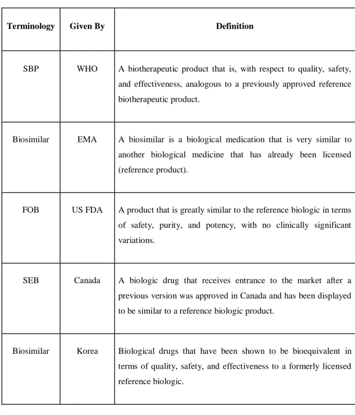
The comparable biologic drug products are recognized as similar biotherapeutic products (SBPs) by the World Health Organization (WHO), biosimilars by the European Medicines Agency (EMA) of the European Union (EU), follow-on biologics (FOBs) by the US Food and Drug Administration (FDA), and subsequent-entry biologics (SEBs) by Health Canada. In few events, the term “biosimilar” has been utilized, and hence it is quite essential to analyze variations in biosimilar definitions in various regions (Table 1).
27
Table 1: Definitions of biosimilar according to different country’s regulatory bodies (Biosimilar Medicines: Overview | European Medicines Agency, n.d.; Kabir et al., 2019).
Terminology Given By Definition
SBP WHO A biotherapeutic product that is, with respect to quality, safety, and effectiveness, analogous to a previously approved reference biotherapeutic product.
Biosimilar EMA A biosimilar is a biological medication that is very similar to another biological medicine that has already been licensed (reference product).
FOB US FDA A product that is greatly similar to the reference biologic in terms of safety, purity, and potency, with no clinically significant variations.
SEB Canada A biologic drug that receives entrance to the market after a previous version was approved in Canada and has been displayed to be similar to a reference biologic product.
Biosimilar Korea Biological drugs that have been shown to be bioequivalent in terms of quality, safety, and effectiveness to a formerly licensed reference biologic.
According to the various definitions mentioned above, it is evident that in the definition of biosimilars, there are three universal characteristics:
28 1. The biosimilar drug needs to be a biological drug;
2. The reference biologic requires it to be a previously approved biological product;
3. It is absolutely necessary to exhibit high bioequivalence in quality, effectiveness and toxicology (Wang & Chow, 2012).
In order to confirm biosimilarity to a reference biologic, the US Food and Drug Administration (FDA) applies a totality-of-the-evidence approach that involves analytical research, animal studies, and clinical trials to make a comparison of human pharmacokinetics (PK), pharmacodynamics (PD), clinical effectiveness, and safety, including immunogenicity. (Cuellar et al., 2019).
Although it is quite necessary to acknowledge the fact that biosimilars are not identical to their generic versions, and thus therapeutic equivalence is not exhibited by default. The term
“generic” medication is generally adopted in order to refer to drugs composed of small molecules which are structurally and therapeutically equivalent to their respective reference drug. In contrast, biologics are more difficult to be characterized structurally. In terms of size, biologics are exceedingly bigger than chemically synthesized drugs. They contain hundreds of amino acids which are combined in a biochemical process following a definite sequence. Consequently, biologics usually own several secondary and tertiary structural and also post-translational modification alterations. Glycation, oxidation, glycosylation, sulfide crosslinking, etc. which fundamentally prompt structural alterations, can be found within the identical lot of biological products. Since small variations are quite hard to eliminate, extremely similar biosimilars and their reference products do not have the similar therapeutic effectiveness. Due to the complex large molecular structure, there are various challenges in the manufacture of these biotherapeutic products in comparison with conventional small molecule generic drugs.
Biosimilar market is well established in the United States and European countries (Kabir et al., 2019). Aside from Europe and the United States, biosimilars have been launched as alternatives to biological pharmaceuticals in Japan, Australia, and a few other nations. The market for biosimilars is expected to expand significantly, with advantages in terms of new advanced technologies, market competition, patent expiration, the development of reliable, quality drugs at reasonable costs, and global demand all contributing to the expansion of the biosimilar market.
29
The success resulting from biosimilars can greatly prompt the sustainability of pharmaceutical business in the upcoming decade (Rehman et al., 2018).
5.1.1 Advantages of Biologics and Biosimilars
● Biologics is essentially capable of binding with target sites which are most likely tricky or unfeasible for small molecule drugs.
● Biologics are capable of providing improved economical return in comparison to that of small molecule drugs (Kabir et al., 2019).
● As the clinical evaluation and biosimilar approval processes are simplified along with an efficient manufacturing process, the overall costs of biosimilars development tend to be lower than reference products. Because of the price competition, biosimilar’s introduction can greatly lessen the expenses of originator biological products.
● By reduction of the costs, biosimilars possess the potential of allowing the budget reallocation to novel treatments or reinvestment (Cuellar et al., 2019).
● The availability of biosimilars is considered to be a way to expand access to biotherapeutic products as they are fundamentally providing more treatment options (Kang et al., 2020).
● Biosimilars have the promise to enhance the use of biologics (Health Care System Benefits | Pfizer Biosimilars, n.d.).
● The capability of biosimilars in increasing the efficiency of the healthcare system is incredible. Hence, it is assumed that biosimilars will grant a positive impact on the public health and healthcare system (Santos et al., 2019).
5.2 Manufacturing Process of Biologics and Biosimilars
Manufacturing process of both biologics and biosimilars involves a step wise process which includes cell line development, cell culture recovery and purification of the product. The whole process can be elucidated by these subsequent 4 steps:
i. Cell line development: A specific cell line requires to be engineered that contains the required gene which will function for the transcription of the preferred biologic.