Review Article
Prolonged Survival in a Non-Small Cell Lung Cancer
with Brain Metastasis Treated with a Brain Mass
Evacuation and Ge
fi
tinib
Mardiah Suci Hardianti, Johan Kurnianda
Division of Hematology and Medical Oncology, Department of Internal Medicine Sardjito General Hospital/ Universitas Gadjah Mada, Yogyakarta
INTRODUCTION
The incidence of brain metastasis (BM) is reported to be increase about 20-40% over the last few years in patients with non-small-cell lung cancer (NSCLC).1 The use of epidermal growth factor receptor (EGFR) tyrosine kinase inhibitor (TKI) in NSCLC patients with BM, especially those harboring EGFR mutations have been reported.
CASE REPORT
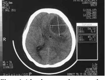
A 56-year-old woman with the initial major complaint of persistent and progressive headache within the last 4 months. Within the last 1 year before she sometimes complained of mild to moderate headache that still allowed her to work. She had seeked for medication to several private hospitals before then reffered to our hospital. A head computed tomography (CT)-scan confi rmed multiple lesion in the frontal area (January 29 2013) (Figure 1). She was admitted into our hospital with worsening symptoms of consciousness and unable to communicate either passively or actively since the last days before hospitalization. Other imaging finding was a suspected mass in
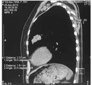
the lung eventhough the patient had never complained for any respiratory problems (Figure 2)
Figure 1. A head computed tomography (CT)-scan confi rmed multiple lesion in
the frontal area
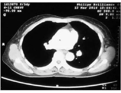
(Figure 3). An ultrasound guided fi ne needle aspiration confi rmed a diagnosis of Non Small Cell Carcinoma. The EGFR mutation analysis from the fine needle aspiration revealed a deletion at exon 19.
Figure 3. Lung mass of the left medial lobe (3.3x2cm)
She also felt that the headache got progressive in the last one month. Evaluation of head CT Scan showed multiple brain metastasis of the frontal area (Figure 4).
The administration of EGFR-TKI (Gefi tinib) was started in May 23 2013. The
following imaging for evaluation showed improvements of the brain and lung (Figure 5a and b).
The patient kept on consuming Gefi tinib for 16 months (May 2013- September 2014). During this period the patient mostly had a good cognitive function, and good daily functional and social life. However, her husband informed that she recently sometimes complained of slight headache and forgetfulness. Head MSCT was done with result of multiple brain metastasis of the left frontal lobe with midline
Figure 2a and b. Suspected lung mass of the left medial lobe (2.5x2cm)
Figure 4. Multiple brain metastasis in the frontal area. Shifting of the midline
shifting to the right (Figure 6). Thorax MSCT resulted in stable disease for partial response (d 0.7cm) as compared to the result in September 2013 (Figure 7).
DISCUSSION
Non-small-cell lung cancer (NSCLC) is still the leading cause of cancer-related death worldwide, mainly due to its high incidence and late diagnosis. The 20–40% increase in the incidence of brain metastasis (BM) in NSCLC patients reported over the last few yearsis largely the result of advances in neuroimaging and prolonged survival achieved using new treatments.1 BM has long been a major issue in clinical practice, since its symptoms lead to a signifi cant impairment of quality of life and a rapid decline in the patient's clinical condition. If untreated, the prognosis of BM patients is poor, with a median survival of 1–2 months.1
In this case the initial complaints of the patient was persistent headache which gradually progressed leading to lack of consciousness and motoric aphasia. The symptoms of mild to moderate headache had started approximately
Figure 5a. Improvement of the perifocal edema and lesion in the right frontal area (when compared to the result in September 2013), 5b. Two months after routine
Gefi tinib administration.
Figure 6. Multiple brain metastasis of the left frontal lobe with midline shifting
to the right
Figure 7. Compared with September 2013: stable disease for partial response
from one year before getting worsened in the last 4 months.
Several treatments for BM include whole-brain radiation therapy (WBRT), long considered the cornerstone of treatment, is effective in achieving local disease control and a survival prolongation of 4–6 months, with an improvement or, at least, a stabilization in symptoms, but it is frequently burdened with signifi cant comorbidities and side effects. In the presence of one to three brain lesions, the treatment of choice may be gamma knife, radiosurgery or surgical removal, which is known to prolong survival.2
In this case a salvage craniotomy for mass evacuation was done due to severe clinical symptoms of lack of consciousness and motoric aphasia.
The efficacy of chemotherapy in the treatment of patients with BM is limited, with data reported in various studies showing response rates ranging from 15 to 50%. Interestingly, following chemotherapy for BM, the response rates achieved are comparable with those obtained in systemic disease. Nonetheless, the median survival remains poor, ranging from 4 to 6 months.3
The blood–brain barrier (BBB) is an anatomic structure consisting of endothelial vessel cells, astrocytes and pericytes with tight junctions (TJs) and a number of carrier proteins which controls and limits the passage of molecules to the brain. In the presence of an intact BBB, only small lipophilic molecules (molecular weight [MW] <400 Da) can cross the BBB by diffusion, while the passage of other molecules is regulated by the carrier proteins. The loss of TJs in the tumor vascular system in macroscopic intracranial lesion does not necessarily incur the loss of other biological BBB components, such as
detoxifi cation and drug resistance mechanisms, which may remain effective and compromise drug ¬concentrations in BM.4
Epider mal growth factor receptor (EGFR)-dependent pathway plays an important role in the development and progression of human epithelial cancers, including NSCLC. EGFR is expressed on the cell surface of a substantial percentage of NSCLCs. Activation of the EGFR pathway is able to promote tumor growth and progression, stimulating cancer cell proliferation, production of angiogenic factors, invasion and metastasis, and inhibiting apoptosis.5 EGFR works through two different
downstream signaling pathways, which are activated by the phosphorylation of the ATP-binding domain of the receptor: the MAPK cascade, which activates different genes linked to cell proliferation and survival; and the PI3K–AKT cascade, in which phosphorylated AKT inactivates proapoptotic proteins. TKIs inhibit the phosphorylation and tyrosine kinase activity of the intracellular domain of EGFR through competitive binding to this site, thus preventing the activation of downstream signaling.6 In general, activating EGFR
mutations are more commonly observed in patients with adenocarcinomas and no prior history of smoking, as well as in females and those of Asian descent.
responses in 210 of 268 patients (78%). These response rates observed in patients with EGFR-mutant tumors receiving TKIs were much higher than what had been described in patients treated with standard platinum-doublet chemotherapy.7 By contrast, the exon
20 T790M mutation is associated with acquired resistance to TKI therapy.8
In this patient the result of EGFR mutation analysis from the fine needle aspiration revealed a deletion at exon 19 suggesting the administration of Gefi tinib.
Lung cancers harboring mutations in the epidermal growth factor receptor (EGFR) respond to EGFR tyrosine kinase inhibitors, but drug resistance invariably emerges. All drug-resistant tumors retained their original activating EGFR mutations, and some acquired known mechanisms of resistance including the EGFR T790M mutation or MET gene amplifi cation. Some resistant cancers showed unexpected genetic changes including EGFR amplifi cation and mutations in the PIK3CA gene, whereas others underwent a pronounced epithelial-to-mesenchymal transition.9
In this case, the underlying cause of the progression was not yet defi ned. Nonetheless any molecular changes is possible. Further molecular analysis should be done not only to defi ne the mechanism of resistance but also to determine further treatment.
REFERENCES
1. Wen PY, MB P, Loeffl er J. Metastatic Brain Cancer.Wen PY, MB P, Loeffl er J (Eds). Lippincott Williams and Wilkins, PA, USA, 1999.
2. Gaspar LE, Mehta MP, Patchell RA et al. The role of whole brain radiation therapy
in the management of newly diagnosed brain metastases: a systematic review and evidence-based clinical practice guideline. J. Neurooncol . 2010. 96(1), 17–32. 3. Bartolotti M, Franceschi E, Brandes AA.
EGF Receptor Tyrosine Kinase Inhibitors in the Treatment of Brain Metastases From Non-small-cell Lung Cancer. Expert Rev Anticancer Ther . 2012. 12(11):1429-1435.
4. Muldoon LL, Soussain C, Jahnke K et al. Chemotherapy delivery issues in central nervous system malignancy: a reality check. J. Clin. Oncol. 2007. 25(16), 2295–2305.
5. Gridelli C, Ciardiello F, Gallo C, Feld R, Butts C, Gebbia V, Maione P et al. First-line erlotinib followed by second-First-line cisplatin-gemcitabine chemotherapy in advanced non-small-cell lung cancer: the TORCH randomized trial. J Clin Oncol . 2012 Aug 20;30(24):3002-11
6. Maemondo M, Inoue A, Kobayashi K, Sugawara S, Oizumi S, Isobe H, Gemma A et al. Gefi tinib or Chemotherapy for Non– Small-Cell Lung Cancer with Mutated EGFR. N Engl J Med. 2010;362:2380-8. 7. Sequist LV, Bell DW, Lynch TJ, Haber
DA. Molecular predictors of response to epidermal growth factor receptor antagonists in non-small-cell lung cancer. J Clin Oncol. 2007 Feb 10. 25(5):587-95.
8. Pao W, Miller VA, Politi KA, et al. Acquired resistance of lung adenocarcinomas to gefitinib or erlotinib is associated with a second mutation in the EGFR kinase domain. PLoS Med. 2005 Mar. 2(3):e73.