Molecular based detection for drug resistance in mycobacterium
tuberculosis
Sumanto Simon*, Inggriani Listiawan**
Abstrak
Multi drug resistant – tuberculosis (MDR-TB) masih merupakan masalah yang serius, terutama bagi negara-negara yang sedang berkembang. Untuk melakukan suatu tindakan pengobatan yang tepat dan mencegah terjadinya resistensi obat lebih lanjut, maka deteksi dini atas isolat klinis Mycobacterium tuberculosis sangat penting. Selama ini untuk mengidentifikasi isolat-isolat tersebut digunakan metode konvensional yaitu media solid, dan akhir-akhir ini juga telah diperkenalkan suatu metode secara manual dan otomatis (Bactec atau MB/BacT) yang menggunakan metode cair, namun hasil pemeriksaan memerlukan waktu sekitar 2 sampai 4 minggu. Penggunaan tes molekul berbasiskan genetika sanggup mengidentifikasi gen yang bermutasi yang menyebabkan resistensi obat; misalnya resistensi terhadap rifampisin, dalam 1 hari kerja. Salah satu pendekatannya ialah menggunakan analisis molekul untuk mendeteksi mutasi yang berkaitan dengan resistensi obat INH dan rifampisin. Pada kasus INH, mutasi terjadi pada gen katG, inhA, kasA dan ahpC yang merupakan gen-gen yang bertanggungjawab terhadap sebagian besar dari M. Tuberculosis yang resisten INH, sedangkan mutasi-mutasi dari rpoB bertanggungjawab terhadap M. Tuberculosis yang resisten RIF. (Med J Indones 2003; 12: 259-65)
Abstract
Multi- drug resistant tuberculosis continues to be a serious problem, particularly among some developing countries. Early detection of drug resistance in clinical M. tuberculosis isolates is crucial for appropriate treatment and to prevent the development of further resistance. Compared to conventional methods using solid media, the introduction of manual and automated methods (BACTEC or MB/BacT) for susceptibility testing in liquid media has resulted from 4 to 6 weeks to 3 to 15 days. The identification of resistance mutations, e.g., the genetic basis for RIF resistance, enables the development of molecular test that allows the detection of resistant strains within 1 day. One approach is the use of molecular analysis to detect mutations that are associated with resistance to drugs including INH and RIF. In the case of INH, mutations of the katG, inhA, kasA, and ahpC genes are responsible for the majority of INH-resistant M. tuberculosis, whereas mutations of rpoB are responsible for RIF-resistant M. tuberculosis. (Med J Indones 2003; 12: 259-65)
Keywords: Mycobaterium tuberculosis, molecular analysis, rapid detection, MDR-TB
Despite the recent promotion of directly observed therapy-short course (DOTS) by World Health Organization (WHO) in many countries, multi drug resistant-tuberculosis (MDR-TB) continues to be a 21st century problem for TB control programs.1 Multiple-drug-resistant Mycobacterium tuberculosis is a major concern to health authorities worldwide.2,3 Unlike the antibiotic resistance in many bacterial species, which is acquired by gene transduction, conjugation, or transformation, the drug resistance in
M. tuberculosis is genomically based. Resistance to first-line anti tuberculosis drugs has been linked to mutations in nine genes, viz., katG, inhA, aphC, and
kasA for isoniazid resistance, rpoB for rifampicin resistance, rpsL, and rrs for streptomycine (SM) resistance, embB for ethambutol resistance, and pncA
for pyrazinamide resistance.4 Mutations identified in these genes have been associated with drug resistance based on their absence in drug susceptible isolates.4
Multiple-drug resistance results from the accumulation of mutations in different genes.5,6
In the past decade, molecular techniques have been developed to allow the amplification and detection of minute amounts of nucleic acid sequences from tissues
*
Department of Bio-medic Faculty of Medicine, University of Catholic Atma Jaya, Jakarta, Indonesia
**
or body fluids. These nucleic acid amplification methods can create millions of identical copies of a DNA or RNA “target” sequence in a few hours. The ability to determine whether specific DNA or RNA sequences are present in clinical samples using molecular technology has dramatically changed our approach to the diagnosis of many diseases. Molecular based techniques have been used in the diagnosis of infections due to slow-growing or fastidious micro-organisms, detection of infectious agents that cannot be cultured, epidemiological surveillance, and rapid identification of anti-microbial resistance. Radio-isotope based molecular-biology methods have been demonstrated to have comparative advantages in being sensitive, specific, cost-effective, and suitable for application to large-scale molecular surveillance for drug resistance.7
Epidemiology
Around 8 million people are infected by Mycobaterium tuberculosis every year, of which about 3 million occur in South-east Asia and 1,5 million in sub-Saharan Africa. Tuberculosis kills 2 million people each year. Overall, one-third of the world’s population is currently infected and, of these, 5-10% develop active tuberculosis to become infectious during their life- time. TB is a leading cause of death among people who are HIV-positive. In Africa, HIV is the
single most important factor determining the increased incidence of TB in the last ten years.1 In Indonesia, there are no accurate data available but it should be very high in incidence and prevalence of TB, since Indonesia is the country in the third rank of TB-burden after China and India.
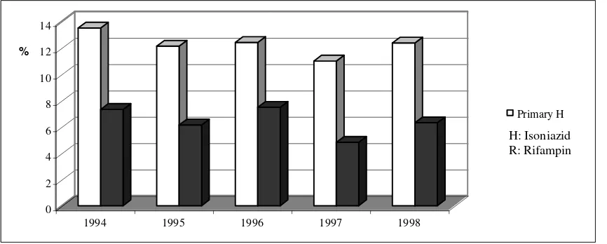
Multi-drug resistant tuberculosis (MDR-TB), due to mycobacterium tuberculosis resistant to at least INH and rifampicin, the two most effective and commonly- used anti-tubercular drugs, is rising at an alarming rate. Resistant in TB develops under the selection pressure exerted by the use of these drugs. It is classified as acquired resistance, when drug-resistant mutants are selected as a result of ineffective treatment; or as primary resistance, when a patient is infected by a source case with a resistant strain. According to a recent WHO/IUATLD survey on TB drug resistant from 35 countries, the weighted mean of primary resistance to any drug is 18% and the global prevalence ranges from 0-22.1%. WHO Collaborating Centre For TB / Persahabatan Hospital Jakarta, Indonesia conducted a five year (1994-1998) survey on TB drug resistant and reported primary resistance to INH and Rifampicin is 13% and 7%, consecutively.8
About 98% of all TB death occurs in developing country, where surveillance for resistance of M. tuberculosis isolates to anti-TB drugs is uncommon.9
Figure 1. Pattern of Primary Resistance to Antituberculosis Drugs WHO Collaboration Centre for TB Persahabatan Hospital Jakarta (1994-1998) 8
0 2 4 6 8 10 12 14
%
1994 1995 1996 1997 1998
Primary H
Primary R
Rapid Drug Susceptibility Testing
Early detection of drug resistance in clinical M. tuberculosis isolates is crucial for appropriate treatment and to prevent the development of further resistance. Compared to conventional methods using solid media, the introduction of manual and automated methods for susceptibility testing in liquid media has resulted in a reduction of turn around times for susceptibility results from 4 to 6 weeks to 3 to 15 days.4,10 The commercial radiometric BACTEC system may also take 2 weeks to provide a result,11 even with expensive equipment and reagents.
Techniques used for Molecular Detection of Resistance to Anti-microbial agents
It is now known that resistance to drugs is due to a number of genomic mutations in specific genes of microbes. These mutations can be used as markers for drug resistance, since isolates susceptible to these drugs lack the corresponding gene mutations. The initial common step in many methods for the molecular detection of drug resistance is the polymerase chain reaction (PCR). This technique is used to amplify the genes in which the mutations associated with drug resistance are to be detected.
PCR
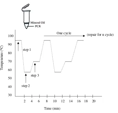
PCR is performed in a thermocycler, which allows the reaction to occur at the various temperatures required. The steps are as follows. The nucleic acid (e.g. DNA) target is extracted from a microbial culture or a clinical specimen of interest. This target DNA is used as the template for amplification. Heating, at about 95 C, is used to separate the extracted double-stranded DNA into single strands (denaturation). Cooling to 50 to 55 C then allows primers, specifically designed to flank the target nucleic acid sequence, to adhere to the target DNA (annealing). The annealing temperature for each primer set was calculated as follows: TM. = 4 (G + C) + 2 (A + T).12 Then the thermostable enzyme Taq Polymerase is allowed to act for some time at 72 C, to allow incorporation of nucleotides (dNTPs), that are provided in the reaction mix, to create new DNA fragments complementary to the target DNA (extension). This completes one cycle of PCR. This process of denaturation, annealing and extension is repeated several times in the thermocycler. At the end of each cycle, each newly synthesized DNA sequence
acts as a template for the next cycle, so that after 25 to 30 cycles millions of copies of the original target DNA are created. The result is the accumulation of a specific PCR product (amplicon) with sequences located between the two flanking primers.13
Standard PCR is followed by amplicon sizing by gel electrophoresis. The amplicon band migrates on the gel based on its size (i.e. number of base pairs). The band can be visualised by staining with ethidium bromide, a DNA intercalating dye, and the size compared with molecular weight markers, which are also run on the gel. The specificity of the band is confirmed by hybridisation with a labelled probe specific and complementary to the amplicon. Hybridisation also increases the sensitivity of detection, as compared to ethidium bromide staining.
The Detection of Mutations
Figure 2. Polymerase Chain Reaction (PCR).14 PCR is a cycling process; with each cycle the number of DNA targets doubles. The
Figure 3: PCR Temperature Cycling Profile.14
Step 1: Heat denature the double-stranded DNA in the presence of primers, the four dNTPs, PCR buffer and a thermo-stable DNA polymerase. Denaturation is normally in the range 93-100 0 C
Step 2: Anneal the oligonucleotide primers to the denatured template by lowering the temperature to 37-65 0C depending on the Tm of the oligonucleotide primers.
Step 3: Extend the primers at 72 0C with a thermo-stable DNA polimerase. Step 1 to 3 constitute one cycle of PCR. This process is then normally repeated (cycled) for at least 20 cycles. At the last cycles the extension time may be increased by several minutes to complete the synthesis of all strands.
PCR amplification followed by DNA sequencing is the most widely used technique for the identification of mutations in developed countries, it is not readily available in routine laboratories in developing countries, and is not currently suitable for analysing large number s of samples for epidemiological surveillance of drug resistance.7
Advantage and limitation of molecular detection of resistance
Genotypic resistance tests are rapid, especially for slow growing organism (e.g. Mycobacterium tuberculosis). They can be applied to organism that cannot be cultured, and to partially treated cases where the growth of the organism is inhibited. Genetic
2 4 6 8 10 12 14 16 18 20
100
90
80
70
60
50
40
30
(repair for n cycle)
One cycle
T
emperatur
e (
C)
step 2
step 3
step 1
methods may be adapted to detection of resistance directly in clinical specimens, and obviate then need for prior isolation of organism by culture. These methods assess the genotype of the organism, whereas conventional techniques assess the phenotype (genotype expression) under artificial or laboratory conditions. Genetic methods do not carry the biohazard risk associated with cultivation of microbes.7,18 However, these methods also have certain limitation. As most of them are based on initial amplification of the gene targets by PCR, the inherent risks of PCR apply to these methods too. Proper precautions are required to avoid amplicon contamination, including the physical separation of areas for handling pre- and post-amplification steps. Even PCR may not be able to amplify the gene from targets from samples that have very few organism. Background sequence information and prior knowledge of mutations associated with resistance is required. Genotypic mutations may not always lead to phenotypic expression levels manifesting as drug resistance.18 However, in this context, it is important to remember that even the data generated by conventional in-vitro methods of drug susceptibility testing do not always correlate to clinical drug resistance.21
The Future
More recently, Victor et al.12 have developed a PCR-based screening method, that allows batch analysis of samples using dot-blot hybridisation with radio labelled probes. The utilisation of this technique, with International Atom Energy Agency (IAEA) assistance, has shown that it is reproducible, technically undemanding, and takes only two working days to provide results from the start of amplification to the final auto-radiography step of the dot-blot hybridization.22 Molecular prediction for drug resistance is optimal for rifampicin, where approximately 98% of RIF-resistant clinical isolates have a mutation in the rpoB gene.13
Moreover, it is very challenging for Indonesia as a country with very high TB burden to conduct a comprehensive, effective, nationwide reaching TB control program.
One of the component in setting up of this TB-control program is one Top National Referral - Laboratory using radioisotope molecular tests for detection of drug resistance in M. tuberculosis.
CONCLUSION
In summary, molecular methods have been used for the detection of drug resistance in Mycobacterium tuberculosis. Several post- PCR strategies are available to rapidly detect mutations, including those that lead to drug resistance. These methods include restriction fragment length polymorphism (RFLP), single strand conformational polymorphism (SSCP), probe- based methods like the dot blot assay. Many of these methods have practical limitation for genetic assessment of drug resistance. Not all mutations result in the gain or loss of a restriction enzyme site. The mutation that does not do so cannot be detected by RFLP. Other oft-used screening procedure, which depend on DNA electrophoretic mobility shifts (e.g. PCR-SSCP) are technically demanding and are not sufficiently sensitive. The use of radioisotope based molecular biology diagnostic procedure for the detection of drug resistance in connection with post-PCR method (e.g. dot blot hybridisation) could be the answer to face this challenge in the developing countries such as Indonesia.
REFERENCES
1. World Health Organization, International Union Against Tuberculosis and Lung Disease. Anti-tuberculosis drug resistance in the world : The WHO/IUATLD Global Project on Anti-tuberculosis drug Resistance Surveillance, Report No.2. WHO/TB/2000.278.Geneva :WHO, 2000. 2. Edlin BR, Tokar JI, Grieco MH, Crawford JT, Williams
J, Sorddillo EM, et al. An outbreak of multidrug-resistant tuberculosis among hospitalized patients with the acquired immunodeficiency syndrome. N Engl J Med 1992; 326:1514-21.
3. van Rie A., Warren RM, Beyers N, Gie RP, Classen CN, Ricardson M, et al. Transmission of multidrug-resistant Mycobacterium tuberculosis strain among non institutionalised, human immunodeficiency virus-seronegative patients. J Infect Dis 1999;180:1174-79. 4. Ramaswamy S, Musser JM. Molecular genetic basis of
antimicrobial agent in Mycobacterium tuberculosis. Tuber Lung Dis 1998;79:3-29.
5. Heym B, Honore N, Truffat PC, Banerjee A, Schurra C, Jacobs WR, et al. Implications of multi-drug resistance for the future of short-course chemo theraphy of tuberculosis : a molecular study. Lancet 1994; 344: 293-8.
6. Morris S, Han Bai G, Suffys P, Portillo-Comez M, Fairchok M, Rouse D. Molecular mechanisms of multiple drug resistance in clinical isolates of Mycobacterium tuberculosis. J Infect Dis 1995;171: 954-60.
8. Aditama TY. Facts & Figures. Mycobacterial Laboratory Persahabatan Hospital WHO Collaborating Center for TB. 2000. Published on world TB Day. March 24, 2000. 9. Nunn P, Felten M. Surveillance of resistance to
anti-tuberculosis drugs in developing countries. Tuber Lung Dis 1994; 75:163-7.
10. Frieden TR, Sherman LF, Maw KL, Fujiwara PI, Crawford JT, Nivin B, et al. A Multi-institutional outbreak of highly drug tant tuberculosis: epidemiology and clinical outcomes. JAMA 1996; 276: 1229-35. 11. Rastogi N, Goh KS, David HL. Drug Susceptibility
testing in tuberculosis: a comparison of the proportion methods using Lowenstein-Jensen, Middlebrook 7H10 and 7H11 agar media and a radiometric method. Res Microbiol 1989; 140 : 405-17.
12. Victor TC, Jordaan AM, van Rie A. Detection of mutations in drug resistance genes of Mycobacterium tuberculosis by a dot-blot hybridisation strategy. Tuber Lung Dis 1999; 79: 343-8.
13. Mullis KB, Faloona FA. Specific` synthesis of DNA in vitro via a polymerase chain reaction. Methods Enzymol 1987; 155: 335-50.
14. Newton CR, Graham A: PCR: Introduction to Biotechniques, Bio Scientific Publishes Limited, United Kingdom 1994:1-38
15. Cockerill FR 3rd, Uhl JR, Temesgen Z. Rapid identification of a point mutation of the Mycobacterium tuberculosis catalase-peroxidase (katG) gene associated with isoniazid resistance. J Infect Dis 1995; 171: 240-5.
16. Telenti A, Imboden P, Marchesi F. Detection of rifampicin-resistance mutation in Mycobacterium tuberculosis. Lancet 1993; 341: 647-50.
17. Heym B, Alzari PM, Honore ST. Missense mutations in the catalase-peroxidase gene, katG, are associated with isoniazid resistance in Mycobacterium tuberculosis. Mol Microbiol 1995; 15: 235-45.
18. Cockreill FR 3rd. Genetic methods for assessing antimicrobial resistance. Antimicrob Agents Chemother 1999; 43: 199-212.
19. Doukhan L, Delwart E. Population genetic analysis of the protease locus of human immunodeficiency virus type 1 quasispecies undergoing drug selection, using a denaturing gradient-heteroduplex tracking assay. J Virol 2001; 75:6729-36.
20. Garcia de Viedma D, del Sol Diaz Infantez M, Lasala F, Chaves F, Alcala L et al. New Real-Time PCR Able To Detect In A Single Tube Multiple Rifamicin Resistance Mutations And High-Level Isoniazid Resistance Mutations In Mycobatedrium Tuberculosis. J Clin Microbiol 2003:988-95.
21. Acar JF, Goldstein FW. Consequences of increasing resistance to antimicrobial agents. Clin Infect Dis 1998; 27 Suppl 1: S125-30.