Vol9, No 1, January - March2000 Diagnosis of dengue infection
Semi-quantitative
dot
immunoassay
for detection
of
IgM
anti-dengue
antibodies
in
human
sera
Agus Sjahrurachman
,Betty
Ernawati,
FeraIbrahim, Mardiastuti
,Tjahjani
Mirawati
Sudiro,
Pratiwi
SudarmonoAbstrak
Diagnosis laboratorik untuk infeksi virus dengue berperan penting dalam pengelolaan kasus dan pemberanta.san penyakit. Sayangnya,
cara diagnosis laboratorik yang ada saat ini sebagian besar lambar hasilnya dan sukar pengerjaannya. Dalam makalah ini dilaporkan
hasil sementara
uji coba
forntat
imunoesei noktah yang kami kembangkan untuk mendeteksi antibodi anti-dengueIgM
secarasemikuantitatif menggunakan vints clengue terlabel biotin. Hasilnya menurlukkan bahwa cara baru ini nttunpu nrembeikan hasil dalam waktu 4,5 jam dan cukup sensitif, klwsusnya nttuk infeksi sekunder.
Abstract
Laboratory diagnosis of dengue infection plays an important role
for
case nlanagenrent and disease control. Unfortunately, many of the available laboratory tests are not appropriate in tet'ms of rapidity and .simplicity. H ere, we report a new format of semiquantitative dot immunoassayfor
detectionof
IgM-anti dengue antibodies in humansera
entltloyittg biotinylated-dengue antigens which is simple in term of methodoLogy and rapid in ternt of the lest result. Tlrc results indicated that tlrc new testis
sensitivefor
diagnosis of secondarydengue infection. The test result can be obtained withinfour and a halfhours. Key w o rds : bio t in1, I a t e d - de n gue, dia gno s i s, inununoas s ay
Dengue infection
is
prevalent
in
tropical rcgion of
Asia, America
andAfrica.'-"
Sporadic outbreaks havealso occurred
in
subtropical regions
of
Australia,
America,
Asia
andAfrica.l'a In
other words,
dengueinf'ection
is
one among other major
public health
problems
in
many developing countries. On
theother
hand,it
iswell known
thatdefinite
diagnosis of dengueinfection is hardly
determined
solely on
the basis
of
clinical
signs and symptoms. Laboratory
investiga-tions
is
required
in majority of cases.a')
Several
laboratory
approaches to diagnose denguein-fection
in
suspected caseshave
been developed.4'6-10Virus
isolation
and polymerase
chain reaction
havebeen proved
to
be themost valuable
methodsfor the
viremic phase
of
acute stage.4'8'10
Ho*"ver,
thosemethods are
not
appropriate
to be routinely
used
in
developing countries where laboratory facilities
arerelatively insufficient. Simpler laboratory
method
isneeded
for
suchdeveloping countries. Here
wereport
anew
andsimple laboratory method to
diagnoseden-Department of Microbiology, Faculty of Medicine, University cif Indonesia, Jakarta, Indonesia
gue infection
in
suspected casesemploying
biotiny-lated-denguc
fbr IgM-anti
denguedot
immunoassay.METHODS
Serum
samples
Serurn samples were
obtained
fiorn
patients
clinically
diagnosed as dengue
haemorrhagic
fever
and patientssuffering
fiom f'ever
without any
clinical symptoms'
and signs
of
respiratory, skin, gastrointestinal
nor
neurological
disorders.Conlirmation of
dengueinfec-tion
was done by haemagglutination
inhibition (HI)
test employing
4-8 HAU/0.025
ml of
four types
of
dengue antigenson
a microtiterplate
based onClarke
and
Casals
method./
Classification
of
immune
response
to dengue
was done according
to
W.H.O
critcria.4
Sera fiom volunteerswithout fever
within the
last three
months were
alsocollected.
Biotinylation of
dengue
viruses
Dengue
I
Mochizuki
strain,
dengue 2New
Guinea
C10
Sjahrurachman et alviruses
infected
C6136monolayer
cell culture fluid
was
clarified
from
cellular
debris
by low
speedcentrifugation.
Viruses
in
supernatant
fluid
were
precipitated by polyethylene
glycol
andcentrifuged
at10,000
rpm
for
30
minutes.
Viral
pellet
were
resuspended
in
phosphate
buffer saline
and
biotiny-lated
bybiotinyl-e-amidocaproic
acidN-hydroxy
suc-cinimide ester (Boehringer Mannheimn Co.)
for 3
hours at room
temperaturie aspreviously d"scribed.ll
Dot
Immunoassay employing biotinylated-dengue
virus
(BDIA
)BDIA
was performed as describedpreviously.l2
Bti"f-ly,
onemicrogram
of
anti
humanIgM
wasblotted
ona piece
of
nitrocellulose
paper(Hybond
C,Amersham
Co) which
was
blocked with 2
7o skimmedmilk
andair dried.
Two
hundred
andfifty
microliter
of
I
:
100time diluted
test
serum wasthen applied
on the paperand incubated
at
room
temperature
for
two hours.
Reactions
of
IgM
and amixture
ofbiotinylated-dengue
as
well
as
the
mixture
of
biotinylated-dengue
andhorse-radish peroxidase-labelled streptavidin
weredone
atroom
temperature
for
one hour
each.Finally,
5-chloronaphthol
substrate
was
added
for
color
development.
After 30
minutes at
room
temperature, theintensity
of color (optical
density)
on
thedot
was measuredat
490nm wavelength
with
a portablecom-puting
reflection
densitometer (Tobias
AssociatesInc,
USA). The result
of
the test was also
observed by
naked eyes.
RESULTS
Optical density (
OD
) of
BDIA
One hundred
andforty six
paired patients sera
which
had been subjected to
HI
tests werecollected and tested
by BDIA.
They were
42
paired
serafrom
patientswithout
evidence
of
dengueinfection
;
23paired sera
showing
primary
dengue infection,
48
paired
serashowing acute secondary
dengue
infection and
33paired
serashowing
recent secondary
infection.
TheBDIA
results showed that
OD
490
from
the
seraobtained
from healthy
subjects'and non-dengue
sub-jects
were
very
low
compared
to those from
denguepatients
as shown
on Table
I
&
Figure
1.
Further,
observed
by
nakedeyes
all of the
blot
of
the serafrom
healthy
subjects andalmost all
serafrom
non denguesubjects
were colorless.
As
to the
blot
of
serafrom
dengue subjects,
our observation indicated that
OD
490
of
the sera takenfrom
convalescent stage patientswere higher than
sera from
acute stagepatients'
Table
l.
Optical density of BDIA on patient seraMed J Indones
Group of
subject
Stage OD490 value(Mean + SD)
Non-Dengue
(n=
42)Acute, primary dengue
(n=
23)Acute, secondary dengue
(n = 48)
Recent, secondary dengue
(n = 33)
Healthy person
(n
= l0)
Acute
Convalescence Acute
Convalescence Acute Convalescence Acute Convalescent
0.09 + 0.027 0.09 + 0.039
0.1
l8 + 0.077
0.165 + 0.102 0.148 + 0.079 0.191 + 0.083
0.168 + 0.076 0.176 + 0.076 0.06 + 0.026
0.t
E
o.sÀ o.r
3
-
0.3-æo5 .3.
7J-!o
:t:
a
a
o
e
r!r
+Sf-":l'
aaaaaa'aaa
a
!'
q1*
3I
o
I
-i-aa a
aa
;o
oo.
a
0.1 .a'olr
a o t..'.1'rlr8r
Figure
l.
Serum specimens collected from primary denguein-fection
( IaneI
and 2 ), secondary dengue ( Iane 3 and 4 ),non-dengue infection ( Iane 5 and 6 ) and heaLthy persons (lane 7 ). Lane 1,3 and 5 indicate sera taken at acute stage
and lnne 2,4 and 6 indicate sera taken at convalescent stage
of diseases. Optical density of the blot was measured by a
handy densitometer. Bar indicates mean value of optical
density.
Positivity of
BDIA
Mean OD490 value of healthy
personsplus
two
stand-arddeviation
was used as thecut
offîalue
.Using this
cut
off,
the
specimenshowing
the
OD
value
of
mini-mum 0.1
I
was scored aspositive while
the
specimenshowing
theOD value
of
less than 0. II
was scored asnegative
.Of
23 pairedseraof
primary
dengue, 30.4Vo [image:2.595.329.574.108.498.2]Vol9, No
l,
January - March2000sera
of
secondary dengue, 77.l
Voof
the acute sera and 91.7 Voofthe
convalescent seracould
be diagnosed aspositive
by
BDIA.
Asfor
recent denguecases,72.'l
7oof
acute
and75.7
7, convalescent
serawere
scored aspositive while only
19 .0 7oof
theacute
and 23.8 Voof
theconvalescent
sera showedpositive results for
non-dengue
cases.Details
of
data
and
typical results of
[image:3.595.82.313.212.687.2]BDIA
areshown
onTable 2
andFigure 2'
Table 2. Positivity
of BDIA to
detectIgM
anti-dengue in human seraGroup of
subject
Stage BDIANo. positive I total (Eo)
Diagnosis ofdengue
infection
II
Relationship between
duration
of
fever
and
DIA
result
In primary infection, only
afew of
the
cases showedpositive
BDIA
results when the sera weretaken
at
day5 of
fever
or earlier. Proportion of positive
DIA
in-creased
on the
serataken
atmore than
5
day after
the onsetof
fever.However,
in
secondaryinfection
hlgh€r
proportion of positive
BDIA
could
be observed
eventhough the
sera
were
taken
before
5
days
of
fever.
Details
of
data are shown onTable
3 andTable
4.Table 3. Conelation between duration
of
fever and BDIA result in primary infectionNon-Dengue
(n=
24) Primary dengue(n = 23) Acute, secondary dengue (n = 48)
Recent, secondary dengue (n = 33)
Acute
Convalescence Acute
Convalescence
Acute
Convalescence
Acute Convalescent
8t42 (19.0 %) 10142 (23.8 Vo) '7123 (30.4 Vo) 18123 (78.3 Va)
37148 (77.1 Va)
44148 (91.7 7")
24133 (72.7 7o)
25133 (75.7 Vo)
I 2 3 4 5
>5
Unknown
Ol4 (0 Vo) 011 (0 Vo) 214 (50 Vo) otz (0 %)
ll2
(50 Vo)lll
(100 Vo) 319 (33.3 %)0/0 0/0 0/0 012 (O Vo)
lll
(t0O Vo)l2ll2
(1,00 Vo) 5/8 (62.5 Vo) Dayof
Proportion of casesfever
showing positive DIAon acute sera
Proportion of cases showing positive DIA on convalescent
[image:3.595.339.580.258.604.2]sera
Table 4. Correlation between duration of fever and BDIA result in secondary infection
Day
of
Proportion of casesfever
showing positive DIAon acute sera
Proportion of cases showing positive DIA on convalescent sera
1
2 3 4 5
>5
Unknown
ll2
(50 7a) 315 (60 Vo) 7t9 (77.8 %) 717 (1OO Vo) 5/5 (71.4 Vo) 819 (88.9 Vo) 619 (66.7 Vo)0/0 0/0 0/0 0/0
1 11 (lOO Va)
38141 (92.7 Vo) 5/6 (83.3 Vo)
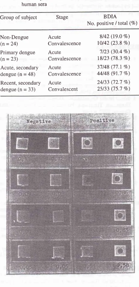
F i g ure 2. S e ra fr o m den g u e - inf e c t ed and de n gue non - infe c I e d
persons were subjected to BDIA. Optical density of the result
was measured with handy densitometer. Number below the
dots indicate optical density value.
DISCUSSION
Several
laboratory diagnostic
techniques
for
dengueinfection
areavailable.
Polymerasechain reaction
andvirus isolation
are among the most valuable techniquesin
termof
academic intJrest.a'8'10Positive result of
the test isindisputable.
Those techniques, however, arenot
suitable
for
developing countries where
laboratory
facilities
are
relatively insufficient.
Further,
thosetechniques are
time
consuming
and isnot appropriate
for
field
work especially in
remote
area. Becauseof
diag-12
Sjahrurachman et alnosis
of
dengue infection. Unfortunately,
however,
interpretation
of
theHI
testrequire
apair
sera taken atleast a
week
apart.Therefore, though
HI
testis
practi-cal
in
term
of
the
methodology, conclusion
of
theresult often cannot
bedrawn
sinceafter
oneweek
theinfected
casesmay
already be cured
or
passed away.On
theother
hand,it
is well known
that
in
anyinfec-tious
diseases,humoral immune
responseis
evoked.Further,
IgM
antibodies
is
one among other immune
response
that
appearearlier
in
the course
of
the
dis-eases.
"
Detection
of
lgM-anti
denguein
human
seratherefore
would be
an
interesting
target
to
beelaborated.
Here, we report a preliminary study
ondetection
of
an
IgM-anti
dengue
in
human
using
biotinylated-dengue.
The results indicate
thatIgM-anti
denguetiter in
den-gue
casesis
higher
compared
to
non-dengue
cases.Further,
IgM-anti
denguein
the
seraof
acute denguetaken
at
convalescent stage
is
higher than
at
acute stage,indicating
that
the humoral
immune
responsesto
the infecting
agents
is
switched on. On the
other
hand
lgM-anti
denguein
the
seraof
recent
infection
taken
from
acute andconvalescent
stagos arenot
dif-ferent, indicating that
the
humoral immune
responsehas
reached
a
plateau
phase
or viral
replication in
human
body
hasrecently
ceased. The results alsoindi-cate
that
lgM-anti
dengue
in
non-dengue cases
ishigher
as compared tohealthy
persons. The latestfind-ing
reflect
thatmajority of
healthy
personsenrolled
in
this study
are
immunologically virgin
to
dengue
an-tigens
while
thoseof
non-dengue cases has beenin
thepast exposed
to
dengue
antigen
as
indicated
by
the presenceof
anti
o*rJ:;ni:iaglutinin
tn
,n"t:r,";'I
emic
area
engueersist
for
Employing
cut
off
value
of
OD
0.1l,
apossibility for
using
BDIA
asdiagnostic
tool
is explored. The result
indicate that sensitivity
of BDIA
on
acute
serafrom
primary infection
is
low.
The
sensitivity
of
BDIA
sharply
increase
on
convalescent sera. Considering
that
majority
of
acute serafrom primary infection
aretaken
before day
5of
fever
,our
findings
isconsistent
with
previous findings which indicate that IgM-anti
dengue
in
seraof primary infection
could
be detectedwhen
the serais
takenby or
after day
5of
fever.4Compared
to primary infection, proportion
of
secon-dary infection
cases
showing
positive
BDIA
arehigher, reaching
9I.7
7o at convalescent stage.Further,
IgM-anti
dengue
in
secondary
infection
can
beMed J Indones
detected
earlier
as compared toprevious reports.l5
Theability
toearlier
detectIgM
anti
dengueby our
system, compared toprevious
reports, probably relate
to
thedifference
in sensitivity of
the
assay system.CONCLUSION
BDIA
is asimple
test andrequire
shortertime
than theHI
test.Further,
BDIA
has ahigh
sensitivity
especially
for
secondary dengue.Therefore,
BDIA might
beuse-ful
asroutine diagnostic method especially
for
secon-dary denguein developing countries.
Additional
study
involving larger
number
of
specimens
is required
toconfirm
the presentfindings.
REFERENCES
l.
GublerDJ.
Arboviruses as imported diseases agents : Theneed for increased awareness. Arch
Virol
1996l, llS:21-32.2. Gubler
DJ.
Resurgent of vector-borne diseases. EmergIn-fect Dis 1998; 4:442-50.
3. Lam SK. Emerging infectious diseases-South East Asia.
Emerg Infect Dis 1998; 4:145-'l .
4. World Health Organization. Dengue haemorrhagic fever.
Diagnosis, Treatment, Prevention and
Control.
WHOCeneva, 1997; p l-68.
5. Halstead SB. Dengue in health transition. Kaoshiung. J Med
Sci 1994; l0S:2-14.
6. Cardosa
MJ,
Tio PH. Dot enzyme immunoassay : analter-native diagnostic aid for dengue and dengue haemorrhagic
fever. Bull WHO 1991;
l2l:741-5.
7. Clarke
DH,
Casals J. Technique for haemagglutination andhemagglutination inhibition with arthropod-bome viruses.
Am J Trop Med Hyg 1958; 7:561-73.
8. Henchal EA, Putnak JR. The dengue viruses. Clin Microbiol
Rev 1990; 3:3'16-96.
9. Lam SK, Devi S, Pang
T.
Detectionof
specificIgM
indengue infection. SouthEast Asian J Trop Med Publ Health
1987 ; 18 :532-8.
10. Lanciotti RS, Calisher CH, Gubler DJ, Chang GJ, Vomdam
AV.
Rapid detection and typingof
dengue viruses fromclinical samples by reverse-transcriptase polymerase chain
reaction. J Clin Microbi ol 1992; 30:545-51 .
I
l.
Agus S, Tallei T, Betty E, Amin S, Pratiwi S. Biotin-labeleddengue virus for detection of
IgM
anti dengue antibodies.Indon J Clin Microbiol 1995 ; 7 :25-30.
12. Agus S, Betty E,'ljahjani MS, Amin S, Pratiwi S.
Develop-ment
of
dot enzyme immunoassayfor
detectionof
IgMantidengue. Indon J Publ Health 1996;3:208-14.
13.
Heinzell
FP,
RootRK.
Antibodies.In:
Mandell
GL,Douglas Jr RG, Bennett JE, editors. Principles and Practice
of
Infectious Diseases. NewYork:
Churchill LivingstoneInc;1990.
p.4l-61.
14. Suroso. Kebijakan Nasional pada Demam Berdarah dengue.
Cermin Dunia Kedokteran 1992; 8 I : l4-6.
15. Laboratory diagnosis of dengue virus infection.