Effects of Centrifugation and Lipid Removal on the Cryopreservation
of
in Vitro Produced Bovine Embryos at the Eight-Cell Stage
M. Murakami,* T. Otoi,† C. Sumantri,* and T. Suzuki*
*United Graduate School of Veterinary Sciences, Yamaguchi University, Yamaguchi 753, Japan; and†Tokushima Prefectural Beef Cattle and Swine Experiment Station,
Anan, Tokushima 774, Japan
The effects of intracellular lipid polarization and lipid removal treatments on the postthawed in vitro
development of frozen bovine embryos at the 8-cell stage were studied. As the first step, bovine presumptive zygotes were centrifuged at 16,000gfor 20 min for the cytoplasmic lipid polarization and their lipid layers were removed by micromanipulation in order to examine the influence of these treatments on the developmental capacity of bovine zygotes. As the second step, bovine embryos developed to the 8-cell stage following centrifugation treatment at various forces (8000, 12,000, and 16,000g) or lipid removal treatment at the 1-cell stage were frozen in 1.8 M ethylene glycol10.05 M trehalose supplemented with 5% polyvinylpyrrolidone in a one-step procedure. There were no significant differences among the control (nontreatment), lipid-polarized, and lipid-removed groups with respect to the developmental capacity of fresh nonfrozen zygotes (experiment 1). The rates of survival and development to the blastocyst of frozen-thawed 8-cell embryos increased slightly with increasing force of centrifugation (experiment 2). The rate of development into blastocysts of the frozen-thawed 8-cell embryos was significantly higher in the groups that underwent centrifugation (at 16,000gfor 20 min;P,
0.05) or lipid removal (P,0.01) treatments than the control (intact) group. However, there were no significant differences among the groups with respect to the rate of development to the expanded/hatched blastocyst stage. In addition, the mean cell numbers of embryos developed into blastocysts (day 8) derived from frozen-thawed 8-cell embryos tended to be low in the centrifugation and lipid removal groups compared to the controls (experiment 3). These results suggest that although the centrifugation with or without lipid removal treatments has no detrimental effects on the developmental capacity of bovine zygotes, the freezing tolerance of bovine 8-cell embryos was not improved by these treatments. © 1998 Academic Press
Key Words:intracellular lipid; polarization; removal; freeze; bovine embryo.
The effective cryopreservation of in vitro produced (IVP) bovine embryos at the early cleavage stage would not only provide a greater availability of materials for basic research and an improved manageability of theirin vitro cul-ture and subsequent embryo transfer, but would also broaden the range of practical applications of genetic engineering, such as the preservation of donor blastomeres for the purpose of cloning by nuclei transfer (16). It would also allow the preservation of embryos for production of chi-meric calves by aggregation of the 8-cell stage embryos (1). Moreover, in regard to the cur-rently available cryopreservation methods, freezing of the embryos might be much simpler in the cleavage stage rather than in earlier stages
such as immature and mature oocytes, which have shown relatively low survival rates after freezing and thawing (11, 12, 14). However, it has been suggested that IVP bovine embryos have a higher sensitivity to low temperature compared with in vivo derived embryos (7). Bovine embryos have been shown to contain a large number of lipid droplets at the early cleav-age stcleav-ages, and the decline of lipid droplets at the blastocyst stage is coincident with a loss in sensitivity to cooling (8, 15). Leibo et al. (7) suggested that intracellular lipids were at least partially responsible for the increased chilling and freezing sensitivity exhibited by IVP bo-vine embryos at the compact morula stage.
It has also been demonstrated that the toler-ance of porcine embryos at the early cleavage stage to cryopreservation can be highly in-creased after decreasing their relatively high Received September 1, 1997; accepted February 12,
1998.
206 0011-2240/98 $25.00
content and/or high composition of cytoplasmic lipid droplets by aspirating the lipids with a micropipette; the embryos continued to develop to blastocysts in vitro and to live offspringin vivo (10). However, this procedure harbors some operational problems for routine applica-tion, such as its complexity, laboriousness, and requirement of micromanipulation skills. Otoi et al. (12) recently suggested that polarization of cytoplasmic lipid droplets by centrifugation may improve the freezing tolerance of the spin-dles or organelles in mature bovine oocytes. We therefore suspected that, by centrifuging the bo-vine zygotes at higher speed and for longer times compared with other conventional levels (4, 9, 10, 16), the redistribution throughout the cytoplasm of the polarized lipid layer might be delayed and consequently the freezing tolerance of bovine embryos at the early cleavage stage could be enhanced by this treatment.
To test this hypothesis, the intracellular lipids of bovine zygotes were polarized by centrifuga-tion at various speeds, and the sensitivity of the embryos at the 8-cell stage to freezing was then assessed. In addition, the effect of a lipid re-moval treatment on the cryopreservation of bo-vine 8-cell embryos was investigated.
MATERIALS AND METHODS
Zygote Preparation
The methods used for thein vitromaturation, in vitrofertilization (IVF), and subsequent cul-ture in the experiments were modifications of the procedure described by Boedionoet al.(2). Ovaries were collected from cows at a local abattoir and were brought to the laboratory in physiological saline [0.9% (w/v) NaCl] at 25 to 30°C within 3 h. The cumulus– oocyte com-plexes (COCs) in follicular fluid (5 to 10 per ovary) were aspirated from follicles (2–7 mm in diameter) with a 5-ml syringe fitted with an 18-gauge needle. After being washed twice with modified phosphate-buffered saline (Embryo-teck, Nihonzenyaku, Fukushima, Japan), the COCs were cultured for 21 to 23 h in maturation medium [25 mM Hepes TCM-199 with Eagle’s salts (Gibco, Grand Island, NY) supplemented
with 5% superovulated cow serum (SCS), 0.01 mg/ml follicle-stimulating hormone (Denka Pharmaceutical Co., Kawasaki, Japan), 20 mM taurine (Wako Pure Chemical Industries Ltd., Osaka, Japan), and 50 mg/ml gentamicin
(Sig-ma Chemicals, St. Louis, MO)] at 38.5°C under 5% CO2in air.
Frozen semen was thawed in a water bath (37°C) and washed with 5 mM caffeine (Sigma) in Brackett and Oliphant (BO) medium (3) by centrifugation at 500gfor 5 min. The resultant sperm pellet was resuspended in BO medium supplemented with 2.5 mM caffeine, 3 mg/ml bovine serum albumin (BSA, fraction V; Sig-ma), 20 mM taurine, and 20 mg/ml heparin (Shimizu Pharmaceutical Co., Ltd., Shimizu, Japan) to a final concentration of 53106/ml. A 100-ml aliquot of the sperm suspension was
covered with mineral oil (Sigma). COCs that had been cultured for maturationin vitro were transferred into sperm microdrops (10 –15 oo-cytes per microdrop) for insemination. After incubation for 5 h, the COCs were transferred and washed several times in culture medium [TCM-199 supplemented with 5% SCS, 5
mg/ml insulin (Wako), 20 mM taurine, and 50 mg/ml gentamicin] and then cultured for an
additional 13 h.
Lipid Removal from Zygotes
After IVF and culture, the zygotes were in-cubated with 300 IU/ml hyaluronidase (Sigma) in culture medium for 20 min, and their adher-ent cumulus cells were then removed by pipet-ting. After being rinsed in fresh medium, the denuded zygotes were then transferred into 1.5-ml test tubes (Sarstedt, Germany) contain-ing 500 ml of the culture medium with 7.5 mg/ml cytochalasin D (Sigma). After being
cul-tured for 10 min, the tubes were centrifuged at 16,000gfor 20 min at 38.5°C by a high-speed refrigerated centrifuge (MR-150; Tomy Seiko, Tokyo, Japan) in order to polarize the intracel-lular lipid droplets. In some zygotes, an esti-mated 90% of the resultant lipid layer was then aspirated using a beveled suction pipette (30
mm in diameter) attached to a micromanipulator
Ca21 or Mg21 [DPBS(
2); Gibco] supple-mented with 5% SCS, 5mg/ml insulin, 20 mM
taurine, and 50mg/ml gentamicin.
Freezing and Thawing
The solution applied as a carrier for the cryo-protectant was DPBS (Gibco) supplemented with 3 mg/ml BSA. After 2 days of IVF (onset of IVF, day 0), embryos at the 8-cell stage were selected and then immersed directly into 1.8 M ethylene glycol (EG; Wako) containing 0.05 M trehalose (T; Wako) and 5% polyvinylpyrroli-done (PVP; Denka) by a one-step method for 5 min at room temperature (25°C). Following this exposure, approximately 0.1 ml of the embryo-cryoprotectant solution was loaded into a 0.25-ml plastic straw (FHK, Tokyo, Japan). The straws (10 to 30 embryos per straw) were placed horizontally in the ethanol bath of a program-mable freezer (ET-1; FHK) kept at 0°C and cooled to27°C at a rate of 1°C/min. They were seeded at 27°C, held for 10 min, cooled at a rate of 0.3°C/min to 230°C, and then plunged into liquid nitrogen (LN2). After being
main-tained at 2196°C for 1 to 2 weeks, the cryo-preserved straws were placed in air for 5 s and then plunged into a 37°C water bath for 10 s. The contents were subsequently transferred di-rectly to 2.5 ml of the culture medium and washed three times to dilute the cryoprotectant. Survival Evaluation
Frozen-thawed embryos were evaluated mor-phologically following incubation of the embryos on cumulus layer in the culture medium for 24 h (on day 3). The number of embryos that had normal-shaped blastomeres and the number that had cleaved to more advanced stages were re-corded as the criteria of survival. The embryos that had irregularly shaped and shrunken blas-tomeres with dark yellow cytoplasm were consid-ered to be degenerated. These embryos were cul-tured for an additional 6 days (until day 9) to examine their ability to develop into blastocysts. Experiment 1
As the first step, the influence of centrifuga-tion treatment and subsequent lipid removal
treatment on the developmental capacity of bo-vine zygotes was examined. The zygotes were centrifuged at 16,000gfor 20 min, and then the lipid layer within the cytoplasm was removed from some of the centrifuged zygotes as de-scribed above. These treated zygotes were sub-sequently cultured with cumulus cells in the culture medium. The frequencies of cleavage and development to blastocysts of the zygotes in these treated and control (intact) groups were evaluated on days 2 and 7 to 9, respectively.
Experiment 2
The influence of the force of centrifugation on the developmental potential of frozen-thawed embryos which developed to the 8-cell stage following centrifugation treatment at the one-cell stage was examined. Zygotes were cen-trifuged at 8000, 12,000, or 16,000gfor 20 min and subsequently cultured with cumulus cells in the culture medium. After incubation, the em-bryos that developed to the 8-cell stage were selected on day 2 and then frozen with 1.8 M EG containing 0.05 M T and 5% PVP by the one-step method. The frozen embryos in each treatment were thawed and then cultured with cumulus cells. The survival and development to blastocysts of the frozen-thawed 8-cell embryos in these treated and control (intact) groups were evaluated on days 3 and 7 to 9, respectively.
Experiment 3
and control (intact) groups were evaluated on days 3 and 7 to 9, respectively.
In experiments 2 and 3,in vitro developmen-tal capacity into blastocysts of the fresh intact 8-cell embryos was also evaluated concurrently.
Count of Cell Numbers
As a supplement to the third experiment, 10 to 20 frozen-thawed embryos which developed into blastocysts on day 8 were collected ran-domly from each treatment group, treated with 0.25% pronase (Sigma) in culture medium for 1–2 min to dissolve the zona pellucida, rinsed in fresh medium, and then incubated in DPBS containing bisbenzimide (Hoechst 33342, 10
mg/ml; Sigma). After 20 min of incubation, the
embryos were pressed onto a glass slide and their cell nuclei were then counted under a fluorescent microscope (Optiphot, Nikon, To-kyo, Japan).
Statistical Analysis
The treatment effects were compared by x2
analysis and the Fisher exact probability test. Duncan’s multiple range test was used to com-pare the mean cell numbers of the blastocysts. Differences at a probability value (P) of 0.05 or less were considered significant.
RESULTS
Experiment 1
As shown in Table 1, there was no significant difference among the treatment groups with re-spect to the rates of cleavage and development to both the blastocyst and the expanded/hatched blastocyst stages of the zygotes.
Experiment 2
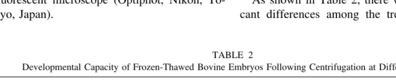
As shown in Table 2, there were no signifi-cant differences among the treatment groups TABLE 1
Developmental Capacity of Bovine Zygotes after Various Treatments
Treatments
No. of zygotes examined
No. of zygotes cleaved (%)
No. of zygotes developed to (%)
Blastocyst E./H. blastocysta
Controlb 138 105 (76.1) 58 (42.8) 53 (38.4)
Centrifugation 146 112 (76.7) 51 (34.9) 44 (30.1)
Lipid removal 122 103 (84.4) 50 (41.0) 39 (32.0)
Note.Three experiments were performed in each group.
aE./H. blastocyst, expanded/hatched blastocyst.
bControl, zygotes that were simply cultured and are not treated in any way.
TABLE 2
Developmental Capacity of Frozen-Thawed Bovine Embryos Following Centrifugation at Different Forces
Centrifugation (g)
No. of embryos examined
No. of embryos survived (%)
No. of embryos developed to (%)
Blastocyst E./H. blastocysta
Controlb 78 53 (67.9) 19 (24.4) 13 (16.7)
8,000 84 63 (75.0) 20 (23.8) 13 (15.5)
12,000 70 54 (77.1) 21 (30.0) 11 (15.7)
16,000 78 61 (78.1) 24 (30.8) 11 (14.1)
Note.Three experiments were performed in each group.
aE./H. blastocyst, expanded/hatched blastocyst.
with respect to the rates of survival and devel-opment to both the blastocyst and the expanded/ hatched blastocyst stages of the frozen-thawed 8-cell embryos. However, in the centrifugation groups, the rates of survival and development to the blastocyst tended to increase with increasing force of centrifugation, but the rates were not significantly different among the groups. Experiment 3
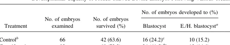
As shown in Table 3, there was no significant difference among the treatment groups with re-spect to the survival rate of the frozen-thawed 8-cell embryos. However, the rates of develop-ment to the blastocyst stage of the centrifuga-tion (P , 0.05) and lipid removal (P , 0.01) groups were significantly higher than those of the control group. Nevertheless, there were no significant differences among the groups with respect to the rate of development to the ex-panded/hatched blastocyst stage. The mean cell number of frozen-thawed embryos which devel-oped into blastocysts on day 8 was significantly lower (P , 0.05) in the centrifugation group and also tended to be lower in the lipid removal group, compared to the control.
In experiments 2 and 3, 62 of 97 fresh intact 8-cell embryos (63.9%) developed to the ex-panded/hatched blastocyst stage.
DISCUSSION
Several investigators have recently demon-strated that the removal of the intracellular lipid droplets prior to cryopreservation improves the
survival of both porcine (10) and IVP bovine embryos (16) at the early cleavage stage and even that of IVP bovine blastocysts (4) at sig-nificant levels. However, polarization alone or the partial removal of the lipids from porcine embryos have proven to be inferior to full lipid removal in terms of the chilling tolerance (9). It was also demonstrated that centrifugation treat-ment prior to cryopreservation did not improve the frequency of development to blastocysts of both frozen-thawed bovine oocytes (12) and embryos at the early cleavage stage (16). It has been suggested, in porcine embryos, that the high concentration and variable distribution of intracellular lipids in the blastomeres may in-duce heterogeneous intracellular ice nucleation or lipid-phase changes that contribute to the high sensitivity of the embryos to cryopreserva-tion (5). Further, preimplantacryopreserva-tion mouse em-bryos, which have few lipid droplets, can be successfully frozen at all stages of development (8, 17). Therefore, although initially high-speed centrifugation for a long period at the zygote stage was attempted to reduce the freezing sen-sitivity of IVP bovine embryos at the 8-cell stage, the lipid removal by micromanipulation was considered to be indispensable to obtain sufficient freezing tolerance of these embryos.
In the present study, although an obvious difference was not observed among the embryos centrifuged at various speeds in regard to the distribution of the polarized lipids to the blas-tomeres (data not shown), there was a little TABLE 3
Developmental Capacity of Frozen-Thawed Bovine Embryos Following Various Treatments
Treatment
No. of embryos examined
No. of embryos survived (%)
No. of embryos developed to (%) Mean cell number of blastocysts
(6SD) Blastocyst E./H. blastocysta
Controlb 66 42 (63.6) 16 (24.2)c 10 (15.2) 87.4
618.6A
Centrifugation 82 60 (73.2) 34 (41.5)de 12 (14.6) 60.5
630.0B
Lipid removal 67 53 (79.1) 32 (47.8)e 12 (17.9) 66.3
623.2AB
Note.Values with different superscripts within a column are significantly different [c, d (P,0.05); c, e (P,0.01); A, B (P,0.05)]. Five experiments were performed in each group.
aE./H. blastocyst, expanded/hatched blastocyst.
evidence to suggest that the polarization of lip-ids in the cytoplasm of zygotes by higher cen-trifugal force may improve the freezing toler-ance of bovine 8-cell embryos developed after the centrifugation treatment of the zygotes (Ta-ble 2). The developmental capacity into blasto-cysts of the frozen-thawed 8-cell embryos was improved by the treatments of centrifugation (at 16,000g for 20 min) or subsequent lipid re-moval at the 1-cell stage. However, with respect to the developmental potential to the expanded/ hatched blastocyst stage, no difference was found among these treated and intact 8-cell em-bryos after freezing and thawing (Table 3). It was observed in this study that the frequencies of frozen-thawed embryos forming blastoceles and containing some degenerated blastomeres were relatively higher in the centrifugation and lipid removal groups (data not shown). Such a type of embryo was also regarded as being at the blastocyst stage. It was also found that the mean cell number of frozen-thawed embryos that developed into blastocysts (day 8) tended to be low in both the centrifugation and the lipid-removed groups compared to the control (Table 3). Hence, it appeared that these post-thawed embryos were unlikely to expand or to hatch because of their low cell numbers. The two limitations cited above seem to reflect certain restrictions of the developmental capacity of both treatment groups despite their high devel-opment rates into blastocysts compared to the controls after freezing-thawing. Since both the lipid-polarized and the lipid-removed zygotes maintained their normalin vitrodevelopmental ability after such treatments (Table 1), the de-generation of the blastomeres and the resultant restriction of their further developmentin vitro are thought to be attributable to their injury during freezing and thawing.
Leibo and Oda (6) reported that when PVP was combined with a relatively low concentra-tion of EG, it enhanced the cryoprotective prop-erties of EG solutions, yielding high survival rates of mouse zygotes and embryos frozen either slowly or rapidly. Although the mecha-nism of the large polymer PVP (molecular weight, average 30,000) is not clear, Suzukiet
al.(13), using biopsied bovine blastocysts, sug-gested that PVP might play a beneficial role by forming a protective coating around the mem-brane of the cells during freezing and thawing. In the present study, since small incisions were made at the zona pellucida of the embryos dur-ing the lipid aspiration, a cryoinjury of the em-bryos with the lipid removal treatment (as in the zona-free embryos during freezing and thaw-ing) is expected. Moreover, Suzuki et al. (14) demonstrated that 1.8 M EG plus 0.05 M T supplemented with 5% PVP had the most ben-eficial effect for the slow freezing and thawing of bovine germinal vesicle oocytes. Therefore, this solution was employed for the cryopreser-vation of bovine 8-cell embryos using a direct exposure and rehydration process as previously described. As a result, even in the control intact 8-cell embryos, a relatively high development rate to expanded/hatched blastocyst stage was observed after freezing-thawing [16.0% (23/144); Tables 2 and 3], but this rate was still signifi-cantly lower (P , 0.01) than that of the fresh intact 8-cell embryos [63.9% (62/97)].
In conclusion, the present results indicate that centrifugation with or without lipid removal of zygotes has little or no effects on the ability of the bovine 8-cell embryos to develop in vitro following cryopreservation, but the quality of the embryos is lower. In the future, the optimum freezing and thawing procedure including cryo-protectants must be developed for the efficient cryopreservation of bovine early stage embryos.
REFERENCES
1. Boediono, A., Ooe, M., Yamamoto, M., Takagi, M., Saha, S., and Suzuki, T. Production of chimeric calves by aggregation of in vitro-fertilized bovine embryos without zonae pellucidae.Theriogenology
40,1221–1230 (1993).
2. Boediono, A., Takagi, M., Saha, S., and Suzuki, T. Influence of Day-0 and Day-7 superovulated cow serum during development of bovine oocytes in vitro.Reprod. Fertil. Dev.6,261–264 (1994). 3. Brackett, B., and Oliphant, G. Capacitation of rabbit
spermatozoa in vitro. Biol. Reprod. 12, 260 –274 (1975).
vitro and on the freezing tolerance of blastocysts.
Theriogenology45,166 (1996). [Abstract] 5. Dobrinsky, J. R. Cellular approach to cryopreservation
of embryos.Theriogenology45,17–26 (1996). 6. Leibo, S. P., and Oda, K. High survival of mouse
zygotes and embryos cooled rapidly or slowly in ethylene glycol plus polyvinylpyrrolinone. CryoLet-ters14,133–144 (1993).
7. Leibo, S. P., Pollard, J. W., and Martino, A. Chilling and freezing sensitivity of ‘‘reassembled’’ in vitro-derived bovine embryos. Theriogenology 43, 265 (1995). [Abstract]
8. Mohr, L. R., and Trounson, A. O. Structural changes associated with freezing of bovine embryos.Biol. Reprod.25,1009 –1025 (1981).
9. Nagashima, H., Kashiwazaki, N., Ashman, R. J., Gru-pen, C. G., Seamark, R. F., and Nottle, M. B. Re-moval of cytoplasmic lipid enhances the tolerance of porcine embryos to chilling.Biol. Reprod.51,618 – 622 (1994).
10. Nagashima, H., Kashiwazaki, N., Ashman, R. J., Gru-pen, C. G., and Nottle, M. B. Cryopreservation of porcine embryos.Nature374,416 (1995). 11. Otoi, T., Yamamoto, K., Koyama, N., and Suzuki, T. In
vitro fertilization anddevelopment of immature and mature bovine oocytes cryopreserved by ethylene glycol with sucrose. Cryobiology 32, 455– 460 (1995).
12. Otoi, T., Yamamoto, K., Koyama, N., Tachikawa, S., Murakami, M., Kikkawa, Y., and Suzuki, T. Cryo-preservation of mature bovine oocytes following centrifugation treatment. Cryobiology 34, 36 – 41 (1997).
13. Suzuki, T., Saha, S., Sumantri, C., Takagi, M., and Boediono, A. The influence of polyvinylpyrrolidone on freezing of bovine IVF blastocysts following biopsy.Cryobiology32,505–510 (1995). 14. Suzuki, T., Boediono, A., Takagi, M., Saha, S., and
Sumantri, C. Fertilization and development of fro-zen-thawed germinal vesicle bovine oocytes by a one-step dilution method in vitro.Cryobiology33,
515–524 (1996).
15. Trounson, A. O., Willadsen, S. M., Rowson, L. E. A., and Newcomb, R. The storage of cow eggs at room temperature and at low temperatures. J. Reprod. Fertil.46,173–178 (1976).
16. Ushijima, H., Yamakawa, H., and Nagashima, H. Cryo-preservation of bovine IVM/IVF bovine embryos at early cleavage stage following removal of cytoplas-mic lipid droplets.Theriogenology45,159 (1996). [Abstract]