Mavridis, et al Cardiac complication of pneumothorax
55
Can a pneumothorax break your heart?
ABSTRAK
Kardiomiopati Takotsubo atau apical balooning merupakan kondisi yang ditandai dengan disfungsi ventrikel kiri temporer, yang umumnya mengenai wanita usia pascamenopause
setelah suatu stress emosi atau fisik. Kami melaporkan
kasus seorang wanita usia 63 tahun yang datang dengan keluhan sesak napas berat disertai pneumotorak sekunder sisi kanan, yang pada awalnya di tatalaksana dengan slang
torakostomi. Walaupun pneumotorak membaik, sesak napas tidak hilang. Pada pemeriksaan didapatkan elevasi segmen ST dan peningkatan troponin I. Pasien selanjutnya dirujuk untuk kateterisasi jantung, namun tidak ditemukan adanya stenosis koroner yang bermakna. Selanjutnya didiagnosis sebagai kardiomiopati Takotsubo. Gambaran EKG membaik dalam 3 hari. Selanjutnya dilakukan reseksi apeks dengan torakoskopi diikuti pleurektomi parietal parsial. Walaupun kardiomiopati Takotsubo merupakan kejadian yang jarang,
hal ini selalu harus dipikirkan sebagai komplikasi potensial
dari pneumotorak.
Keywords: apical ballooning, spontaneous pneumothorax, tako-tsubo cardiomyopathy, thoracoscopic, tube thoracostomy pISSN: 0853-1773 • eISSN: 2252-8083 • http://dx.doi.org/10.13181/mji.v24i1.1179 • Med J Indones. 2015;24:55-8
• Received 06 Jan 2015 • Accepted 25 Feb 2015
Correspondence author: Stylianos Mavridis, [email protected]
C a s e Re p o r t
Copyright @ 2015 Authors. This is an open access article distributed under the terms of the Creative Commons Attribution-NonCommercial-ShareAlike 4.0 International License (http://creativecommons.org/licenses/by-nc-sa/4.0/), which permits unrestricted non-commercial use, distribution, and reproduction in any medium, provided the original author and source are properly cited.
Stylianos Mavridis, Hans-Georg Gnauk, Silvio Horn, Peter Adeberg, Martina Schumacher, Roland H. Wagner
Department Vascular and Thoracic Surgery, Ernst von Bergmann Clinic, Potsdam, Germany
Medical Journal of Indonesia
ABSTRACT
56 Med J Indones, Vol. 24, No. 1 March 2015
Pneumothorax is defined by the presence of air
in the pleural cavity and can be spontaneous, traumatic or iatrogenic. Spontaneous
pneumothorax (SP) can be classified as primary
or secondary. The primary SP is common among young population especially thin, tall males without previous lung disease, whereas the secondary spontaneous pneumothorax is often in the elderly with underlying parenchymal lung disease such as chronic obstructive pulmonary disease (COPD), bullous emphysema, pulmonary tuberculosis and other pulmonary infections,
cystic fibrosis, idiopathic pulmonary fibrosis,
lymphangioleiomyomatosis, etc. Treatment of SP ranges from simple observation, needle aspiration, tube thoracostomy or chemical pleurodesis to video-assisted thoracoscopy or thoracotomy.1-4
Sato and Dote back in 1991 were the first to
describe a rapidly resolving left ventricular dysfunction as Takotsubo cardiomyopathy. It is also called apical ballooning, broken heart or acute stress cardiomyopathy. It usually strikes postmenopausal females after acute endogenous (emotional) or exogenous stresses (trauma, surgery etc.) and mimicks the symptoms and
electrocardiogram findings of an acute myocardial infarction. No significant coronary artery disease is
detected in heart angiogram and causes an apical ballooning with compensatory basal hyperkinesis. Increased plasma catecholamine levels or changes in autonomic control of the cardiovascular system have been proposed as pathophysiological mechanisms.5-8 We report the unusual case of a
Takotsubo cardiomyopathy complicating secondary spontaneous pneumothorax.
CASE ILLUSTRATION
A 63 year old cachectic female (body mass index of 18.8), heavy smoker with a past medical history of severe chronic obstructive pulmonary (gold stage III) and hypertension was referred to the emergency department of a neighboring town due to progressive dyspnea worsening the last week. At the night prior to admission she experienced intense shortness of breath accompanied by heavy agony. Chest pain or fever were denied.
At time of admission she showed normal blood pressure, breathing rate and body temperature, rhythmic heart frequency of 116/min, and an
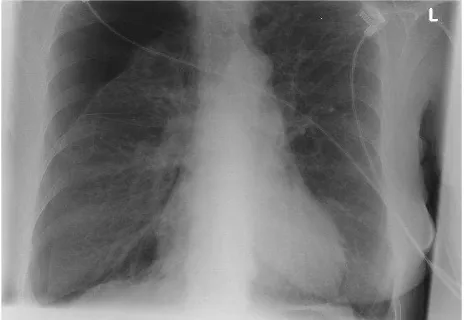
oxygen saturation of 89,5% with 2L of oxygen. C-reactive protein concentration and serum Troponin I levels were elevated to 2.02 μg/L and 24.2 mg/L, respectively. Patient electrocardiogram displayed a sinus tachycardia with a ST-segment elevation in leads V2 and V3. Chest radiograph revealed a right-sided large apical pneumothorax
without mediastinal shift (figure 1). A small bore
8 French thoracic catheter was inserted in the 6th intercostal space and attached to underwater seal. The chest X-Ray taken afterwards demonstrated the complete reexpansion of the right lung. The patient presented progressive subcutaneous emphysema so consequently the drain was placed on suction. Despite the fact that pneumothorax resolved, shortness of breath persisted and therefore urgent cardiac angiography was performed through a right femoral approach. This
ruled out a significant coronary artery stenosis,
while left ventriculography demonstrated the typical apical ballooning with a hypercontraction of the basal segments and an overall left ventricular ejection fraction of 41%. The diagnosis of a Tako-tsubo cardiomyopathy was established and the patient was transferred to the intensive care
unit. No specific treatment was made to maintain
hemodynamics in the acute phase. ST-segment elevation disappeared on the third day and serum Troponin I levels decreased.
After the accidental removal of the small thoracic catheter, a new one was required due to pneumothorax recurrence. This time a 20 French chest tube was preferred.
The air leakage persisted and therefore the patient was transferred to our institution for further treatment. Hence, video assisted
Mavridis, et al Cardiac complication of pneumothorax
57
thoracoscopic surgery through two ports under general anesthesia and single-lung ventilation was performed. At surgery, blebs and one bulla
were indentified at the apex of the right lung and
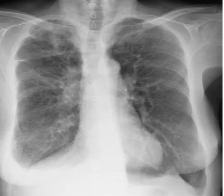
stapled wedge resection of the apex with partial pleurectomy to the level of the diaphragm was
carried out (figure 2). Chest drain was removed
on the third postoperative day with patient’s course being uneventful.
Control echocardiography on the sixth postoperative day demonstrated a normal systolic and diastolic function of the left ventricle with an ejection fraction of 73.7%. Hospital discharge was scheduled on the twentieth day after intensive physiotherapy in a rehabilitation clinic, where an improvement in the six minutes walk test from 171m to 352m was noticed.
DISCUSSION
SP remains a common pathology with an incidence between 18-28/100000 p.a. in males and 1.2-6/100000 p.a. in females. Secondary SP is often from the end of the fourth decade due to progressive intrathoracic pathology in high risk population such tobacco smokers or chronic obstructive pulmonary disease patients, resulting from a ruptured bleb or bulla into the visceral pleura.4,9 Cardiovascular diseases can also occur
in the same high risk group.
On the other hand, Takotsubo cardiomyopathy is a relative new and rare syndrome with an
Figure 2. Post-surgery X-ray
incidence of 0.1% - 2.3% among patients with acute myocardial infarction.5 Yokota was the first
to describe a case of a Takotsubo cardiomyopathy complicating a pneumothorax. In 2002, Akashi, et al reported the second case.10
It presents a transient and reversible left ventricular contractile dysfunction with an acute beginning of symptoms mimickings those of a myocardial infarction.5,11 The name Takotsubo
or apical ballooning derives from the distinctive morphology of the left ventricle during the
end-systole resembling an octopus fishing vessel with
round bottom and narrow neck (the “takotsubo” in Japanese).5,10 Mayo Clinic suggested four
criteria for the diagnosis of the Takotsubo cardiomyopathy, which all have to be met: 1) transitory hypokineses, akinesis, or dyskinesis of the left ventricular mid segments with or without apical involvement; 2) exclusion of an obstructive coronary artery disease or plaque rupture; 3) new pathology in electrocardiogram (ST-segment elevation or T wave inversion), and 4) exclusion of pheochromocytoma or myocarditis. Our
patient fulfilled all of the four criteria mentioned
above. She presented with elevated serum Troponin I levels and her electroctrocardiogram was pathologic with ST-segment elevation. At
heart catheterisation, a significant coronary
epicardial artery disease was excluded and the ventriculography revealed the typical apical ballooning. Further, she had no evidence of pheochromocytoma or myocarditis.
The pathophysiology of the syndrome remains till now unclear. There are several hypotheses involving a coronary artery vasospasm or a microvascular dysfunction or increased catecholamine levels. Exposure to stress (endogenous or exogenous) and elevated sympathetic activity can trigger the Takotsubo cardiomyopathy. Catecholamine oxidation can be toxic and results in calcium overload and direct damage of the myocardial cells.5 In patients
with Takotsubo cardiomyopathy induced by stress, the plasma catecholamine levels are higher compared with patients with myocardial infarction.8 In the present case, our patient, on
day before admission, she experienced terrifying agony after dyspnea exacerbation on the onset of the secondary spontaneous pneumothorax. We thought that this stressfull situation played a catalytic role in the development of
58 Med J Indones, Vol. 24, No. 1 March 2015
the cardiomyopathy. Furthermore, no signs of myocardial spasm were noted during heart catheterisation.
In conclusion, we demonstrated the difficulties
in the differential diagnosis of a Takotsubo cardiomyopathy complicating secondary SP. Population with chronic obstructive pulmonary disease or emphysema presents high risk in the development of a secondary SP or cardiovascular complications such as myocardial infarction. Nevertheless the probability of a Takotsubo cardiomyopathy remains minimal but still a challenging diagnosis.
Acknowledgment
Written informed consent was obtained from the patient for publication of this case report (and any accompanying images). A copy of the written consent is available for review from the editorial
office of Medical Journal of Indonesia.
Conflicts of interest:
The authors affirm no conflict of interest in this
study.
REFERENCES
1. Sousa C, Neves J, Sa N, Goncalves F, Oliveira J, Reis E: 3. Spontaneous pneumothorax: a 5-year experience. J Clin Med Res. 2011;3(3):111-7.
2. Weissberg D, Rafaely Y. Pneumothorax: experience with 1,999 patients. Chest. 2000;117(5):1279-85.
3. Kim SJ, Lee HS, Kim HS, Shin HS, Lee JW, Kim K-II, et al. Outcome of video-assisted thoracoscopic surgery for spontaneous secondary pneumothorax. Koren J Thorac Cardiovasc Surg. 2011;44(3);225-8.
4. Mavridis S, Gnauk HG, Schumacher M, Wagner RH. Spontaneous pneumothorax complicating lung emphysema. So, what’s the catch?. Med J Indones. 2013;22(1):54-6.
5. Nef HM, Möllmann H, Elsässer A. Takotsubo cardiomyopathy (apical ballooning). Heart. 2007;93:1309-15.
6. Waldenborg M, Soholat M, Kähäri A, Emilsson K, Fröbert O. Multidisciplinary assessment of takotsubo cardiomyopathy: a prospective case study. BMC Cardiovasc Disord. 2011;9:11-4.
7. Kurisu S, Ishibashi K, Kato Y, Mitsuba N, Dohi Y, Nishioka K, Kihara Y: Takotsubo cardiomyopathy complicated by QRS prolongation. Intern Med. 2012;51(3):291-4.
8. Chia PL, Foo D. Takotsubo cardiomyopathy precipitated bei pheochromocytoma crisis. Cardiology Journal. 2011;18(5):564-7.
9. Steinhäuslin CA, Cuttat JF. 12. Spontaneous pneumothorax. A complication of lung cancer?. Chest. 1985;88(5):709-13.
10. Akashi YJ, Sakakibara M, Miyake F. Reversible left ventricular dysfunction “takotsubo” cardiomyopathy associated with pneumothorax. Heart. 2002;87(2):e1. 11. Villablanca PA, Sukhal S, Ansari A, Mohammed D. Acute
gastritis-induced Takotsubo’s cardiomyopathy. Clinical Case Reports. 2013;1(2):91-5.
12. Ha JH, Lee H, Park YJ, Kang HH, Lee SH, Moon HS. Takotsubo cardiomyopathy caused by pulmonary tuberculosis: a case report. Tuberc Respir Dis (Seoul). 2014;77(1):24-7.