“I hereby declare that I have read through this report entitle “Power Spectral Analysis of Surface
EMG for Wight Lifting Atheletes” and found that it has comply the partial fulfillment for
awarding the degree of Bachelor of Electrical Engineering (Control, Instrumentation and Automation)”
Signature :
……….
Supervisor name : Wan Mohd Bukhari bin Wan Daud ……….
Date :
POWER SPECTRAL ANALYSIS OF SURFACE EMG OF SURFACE EMG FOR
WEIGHT LIFTING ATHELETES
MUHAMMAD AZRI BIN OMANAP
A report submitted in partial fulfillment of the requirements for the degree of Bachelor
in Electrical Engineering (Control, Instrumentations and Instrumentations) with
Honours
Faculty of Electrical Engineering
UNIVERSITI TEKNIKAL MALAYSIA MELAKA
I declare that this report entitle “Power Spectral Analysis of Surface EMG for Wight Lifting
Atheletes” is the result of my own research except as cited in references. The report has not been
accepted for any degree and is not concurrently submitted in candidature of any other degree.
Signature : ……….
Name : Muhammad Azri Bin Omanap ……….
Date :
ACKNOWLEDGEMENT
I would like to express my grateful to Encik Wan Mohd Bukhari bin Wan Daud for supervising me in this research. I would also give my gratitude, especially to my family for their guidance, advices, and motivation. Also my colleagues and others who involve in assisting me in collecting and analyzing data and figures in the experiment process. Without their continued support and interest, this project would not have been presented here. Nevertheless, I would also like to express my thanks to all of the test subject that involved in this experiment without asking any payment.
ABSTRACT
ABSTRAK
TABLE OF CONTENTS
CHAPTER TITLE PAGE
ACKNOWLEDGEMENT i
ABSTRACT ii
ABSTRAK iii
TABLE OF CONTENTS iv
LIST OF TABLES vi
LIST OF FIGURES vii
LIST OF ABBREVIATIONS ix
LIST OF APPENDICES x
1 INTRODUCTION 1
1.1 RESEARCH BACKGROUND 1
1.2 PROBLEM STATEMENT 2
1.3 OBJECTIVES 2
1.4 SCOPE OF WORK 3
2 LITERATURE REVIEW 5
2.1 THEORY AND BASIC PRINCIPLES 5
2.2 REVIEW OF PREVIOUS RELATED WORKS 12
2.3 SUMMARY AND DISCUSSION OF THE REVIEW 15
3 RESEARCH METHODLOGY 16
3.2 EXPERIMENTAL SETUP 18
3.3 EXPERIMENTAL PROCEDURE 22
3.4 DATA COLLECTION AND FEATURE EXTRACTION OF ELECTROMYOGRAPHY (EMG) SIGNAL
24
3.5 EVALUATION OF FEATURES EXTRATION DATA 25
3.6 REALIABILITY OF THE DATA 26
4 RESULT AND DISCUSSIONS 27
4.1 NOISE ANALYSIS 27
4.2 EMG SIGNAL COLLECTED AND POWER SPECTRAL ANALYSIS
28
5 CONCLUSIONS AND RECOMMENDATION 33
CONCLUSION 33
RECOMMENDATION 34
REFERENCES 35
LIST OF TABLES
TABLE TITLE PAGE
1.1 Example Data Table for EMG Recording for Test Subject 3
1.2 Specification of the Test Subject 4
LIST OF FIGURES
FIGURE TITLE PAGE
1.1 The sEMG device 2
2.1 MUAPs extract from sEMG 7
2.2 Motor Unit 7
2.3 The Depolarization and Repolarization of Motor Unit (MU) 8
2.4 The Action Potential 9
2.5 The depolarization zone on muscle fiber membranes. 9
2.6 The model of a wandering electrical dipole on muscle fiber membranes 10
2.7 Generation of the triphasic MUAP 11
2.8 Motor Unit Recruitment and Firing Frequency 11
2.9 Factor effecting the EMG signal 11
3.1 Matlab Simulink Block Diagram Configuration of Arduino with
EKG/EMG Shield.
18
3.2 Nikon Kohden wet gel disposable electrode 19
3.3 BD Alcohol Swab 20
3.4 Electrode Location 21
3.5 Flowchart of Experimetal Procedure 22
4.1 Data for 2.5 KG (a) Statistic EMG Signal (b) PSD Graph (c) Statistic PSD
30
4.2 Data for 5.0 KG (a) Statistic EMG Signal (b) PSD Graph (c) Statistic
PSD
31
4.3 Data for 10.0 KG (a) Statistic EMG Signal (b) PSD Graph (c) Statistic
PSD
32
LIST OF ABBREVIATIONS
Abbreviation Meaning
sEMG Surface Electromyography
EMG Electromyography
PSA Power System Analysis
MU Motor Unit
MUAP Motor Unit Action Potential
EKG/EMG Shield Electrocardiography Electromyography Shield
RMS Root Mean Square
dB Decibel
mV millivolt
Na+ Sodium Ion
K+ Potassium Ion
Hz Hertz
FFT Fast Fourier Transform
LIST OF APPENDICES
APPENDIX TITLE PAGE
A EKG/EMG Shield 38
B Electrocardiography Electromyography Shield connector 38
C Arduino Mega 2560 Function 39
D Technical specifications for Arduino Mega 2560 39
E Function of each part on Arduino Mega 2560 40
F Passive Electrodes For Electrocardiography Electromiography 40
G EMG signal 41
H Power Spectral 44
CHAPTER 1
INTRODUCTION
Chapter 1, will give a brief explanation of the ‘Power Spectral Analysis of Surface EMG for Weight Lifting Athletes’. Also the research background and motivation of the research will be discuss. Then all problem statement and scope will be determine by the end of this Chapter 1.
1.1 Research background
The Electromyography or EMG is a technique for evaluating and recording the electrical activity produced by skeletal muscles. EMG can be record by various method from low tech and low cost, to a high end tech but high cost method. Basically EMG is perform using an instrument or device call electromyography to produce a record called an electromyogram. An electromyography will detect the electrical potential generated by muscle cells when these cells are electrically or neurologically activated. The signals can be analyzed to detect medical abnormalities, activation level, or recruitment order to analyze the biomechanics of human movement.
Figure 1.1: The sEMG device
1.2 Problem Statement
The EMG signals are used in many clinical and biomedical applications. EMG is used as a diagnostics tool for identifying neuromuscular diseases, assessing low-back pain, kinesiology, and disorders of motor control. EMG signals are also used as a control signal for prosthetic devices such as prosthetic hands, arms, and lower limbs. Thus analyzing the EMG signal is the next step in solving this problem.
1.3 Objectives
The main objectives of this research are:
1. To record the MUAP from sEMG by using developed acquisition system.
1.4 Scope of work
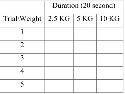
This study scope will be a guideline towards achieving the objectives. The scopes of this study are the muscles that will be used in this study is biceps brachii. The exercise that will be done in this research is by using biceps curl method. The sEMG that will be uses is Olimex Ekg/Emg Shield with Arduino Mega 2560 and also Passive Electrode will be used in this study. The Arduino Mega 2560 will be used as the microcontroller that acts as a data acquisition. The feature extraction that will be used is in the time domain which is mean, root mean square (RMS), standard deviation and variance. The analyze part of feature extraction electromyography (EMG) is using power spectral analysis using Matlab. There are 3 different weight with 5 tries. Table 1.1. There will be 5 person acted as test subject to help generated EMG signal from their hand into the sEMG device. The data collected from these test subject are by doing 10 second arm at rest and 10 second biceps curl. Also the test subject are healthy and no health issues especially nerves issues. Table 1.2.
Table 1.1: Example Data Table for EMG Recording for Test Subject
Duration (20 second) Trial\Weight 2.5 KG 5 KG 10 KG
Table 1.2: Specification of the Test Subject
Specification Age Height Weight Trial Weight Health
Condition
5 Male
20-25
160 cm to 190 cm
50 Kg to 80 Kg
2.5 Kg, 5 Kg, 10 Kg
CHAPTER 2
LITERATURE REVIEW
Chapter 2 will give a brief explanation of the theory and the history of EMG. Further basic principles, review of previous related works, summary and discussion of the review will be discuss. The knowledge of using sEMG and Power Spectral Analysis concept also will be describes.
2.1 Theory and basic principles
EMG is an experimental technique concerned with the improvement, recording and examination of myoelectrical sign or EMG sign create by the physical varieties in the condition of muscle fiber layers. This sign can be utilized as an assessment apparatus for applied research, physiotherapy, rehabilitation, and sports training. It is similar to what are the muscle doing in certain point with specific situations [1, 2].
2.1.1 Weightlifting, Power Spectral Analysis, Motor Unit Action Potential (MUAP).
Weightlifting is a focused quality based game, where competitors switch from the barbell position from the floor to the overhead position when endeavoring a greatest weight single lift. This movement includes entire body muscle power.
The Power Spectral analysis, for example, frequency range and median frequency and made correlations between information got from the influenced and contra-lateral sides of the subjects [3]. Power spectral analysis of surface EMG signals has been utilized to identify possible alterations in the firing frequency as well as action potential shapes. Most EMG enhancers utilize a high pass channel (frequently set at 20-450 Hz) such that the firing frequency range is typically lower than the bandwidth of the filter and thereby limits the ability to identify firing frequencies. Studies have demonstrated that the median frequency of the power spectrum is corresponded to activity potential shape, specifically name the action potential duration. [4]
Figure 2.1: MUAPs extract from sEMG
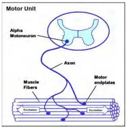
2.1.2 The Motor Unit, (MU).
The Motor Unit consist of Alpha Motoneuron, Axon, Muscle Fibers, Motor Endplates, and Muscle fibers. Figure 2.2.
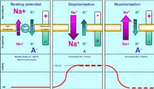
2.1.2 Excitability of Muscle Membranes.
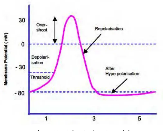
The nerve system will excite a control over muscle membranes. The ion pump in muscle cell create a balance between the internal and external of the muscle membrane. The muscle membrane have 3 stage called the Resting Potential, Depolarization process, and Repolarization. Figure 2.3. [7,8] Resting Potential is where the muscle is not moving, Depolarization is where the muscle moving forward, and Repolarization is where the muscle moving backward [9]. The Na+ ion charges play an important part in this process.
2.1.3 The Action Potential.
The Action Potential also known as Motor Unit Action Potential (MUAP), explain the Depolarization of muscle membrane which is counter by Repolarization immediately after that. Figure 2.4. This process produce about -80mV to +30mV of MUAP [10, 11].
Figure 2.4: The Action Potential
The EMG signal from the Depolarization and Repolarization process will be collect by the sEMG device. Figure 2.5.
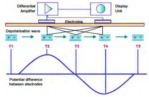
2.1.4 Signal Propagation and Detection
The Depolarization and Repolarization process of the muscle membrane produce MUAP, that will be detect by the electrode and amplify by the sEMG device. This will give EMG signal depend on the specific requirement that will affect the EMG signal. Figure 2.9.
Figure 2.6: The model of a wandering electrical dipole on muscle fiber membranes
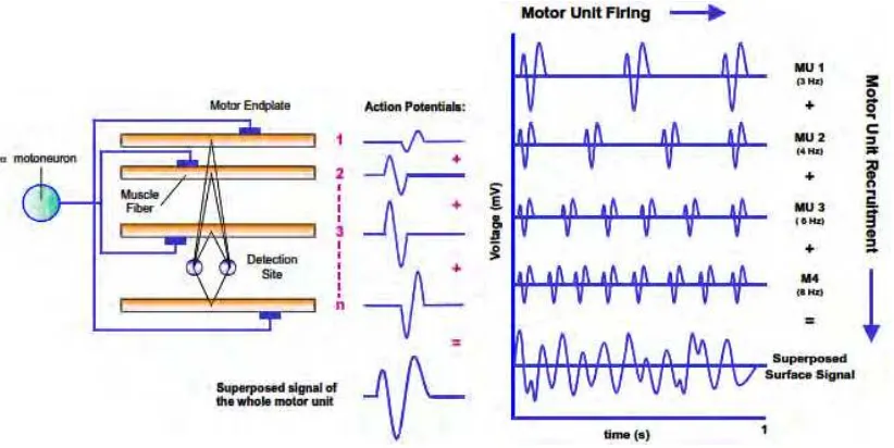
Figure 2.7: Generation of the triphasic MUAP Figure 2.8: Motor Unit Recruitmentand Firing Frequency
2.1.5 Factor Effecting the EMG signal.