The widespread outbreaks of avian influenza (AI) in South-East Asia in the recent years have led to the death of millions of domesticated birds and millions others have to be sacrificed in an effort to eradicate the disease (Swayne and Halvorson 2003; Stegeman and Bouma 2004). During this outbreak, many affected countries suffer a great deal of economic losses brought out by the collapse of their poultry industries (Perkins and Swayne 2002; Perkins and Swayne 2003; Lewis 2006). More importantly, the disease also affects human causing a great concern among health authorities in the world. The availabilities of accurate, simple, safe, and fast diagnostic methods are important for an effort to prevent and to control a future outbreak of AI in both animals and man. Most diagnostic methods developed in the recent years still require expensive facilities and reagents are slow to perform, lack of sensitivity and specificity, unsafe to perform, and unable to determine the virus subtype directly (Gough 2004).
Avian influenza viruses (AIVs) are a group of viruses with great genetic and antigenic diversities in nature. On the basis the antigenic characteristics of their two surface glycoproteins, haemagglitinin (HA) and neuraminidase (NA), AIVs are grouped into many subtypes. As many as 16 HA subtypes that can combine with 9 NA subtypes have been identified (Foucier et al. 2005). Such antigenic diversities have often caused a great difficulty in establishing an appropriate test for an accurate detection of AIV subtypes (Fouchier et al. 2005; Kida dan Sakoda 2006). MAbs which react only with a single epitope on an antigenic structure have been widely used to detect the viral antigen in the infected hosts and also to differentiate closely related viruses (Zheng et al. 2001; Vareckova et al. 2002; Ohnishi et al.
2005). In human influenza virus, for instance, the use of MAbs
Immunological Detection of Avian Influenza Virus in Infected
Ducks by Monoclonal Antibodies Against AIV-H5N1
NYOMAN MANTIK ASTAWA1*, IDA BAGUS OKA WINAYA1, LUH PUTU AGUSTINI2, AND NINING HARTANINGSIH2
1Faculty of Veterinary Medicine, Universitas Udayana, Jalan PB Sudirman, Denpasar 80232, Indonesia 2Biotechnology Laboratory, Disease Investigation Centre Regional VI Denpasar, Jalan Raya Sesetan No. 266,
Denpasar 20223, Indonesia
In order to establish a detection method for avian influenza virus (AIV) infection in ducks, monoclonal antibodies (MAbs) against the virus were produced. The virus used for the production of the monoclonal antibodies was AIV-H5N1 of Indonesian origin. Immortal mouse myeloma were fused with the lymphocytes derived from the spleen of mice immunized with the virus. The MAbs were tested for their specificity by enzyme linked immunosorbent assay (ELISA) and western blotting using formaldehyde inactivated virus and normal allantoic fluid as a negative control. Twelve MAbs which were specific against AIV were isolated and 8 of them were used for detecting of AIV antigen in duck’s tissues. AIV antigen was detected in paraffin embedded tissues of AIV-infected ducks by immunohistochemistry using MAbs. AIV antigen was not detected in ducks, which were confirmed to be AIV negative. In the infected ducks, high intensity of AIV infection was detected in proventricle gland and small intestine. The AIV antigen with a lesser intensity was also detected in lungs, spleen, and bursa of Fabricius, but hardly detected in muscle, brain, and several other issues. This study shows a clear evidence that MAbs produced in this study are applicable for use in immunological detection of AIV in infected duck tissues.
Key words: avian influenza, H5N1, monoclonal antibodies, ducks, virus immunohistochemistry _____________________________________________
________________________
*Corresponding author, Phone: +62-361-223791,
Fax: +62- 361-701808, E-mail: [email protected]
against the HA protein of the virus is reported to have 100% sensitivity and 99.1% specificity in determining the HA subtype of the virus (Vareckova et al. 2002). As in human influenza viruses, MAbs against AIV is very likely to have a similar degree of sensitivity and specificity when used in detecting of AIV antigen in the infected hosts including in determining the virus subtype.
of AIV-H5N1 is very likely to provide a relatively much simpler, quicker, cheaper, and safer diagnostic methods for the detection of AIV antigen in ducks. We have currently been able to produce MAbs against AIV-H5N1 of Indonesian isolate and the applicability of those MAbs for detecting AIV antigen in duck tissues was examined.
MATERIALS AND METHODS
Cells. Myeloma cells (P3-NS1/1-Ag4.1), used for the preparation of hybridomas were obtained from Murdoch University, Australia. The cells were grown in Dubelco’s modified essential medium with 10% newborn calf serum (NBCS) and antibiotics penicillin, 200 IU ml-1, streptomycin 200 µg ml -1.
Virus. Formaldehyde inactivated AIV-H5N1 used in this study was an Indonesian isolate. The virus was isolated in 2005 from chicken with a severe clinical disease and the virus isolate was then designated as A/CK/Bali/2005. The virus was propagated in 10 days-old chicken embryonated eggs and harvested from allantoic fluids. The titer of the virus was determined by HA test (WHO 2002). The virus has been confirmed as H5N1 subtypes and PCR using H5 and N1 primers (data not shown).
Production of Monoclonal Antibodies. MAbs against the Indonesian isolate of AIV-H5N1 were produced by methods similar to those described by Ohnishi et al. (2005). Six to seven week-old female Balb/c mice were immunized with 0.2 ml (equivalent with approximately 27 HA units) virus emulsified in Freund’s complete adjuvant. Fourteen and 28 days after the first immunization the mice were respectively immunized with the same antigen but emulsified in Freund’s incomplete adjuvant. Fourteen, 15, and 16 days after the last immunization, the mice were boosted with the same antigen but without adjuvant. The mice were then sacrificed by cervical dislocation. The spleen was removed and used for the preparation of hybridomas.
As many as 2 x 10⁷ immortal mouse myeloma cells prepared as described above were fused with 108 lymphocytes derived from the spleen of mice immunized with AIV-H5N1. The fusion of the two types of cells was carried out using polyethylene glycol (PEG) 45% (Sigma Co, USA) to produce hybridomas. The hybridomas were then screened by indirect ELISA (Campbell 1991) for the anti-AIV antibodies using formaldehyde inactivated AIV-H5N1 as antigen and normal allantoic fluid as negative control. The hybridomas producing MAbs reacted specifically with the virus were cloned by limiting dilution as described by McKearn (1980) and were then used in the production of MAbs against the AIV-H5N1.
Titration of MAbs. The titer of MAbs in hybridomas’ supernatant fluid was determined by ELISA according to the procedure as described by Campbell (1991). ELISA microtitration plate was coated overnight with formaldehyde inactivated virus diluted in carbonate-bicarbonate coating buffer (15 mM Na2CO3, 35 mM NaHCO3 pH.9.6). Each plate well was coated with 100 ml antigen containing of approximately 1 HA unit of the virus. After three times washes with 0.05% Tween-20 in phosphate buffered saline pH 7.2 (PBST), 100 ml blocking buffer (5% skim milk in PBST)
was added and incubated for another 1 h at 37 °C. A serial two-fold dilution of MAbs was prepared and 100 ml MAb sample from each dilution were added to each well. The plate was incubated for 1 h at 37 °C. After three times washes as above, 100 ml anti-mouse IgG-conjugated with horseradish peroxidase (HRP) (Bio-Rad, USA) diluted 1:2 000 in PBS-T was added to each well. The microplate was then incubated for 1 h 37 °C and washed three times as above. One hundred µl of substrate solution (1 mM 2 2’-azinodi 3-ethylbenzthyazolin-6-sulfonic azide in 0.05% Na citrate, 0.15% Na phosphate, and 0.01% H2O2 was added to each well. After incubation for 15 min at room temperature, the absorbance of the substrate solution in each well was read by multiscan spectrophotometer using 405 nm filter. Titer of MAbs was determined as the antilog the highest dilution giving an absorbance reading of approximately 50% of its optimal reading.
Determination of MAb Isotypes. The immunoglobulin (Ig) class and subclass of the MAbs were determined by indirect ELISA using rabbit antimouse subtyping isotyper kits (Bio-Rad Laboratory, USA) according to the procedures described by manufacturer. ELISA microtitration plate was firstly coated overnight with formaldehyde inactivated AIV-H5N1 as described above. Into each well, 100 µl MAb diluted
1:10 in PBST were added and incubated for 1 h at 37 °C. Following three times washes with PBS, rabbit anti-mouse Ig isotyper from the kit was added to the wells and incubated as above. After 3 times washes, 100 ml affinity purified goat anti-rabbit IgG conjugated with HRP (Bio-Rad, USA, diluted 1:1 000 in PBST) was added and incubated at 37 °C for 1 h. The plate was again washed as above and 100 ml substrate solution (1 mM 2.2‘-azinodi 3-ethylbenzthyazoline-6-sulfonic acid in 0.005 Na citrate, 0.15 Na phosphate, and 0.01% H2O2) was added. The absorbance of the substrate solution was read in Multiscan spectrophotometer with a 405 nm filter.
Table 1 Characteristics of monoclonal antibodies prepared against avian influenza virus subtype H5N1 of Indonesian origin
MAbs Isotypes AIV-H5N1ELISA NA Western blottingAIV-H5N1 NA IHC
AG8 NA: normal alantoic fluid, IHC: immunhistochemistry, -: negative,
+++: strong positive, ND: not ditermined.
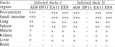
Table 2 Detection of avian influenza virus antigen in infected Ducks by immunostaining staining using monoclonal antibodies
Ducks Infected ducks I Infected duck II
organs AG8DF11EA11EE8 AG8 DF11 EA11 EE8
Proventricle
++: moderate positive, +++: strong positive, +: weak positive, -: negative.
Detection of AIV Antigen in Duck Tissues by Immunoperoxidase Staining. The ducks used in this study were cordially provided by co-assistant students in the Faculty of Veterinary Medicine, Udayana University, Denpasar Bali. All ducks had been tested for AIV infection by the isolation of the virus in chicken embryonated eggs and identification of the virus by haemagglutination/ haemagglutination inhibition (HA/HI) test. The results of the test were then further confirmed by reverse trancriptase-polymerase chain reaction (RT-PCR) using primers specific to H5 and N1 subtypes. Two ducks that were confirmed positive to AIV-H5N1 infection and two ducks confirmed AIV negative (data not shown) were used in this study.
Several organs such as brain, proventricle, small intestine, liver, lung, bursa of Fabricius, spleen, and kidney derived from the ducks were preserved and fixed with 10% buffered formaldehyde. Paraffin embedded organs and thin sections of the organs were prepared by standard methods. Immunoperoxidase staining was then carried out according to the methods similar to those described by Ohnishi et al.
(2005). Thin sections of tissues on microscope slides were de-paraffinized twice in xylol and twice with ethanol absolute. The tissue section was washed twice with PBS and treated with 0.05% trypsin for 1 min at 37 °C. The endogenous peroxidase of the tissues was then inactivated by treatment with 3% H2O2 in PBS for 20 min at room temperature. After blocking with 50% normal goat serum in PBS, MAbs against AIV-H5N1 was added onto the tissue section and incubated for 18 h at room temperature. The bound MAbs were detected by biotinylated goat anti-mouse IgG (Biodesign International) diluted 1:500 in PBS containing 10% normal goat serum and streptavidin-horse radish peroxidase (Sigma Co, USA) diluted 1:100 in PBS. A proper washing procedure was carried out using PBS in between each step. The AIV antigen bound with MAbs was then visualized by adding diazinobenzidine (DAB) substrate (Sigma Co, USA, 50 mg/ 50 ml PBS containing 0.07% H2O2).
RESULT
Characteristic of Monoclonal Antibodies. As many as 12 clones of stable hybridomas secreting MAbs against the AIV-H5N1 of Indonesian isolate were produced. Screening by ELISA using formaldehyde inactivated AIV-H5N1as antigen showed that all of these 12 clones of hybridomas produced MAbs against the virus, not against the normal allantoic fluid. Eight MAbs were further characterized and they were designated as AG8, BC12, CC5, CG1, DD9, DF11, EA11, and EE8. All MAbs reacted strongly in ELISA test using formaldehyde inactivated AIV-H5N1. None of them reacted with normal egg allantoic fluids (negative control). The titer of MAbs varied from 26 to 29 (Table 1). Isotyping of MAbs using rabbit anti-mouse IgG subtyper isotyping kit showed that 3 MAbs (AG8, DF11, EA11) were of IgG1 subclass, 1 MAb (DF9) was of IgM subclass, 3 MAbs (CC5, CG1, DD9) were of IgG3 subclass, and 1 MAb (EE8) was of IgG2a subclass (Table 1).
Western Blotting with MAbs. In western blotting assay, a similar result was observed. All MAbs reacted only with formaldehyde inactivated AIV-H5N1. No MAb reacted with
normal allantoic fluid. Two MAbs (DD9 and CC5) reacted with 2 protein bands with the molecular weight of approximately 76 and 58 kDa, 5 MAbs (AG8, CG1, DF11, EA11, and EE8) reacted with a single protein band of 76 kDa. One MAb (DF9) reacted with a diffuse protein band.
Detection of AIV Antigen in Duck Tissues. Three (CG1, EE8, AG8) produced a good and a strong result when used for the detection of AIV antigen in ducks. One MAb (DF11) did not react with AIV antigen in the duck tissues. AIV antigen was detected in the two infected ducks but not in uninfected ducks. AIV antigen with a high intensity was observed proventricle and in small intestine. AIV antigen at a lesser intensity was also observed in other organs such as lung, spleen, and bursa of Fabricius (Table 2, Fig 1). AIV antigen was difficult to observe in the brain, muscle tissue, and kidney. No clear difference on the distribution of infected tissues was observed between the two infected ducks.
DISCUSSION
Stable anti-AIV-H5N1 MAbs-secreting hybridomas were successfully produced by fusion of immortal myeloma cells with lymphocytes of mice immunized with the virus. The use of formaldehyde inactivated virus for immunization of mice in the preparation of MAbs appeared to be not an important factor for the production of hybridomas stably producing MAbs against AIV. This is evident as all of the isolated hybrodomas consistently produced MAbs against the virus and but not against the normal allantoic fluid. The use of relatively unpurified virus for immunization of mice in the preparation of MAbs has been reported (Wickramasinghe
et al. 1993; Pantophlet et al. 2001).
anti-AIV-H5N1 MAbs. As in immunization, the antigen used in the ELISA test for screening MAbs was formaldehyde inactivated AIV-H5N1. The virus was originally propagated in the allantoic cavity of chicken embryonated eggs. The virus was then harvested from the allantoic fluid of the infected chicken embryo and was therefore expected to contain a plenty of normal allantoic fluid. It was therefore very likely that the immunization of mice with such antigen will stimulate the production of antibodies against both AIV and normal allantoic fluid. This then was confirmed when several MAbs which reacted with normal ascitic fluid were detected by ELISA using the unpurified AIV-H5N1 (data not shown). Such MAbs were excluded by further testing by ELISA using normal allantoic fluid as an antigen.
Isotyping showed that the MAbs produced in this experiment were of IgM, IgG1, IgG2a, and IgG3 subclasses. The information on the isotype of MAbs is important in the selection of techniques used for the purification of MAbs. In addition, the information on the MAbs’ isotype is also important in the selection of techniques to be developed using MAbs. The MAbs with IgG isotype generally produces a more specific and sensitive result and can also be used in a relatively wider range of serological tests then those of MAbs of IgM isotype. In some immunodetection systems, however, antibody with IgM isotype is preferred as it will produce a better and a stronger reaction than MAbs of IgG isotype. The main problem working with IgM is that it is more difficult to purify than IgG. In addition, the availability of MAbs of IgG isotypes will also enable the purification of the MAbs using Protein A or G (de Masi et al. 2005) which is often required for the development of a particular test such as capture ELISA (Ohnishi et al. 2005). In western blotting assay, all isolated MAbs reacted specifically only with AIV-H5N1 antigen. None of them reacted with normal allantoic fluid which was used as an antigen for AIV negative control. The result confirmed that MAbs specific to AIV-H5N1 can be produced by immunization of mice with relatively unpurified virus. The protein bands recognized by MAbs were around 76 kDa, 58 kDa, and several other diffuse bands. The protein band with 76 kDa detected by most MAbs (CG1, AG8, EA11, EE8, BC12) is likely to be uncleaved haemagglutinin (HA0) of AIV-H5N1. The HA protein of AIV is a surface glycoprotein encoded by segment 4 (HA) of the viral segmented RNA gemomes. The protein is initially translated as uncleaved precursor of HA0 protein with the molecular weight of around 76 kDa. It is then post-translationally cleaved by host cellular proteases into two sub units, HA1 (56 kDa) and HA2 (25 kDa) (Skehel and Waterfield 1975;Zhirnov et al. 2002). However, other workers on influenza A virus reported the molecular weight of HA1 varied from 50-61 kDa and HA2 varied from 25-30 kDa (Bucher et al. 1976; Boulay et al. 1987; Jaspers et al. 2005). The protein contains sialic acid which plays an important role in the binding of the virus into the receptor molecules on the surface of susceptible cells (Hulse et al.
2004) and such cleavage step is necessary for the infection of the virus into not react with AIV antigen in the infected ducks, suggesting that this MAbs did not recognized the AIV epitope in formadehyde fixed and paraffin embedded tissues. The reason behind this is unknown. It is possible
that the epitope recognized by this MAb has been destroyed or hidened during the tissue proccecing. When the 4 MAbs were used to immunostain tissues or organs of normal uninfected ducks, none of them produced a positive result. This showed that three of the selected MAbs are applicable for use in development of specific test for the detection AIV infection in ducks. The use of MAbs in the immunochemistry staining for the detection of viral antigen in the infected host has been widely reported (Ohnishi et al. 2005; Astawa
et al. 2006).
In the infected ducks, high intensity of AIV antigen was detected in organs such as preventricle and intestine villi (Fig 1), suggesting that the virus replicates very efficiently in these two gastrointestinal organs. This is in accord with the finding that, in waterfowl, influenza viruses replicate preferentially in the intestinal tract, resulting in excretion of high-titer viruses in the feces (Horimoto and Kawaoka 2001). The combination of the availability of cells bearing the receptor for AIV and the presence of abundant proteolytic enzymes may contribute to the efficient replication of the virus in the intestine and proventricle of ducks. Unlike those which originate from chicken, many AIVs isolated from ducks have a strong binding activity to gangliosides with short sugar chains that were found abundant in duck gastrointestinal tissues (Slemons and Easterday 1978; Gambaryan et al. 2003). In addition, gastrointestinal tract is rich in proteolytic enzymes (Banks and Plowright 2003) which are responsible for post translational cleaving of HA0 of into HA1 and HA2 (Garten and Klenk 1999). The cleavage of HA protein is required for the efficient replication of the virus in the two organs.
The availability of MAbs against AIV-H5N1 has enabled the detection of AIV antigen in duck tissues. The duck used study was previously confirmed to be infected by AIV-H5N1 but the tests used still require expensive facilities and reagents such as PCR. It is also unsafe to perform as isolation of AIV in chicken embryonated eggs and identification by HA/HI test require the use of live virus (Gough 2004). The use of MAbs on formaldehyde fixed and paraffin embedded tissues has made it possible to develop a relatively simpler and safer test which can be performed in laboratory with simple facilities and low biosecurity level. The immunological detection system developed in this experiment also safe to perform on daily basis as it uses formaldehyde fixed tissues which inactivates the AIV. As ducks and other aquatic birds play an important role in the transmission of AIV into susceptible hosts (Matrosovich et al. 1999), the availability of test to detect ducks carrying the virus will be important in preventing the AI outbreaks brought out by this carrier water fowls.
76 kDa 58 kDa
100 7 5 5 0 3 7
2 5 2 1
1 5 100
7 5 5 0 3 7
2 5 2 1
1 5
1 2 3 4 5 6 7 8 9 1 2 3 4 5 6 7 8
a b
Fig 2 Reactivity of monoclonal antibodies with AIV-H5N1 and normal allantoic antigens analysed by western blotting. a: antigen: formaldehyde inactivated AIV-H5N1, and b: normal allantoic fluid. Strip no. 1-8 MAbs: 1: DD9, 2: CC5, 3: AG8, 4: CG1, 5: DF11, 6: EA11, 7: EE8, and 8: DF9, strip no. 9: standard marker.
the cross-reactivity of several MAbs with many different H subtypes. When MAbs react specifically only with AIV of H5 subtype are available then determination of AIV-H5 subtypes can be carried out directly by immunohistrochemistry staining using MAbs. This is important as AIV-H5 virus is one subype that causes most fatal infection in avian species and in mammal including human (Swayne and Suarez 2000). A further investigation is required in order to confirm this suggestion, especially when AIV of many different H subtypes are available for study.
ACKNOWLEDGEMENT
This study was funded partly by the “Hibah Bersaing” grand provided by Directorate of Higher Education, Department of National Education, Republic of Indonesia.
REFERENCES
Alexander DJ. 2000. A review of avian influenza in different bird species. Vet Microbiol 74:3-13.
Astawa NM, Hartaningsih N, Agustini LP, Tenaya WM, Berata K, Widiyanti LPM. 2006. Pelacakan antigen virus penyakit Jembrana pada limfosit darah tepi dengan antibodi monoklonal. Media Kedokteran Hewan 22:154-161.
Banks J, Plowright L. 2003. Additional glycosylation at the receptor binding site of the hemagglutinin (HA) for H5 and H7 viruses may be an adaptation to poultry hosts, but does it influence pathogenicity. Avian Dis 47:942-950.
Boulay F, Doms RW, Wilson I, Helenius A. 1987. The influenza hemagglutinin precursor as an acid-sensitive probe of the biosynthetic pathway. EMBO J 9:2643-2650.
Bucher DJ, Li SSL, Kehoe JM, Kilbourne ED. 1976. Chromatographic isolation of the hemagglutinin polypeptides from influenza virus vaccine and determination of their aminoterminal sequences.
Proc Nat Acad Sci 73:236-242.
Bulaga LL, Garber L, Senne DA, Myers TJ, Good R, Wainwright S, Trock S, Suarez DL. 2003. Epidemiologic and surveillance studies on avian influenza in live-bird markets in New York and New Jersey 2001. Avian Dis 47:996-1001.
Campbell AS. 1991. Monoclonal antibody and immunosensor technology. Amsterdam: Elsevier.
de Masi E, Chiarella P, wilhelm H, Massiimi M, Bullard B, Ansorge W. 2005. High throughput production of mouse monoclonal antibodies using antigen microarrays. Proteomics 16:4070-4081. Fouchier RA, Munster V, Wallensten A. 2005. Characterization of a novel inuenza a virus hemagglutinin subtype (H16) obtained from black-headed gulls. J Virol 79:2814-2822.
Gambaryan AS, Tuzikov AB, Bovin NP, Yamnikova SS, Lvov DK, Webster RG, Matrosovich MN. 2003. Differences between influenza virus receptors on target cells of duck and chicken and receptor specificity of the 1997 H5N1 chicken and human influenza viruses from Hong Kong. Avian Dis 47:1154-1160. Garten W, Klenk HD. 1999. Understanding influenza virus
pathogenicity. Trends Microbiol 7:99-100.
Gough RE. 2004. Diagnosis of avian influenza. In: Proceeding of the 11thInternational Conference of the Association of Institution for
Tropical Veterinary Medicine. Kuala Lumpur, Aug 23-27, 2004. p 144-146.
Horimoto T, Kawaoka J. 2001. Pandemic threat posed by avian influenza a viruses. Clin Microbiol Rev 14:129-149.
Hulse DJ, Webster RG, Russell RJ, Perez DR. 2004. Molecular determinants within the surface proteins involved in the pathogenicity of H5N1 influenza viruses in chickens. J Virol
78:9954-9964.
Jaspers H, Cienewicki JM, Zhang W, Brighton LE, Carson JL, Beck MA, Madden MC. 2005. Diesel exhaust enhances influenza virus infection in respiratory epithelial cells. Tox Sci 85:990-1002. Kida H, Sakoda Y. 2006. Library of influenza virus strains for vaccine
and diagnostic use against highly pathogenic avian influenza and human pandemic. Dev Biol 124:69-72.
Lewis DB. 2006. Avian influenza to human influenza. Annu Rev Med
57:139-159.
Matrosovich MN, Zhou N, Kawaoka Y, Webster R. 1999. The surface glycoproteins of H5 influenza viruses isolated from humans, chicken, and wild aquatic birds have distinguishable properties. J Virol 73:1146-1155.
McKearn TJ. 1980. Cloning hybridomas by limiting dilution in liquid phase: in monoclonal antibodies: new dimension in biological analyisis. In: Kennet RH, McKearn TJ, Bectol KB (eds). New York: Plenum Pr. p 374.
Ohnishi K, Sakaguchi M, Kaji Tohiro. 2005. Immunological detection of severe acute respiratory syndrome coronavirus by monoclonal antibodies. Jpn J Infect Dis 58:88-94.
Pantophlet R, Brade L, Brade H. 2001. Generation and serological characterization of murine monoclonal antibodies against O antigens from Acinetobacter reference strains. Clin Diag Lab Immunol 8:825-827.
Perkins LE, Swayne DE. 2002. Pathogenicity of a Hong Kong-origin H5N1 highly pathogenic avian inuenza virus for emus, geese, ducks, and pigeons. Avian Dis 46:53-63.
Perkins LE, Swayne DE. 2003. Comparative susceptibility of selected avian and mammalian species to a Hong Kong-origin H5N1 high-pathogenicity avian influenza virus. Avian Dis 47:956-967. Skehel JJ, Waterfield MD. 1975. Studies on the primary structure of
influenza virus hemagglutinin. Proc Natl Acad Sci USA 72:93-97. Slemons RD. Easterday BC. 1978. Virus replication in the digestive tract of ducks exposed by aerosol to type-A influenza. Avian Dis
22:367-377. Fig 1 Detection of avian influenza virus antigen in various
duck tissues by immunoperoxidase staining using MAbs anti-AIV H5N1. Infected cells ( ). Uninfeted (purplish blue). Infected small intestine (a), spleen (b), and lung (c). Normal unifected small intestine (d), spleen (e), and lung (f).
a b c
e
Stegeman JA, Bouma A. 2004. Epidemiology and control of avian influenza. In: Proceeding of the 11thInternation confrencne of
the Association of Institution for Tropical veterinary Medicine. Kuala Lumpur, Aug 23-27, 2004. p 255-257.
Swayne DE, Halvorson DA. 2003. Influenza. In: Saif YM, Barnes HJ, Glisson JR, Fadly LM, McDougal LR, Swayne DE (eds). Disease of Poultry Ames. Iowa: Blackwell Publishing Co. p 135-160. Swayne DE, Suarez DL. 2000. Highly pathogenic avian influenza.
Rev Sci Tech 19:463-482.
Vareckova E, Cox N, Klimov A. 2002. Evaluation of the subtype specificity of monoclonal antibodies raised against H1 and H3 subtypes of human influenza a virus hemagglutinin. J Clin Microbiol 40:62220-62223.
[WHO]. 2002. WHO Manual on Avian Influenza Diagnosis and Surveillance of Avian Influenza. Geneva: WHO.
Wickramasinghe R, Meanger J, Enriquez CE, Wilcox GE. 1993. Avian reovirus protein associated with netralization of viral infectivity.
Virology 194:688-698.
Zheng L, Zhang S, Wood C, Kapil S. Wilcox GE, Loughin T, Minocha AC. 2001. Differentiation of two bovine lentiviruses by a monoclonal antibody on the basis of the epitope specificity. Clin Diag Lab Immunol 8:283-287.
Zhirnov OP, Ikizler MR, Wright PF. 2002. Cleavage of influenza a virus hemagglutinin in human respiratory epithelium is cell associated and sensitive to exogenous antiproteases. J Virol