Evaluation of Rapid Detection Kit against Avian Influenza A Virus
and H5 Subtype for Field Sample
Michael Haryadi Wibowo
1,*, Tri Untari
1, Sidna Artanto
1, Krisdiana Putri
2,
Surya Amanu
1, Widya Asmara
11Department of Microbiology, Faculty of Veterinary Medicine, Universitas Gadjah Mada,
Yogyakarta 55281, Indonesia
2Department of Public Health, Faculty of Veterinary Medicine, Universitas Gadjah Mada,
Yogyakarta 55281, Indonesia
Abstract
Avian influenza virus is a poultry viral disease, which causes high economic losses. Various efforts have been made to control the disease. One effort required fast and accurate screening diagnostic test. This study aimed to determine the potential of a rapid test kit, namely AIV/H5 Anigen Rapid Test for the detection of AI virus types A and subtype H5 in the fields. Some tests were carried out, e.g. the potential test, crossreaction test, sensitivity and specificity test. The potential test was done to evaluate detection limits of the kit, by having the test of a serial dilution of AI virus positive control. Crossreaction test was done to detect antigens other than AI virus H5N1, e.g. IB virus of Massachusetts strain, IBV strain 491, Newcastle Disease virus, andEscherichia coli. Sensitivity and specificity test were applied to the field samples which clinically and laboratory were confirmed as H5N1 positive. To confirm the result of rapid test was then being done by reverse transcriptase polymerase chain reaction. Based on these results it can be concluded that Anigen Kit AIV/H5 Ag Rapid Test can detect antigencontaining samples having AI virus HA titer up to 26of type A virus, and up to 25for subtype H5 virus. Anigen Kit AIV/H5
Ag Rapid Test showed no crossreactions with Infectious Bronchitis virus, Newcastle Disease virus, and
Escherichia coli. Anigen A Rapid Test Kit AIV Ag showed a sensitivity of 50% and specificity of 100%, while Anigen Ag Rapid Test Kit AIV/H5 showed a sensitivity of 25% and specificity of 100%.
Keywords: Avian influenza, rapid detection kit, specificity and sensitivity
Introduction
Highly pathogenic avian influenza (AI) H5N1 subtype has been firstly reported to cause the mortality of commercial poultry farms in West Java and Central Java in 2003 (Asmaraet al., 2005; Damayanti et al., 2004; Dharmayantiet al., 2004; Wiyonoet al., 2004; Wibowo et al., 2007). The outbreak was continued to spread to other provinces and it has been declared to be endemic of AI infection in all provinces except Maluku and Gorontalo provinces (NCAIPIP, 2008). In the
year of 2011, AI outbreaks had been reported in Gorontalo, where previously known as free AI cases. The AI infection is still existing sporadically throughout Indonesia, as reported by Wibawaet al. (2012), Hidayantoet al. (2015), and Mahardikaet al. (2016). A further report of H5N1 virus in Indonesia still belong to HPAI virus (Wibowoet al., 2006; Dharmayanti and Indriani, 2007; Wibowo et al., 2013; Srihantoet al., 2015; Susantiet al., 2008).
Avian influenza virus is a member of the familyOrthomyxoviridae, genus Influenza A virus. The viral genome is segmented and consists of singlestrandedRNA, negative sense, and has eight segments gene which responsible for internal and surface proteins. The surface protein consists of hemagglutinin (H/HA), neuraminidase (N/NA), and matrix *Corresponding author:
Michael Haryadi Wibowo
Department of Microbiology, Faculty of Veterinary Medicine, Universitas Gadjah Mada, Yogyakarta 55281, Indonesia
(M2). Internal proteins are nucleoprotein (NP), polymerase complex (PB1, PB2, PA), matrix (M1), NS1, and NS2. Influenza A virus is classified into type A, B, and C based on matrix or nucleoprotein antigenic character. Influenza viruses A are further divided into subtypes based on the antigenicity of protein H/HA and N/NA (Cox and Kawaoka, 1998). The cumulative studies reported that 16 HA and 9 NA subtypes had been recognized (Fourchieret al., 2005). A putative 17thHA was recently discovered from fruit bat (Tonget al., 2012). Avian influenza virus H5N1 subtype has a broad range of hospes, including: variety of avian species (Anonymous, 2003), dogs and cats (Songsermet al., 2006), exotic animals such as leopard and tiger (Kheawacharoen et al., 2004), and humans (Gambaryanet al., 2005).
The outbreak of the disease caused high economic losses due to morbidity and mortality of bird, eradication cost, and exportimport restriction for poultry product. Various attempts have been made to control the disease. The diagnosis could be achieved by in vitro, in vivo, and molecular assay. Molecular diagnosis is the direct detection at the molecular level or nucleic acid genome contained in the sample. Some of the advantages of modern diagnostic techniques are: fast, accurate, sensitive and specific, but requires a more expensive and specific equipment, technically complicated, and standardized reagents (Charltonet al., 2009; Suarez, 2003). One of the control strategies required rapid and precise diagnosis. In the field, the condition of diagnosis could not be performed accurately, due to some constraints. Common field diagnosis is screening to have a quick response. However; the result should be followed by definite laboratory testing.
Rapid test kit has been developed, based on immunochromatography assay. The kit uses cellulose membrane as a carrier and colloidal goldlabeled antibody as a tracer. This method has been widely used as a diagnostic tool for infectious diseases in humans and animals, as well as for detection
of bioactive molecules, hormones, and hapten. Kanget al. (2007) reported rapid immunochromatography method for detecting rabies virus. According to Penget al. (2008), immunochromatography strip model has been used for the diagnosis of AI virus H9 subtype in poultry. Rapid test kit has some advantages especially it is easy to perform, does not need a special skill, can be used immediately in the field situation and possible to differentiate AI virus subtypes. According to Tsuda et al. (2007), the technique also could be used to detect the H5 subtype AI virus in poultry.
One of the AI diagnostic kits is Anigen AIV A/H5 Ag rapid test kit which produced for field AI detection, both A type and H5 subtypes applied directly from feces samples. Technically, the rapid test kit does not need special skills, test result could be observed in short time and could be applied directly to the field condition. This study aimed to evaluate the potential, sensitivity, and specificity of those diagnostic kits.
Materials and Methods
The materials were Anigen A/H5 Rapid AIV Ag test kit, RNA isolation kits, and reverse transcriptasepolymerase chain reaction (RTPCR) kits. Cloacal swabs samples were obtained from chicken with symptoms to AI infection. Positive control of AI virus andEscherichia colihave been characterized by Department of Microbiology, Faculty of Veterinary Medicine, Universitas Gadjah Mada. Newcastle disease virus and Infectious Bronchitis virus were obtained from virus vaccine. Phosphate buffered saline (PBS) was used as negative control. Other materials were viral transport media, cloacal swab, and conical tube.
Limit detection kits
Crossreaction test
To determine the possible reaction with other antigens in the poultry, rapid test kits were examined to the potential cross reaction with Newcastle Disease virus, Infectious Bronchitis virus, andE. coli. Each isolate was tested using the rapid test kit AIV/H5 Ag according to the manufacturer's instructions and was performed with five repetitions.
Field rapid test kits application
The sample used to perform the test of AnigenR rapid test kit AIV A was obtained from Lampung and Yogyakarta, Central Java. Samples taken from Lampung were cloacal swabs and organ samples where the cases had been positively confirmed by RTPCR. Samples from Yogyakarta obtained from cases of AI at a poultry farm by the end of 2010 when this research was being conducted. The case was clinically characteristic to AI infection and already confirmed by RTPCR. Cloacal swabs were collected at day 6 when the case was clinically observed.
A cloacal swabs samples of chickens with AI symptoms were tested with rapid test kits Anigen A/H5 AIV Ag according to manufacturer's instructions. Principally, the cloacal swab was added in the assay buffer, mixed thoroughly and allowing the debris to settle down in a few second. Using the disposable dropper provided by the kits, the supernatant was dropped onto the sample window of the slide provided and allowed to flow by capillary action of the horizontal surface along the chromatography strip. To confirm the result, we also used positive control virus of Indonesian isolate.
Laboratory testing
Sensitivity and specificity of rapid test kits Anigen A/H5 AIV Ag were determined by comparing the results of the rapid test of field sample with RTPCR assay. Cloacal swab samples was placed in the conical tube filled with PBS, antibiotic, and antifungal. The mixtures were centrifuged, and the supernatant was collected and used for RNA isolation.
RNA extraction
Viral RNA was extracted from the supernatant of cloacal swab using RNA extraction kit Invitrogen Pure Link™ Micro to Midi 50xRxn Total RNA Purification System according to the manufacturer’s protocol. To purify total RNA from 0.2 ml supernatant of the cloacal swab, in a 1.5 ml RNAsefree microcentrifuge tube was added 0.2 ml of RNA lysis solution containing 1% (v/v) 2 mercaptoethanol. The mixture was vortexed thoroughly to disrupt and lyse blood cells and followed by a centrifugation at 12,000 x g for 2 minutes at room temperature. The supernatant was transferred to clean 1.5 ml RNAse free microcentrifuge tube and 200μl of 100% ethanol was added. Any precipitate was dispersed by vortexing or pipetting up and down several times. The sample was transferred to the RNA spin cartridge, centrifuged at 12,000 x g for 15 seconds at room temperature. The flowthrough was discarded and 700μl of Wash Buffer I was added to the spin cartridge and centrifuged at 12,000xg for 15 seconds at room temperature. The spin cartridge was placed into a clean RNA Wash Tube and 500μl of Wash Buffer II with ethanol was added to the spin cartridge then centrifuged at 12,000xg for 15 seconds at room temperature. The flowthrough was discarded. This step was repeated once. The spin cartridge was centrifuged at 12,000 x g for 1 minute at room temperature to dry the membrane, and the cartridge was moved into an RNA Recovery Tube. Fiftyμl of RNAse free water was added to elute the RNA and incubated for 1 minute at room temperature then centrifuged for 2 minutes at 12,000xg at room temperature. This step was repeated once, after that the cartridge was discarded and elutes was stored at 4°C.
Thermocycling
3’ and H5R: 5’ccataccaaccgtctaccattcc3’, which has 246 bp expected product size (Ito
et al., 2001). Meanwhile, N1 identification used a primer designed by Payungpornet al., (2004), which has nucleotide sequence: NA F: 5’gtttgagtctgttgcttggtc3’ and NAF: 5’ tgatagtgtctgttattatgcc3’ which has 131 bp expected band. The PCR condition for H5 and N1 were initially done with a reverse transcription step at 50°C for 30 minutes. After a hot start step at 94°C for 5 minutes, amplification was performed as follows: denaturation (94°C for 30 seconds), annealing (50°C for 1 minute for HA, 47°C minute for NA) and extension (68°C for 45 seconds), ended by final extension at 68°C for 5 minutes. Total cycles performed in PCR were 40 times for HA, and 35 times for NA.
Electrophoresis of PCR Product
PCR products were run for
electrophoresis in 1.5% agarose gel in 1x TBE. Electrophoresis tank used MSMIDIDUO, Cleaver Scientific Ltd. The gel and was run at 100V for 40 minutes. The gel was exposed under UV transilluminator at 302 nm wave length to check the result and then followed by determining the size of PCR product fragment by comparing the PCR products with the ladder.
Determine of sensitivity and specificity Sensitivity and specificity test were analyzed by using a 2X2 table (Martinet al., 1988) and comparing the result of the rapid test with RTPCR assay.
Results and Discussion
The potency of Anigen rapid tests as detection kits
The potency of Anigen rapid test kit AIV A/H5 Ag to detect AI antigen were evaluated by serial dilution of AI virus positive control which has been characterized in the Microbiology Laboratory. A positive result was observed a band line appear both in the test sample (T) and control zone (C ) (Figure 1). However, the sample is considered negative if only one line band appears in the control zone (C) of the membrane (Figure 1).
The results showed that AnigenRrapid test kit AIVA Ag is capable of detecting AI virus type A antigen up to 26dilutions. This is equivalent to HA titers 24with a reaction time less than 20 minutes. AnigenRrapid H5 AIV Ag test kit exhibited potential detection of H5 subtypes antigen up to dilution 25, equivalent to HA titer of 25with a reaction time less than 20 minutes. The results showed different results if compared to other rapid tests, such as Vet. Smart AIV kit (manufactured by Pacific Biotech Co. Ltd.). This rapid test reported in Thailand by using Thailand AI virus isolates demonstrated that the potential detection of the kits against AI virus H5N1 subtype up to 107ELD
50/ml (Tantilertcharoen
et al., 2016).
Crossreaction test
To determine crossreactions to other pathogens in chicken, the kits were tested to ND virus, IB virus, andEscherichia coli. The test was performed in five repetitions for each virus and bacterium. The results indicated that the kit showed no cross reaction to those pathogens. This result is similar to Tsudaet al. (2007), who developed an immunochromatographic kit for rapid diagnosis of H5 avian influenza virus infection. In that study, crossreaction of the
kit was evaluated against avian
Paramyxovirus, Infectious Brochitis virus, and other viruses but also against
Staphylococcussp.
Sensitivity and specificity of the test Samples of rapid test kits for typing and subtyping were obtained from cases of AI infection, which clinically characteristic to AI infection and already confirmed by RT
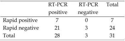
PCR assay. The number of positive and negative by rapid test antigen detection and RTPCR on the same group of the sample are shown in Table 1 and 2. Sensitivity test was calculated by Martinet al. (1988) presented in a 2x2 table.
Typing calculation of sensitivity was 5/10 = 50%, while the specificity was 7/7 = 100%.
Subtyping calculation of sensitivity was 7/28 = 25%, while the specificity was 3/2 = 100%.
Sensitivity test for AI typing kit by the Anigen A Rapid Test kit AIV Ag showed better results compared to AI subtyping kit by Anigen Rapid Test kit AIV H5 Ag. The value of sensitivity was 50% and 25% respectively. Rapid test specificity both typing and subtyping indicated higher results (100%) compared to sensitivity test. Sensitivity and specificity test which was conducted by AI reference laboratory in Geelong, Australia, reported that various rapid test kits indicated similar results to our study (Sellect, pers. comm.).
The results of this study showed that the subtyping sensitivity was low (25%) compared to the similar research on rapid tests which have been conducted in Indonesia by Lothet al. (2008). It was reported that the detection kit Anigen AIV rapid test kit had a higher sensitivity of 69%. Rapid detection test
of Flu DetectTM capture antigen test kit had a sensitivity of 71%. According to Bolandet al. (2006) and the report was cited by Lothet al. (2008) stated that Anigen sensitivity reached 76%, for field application where samples obtained from AI infected chickens in Indonesia. Another study conducted by Chua
et al. (2007) showed that the sensitivity of the 5 types of rapid detection test kits for AI (brandname not specified) generally showed similar results which ranges from 36.3% to 51.4%. According to that study, the result was more variable depending on the type of sample. Fecal sample indicated sensitivity value of 0% to 12.5%, while the cloacal swab samples were 31.5% to 47.5%. Pulmonary organ sample exhibited varied sensitivity results, ranges 34.4% to 81.3%, while the swab brain reaches 40 to 93.3% of sensitivity.
Specificity test calculation according the Martin’s table showed that the test indicated very good result, reaching 100%. The results are relevance to the report of Lothet al. (2008) that specificity of rapid AIV AnigenRreaches 98%. According to the study of others rapid test kit that was Flu DetectTM strip antigen capture test has similar sensitivity of 98%. Specificity tests of the rapid test conducted in Indonesia applied to sample obtained from AI infected chicken reported by Bolandet al. (2006) cited by Lothet al. (2008) was reached 97%.
Analysis for some possible causes of the low sensitivity of the rapid test has been reported by Chuaet al. (2002), stated that the result of the rapid test kit depending on the type of sample. According to that study, feces samples showed the lowest specificity, while cloacal samples exhibited various specificity, ranged from 31.5% to 49.4%. The best specificity obtained from brain swab ranging from 40 to 93.3%, especially in the case of died birds with symptoms of encephalitis. Specimens from chicken origin showed the sensitivity varies between 37.6% to 60.6%, while samples from the dead waterfowl were varies ranges from 30.7% to 49.2%.
Basically, the sensitivity of antigen detection kits depending on the concentration of virus in the sample. The concentration of
Table 1.Anigen Rapid Test kit AIV AAg vs RT PCR (for typing).
antigen is mainly correlated with the titer of virus in the sample. Artificial infection of H5N1 virus in ducks showed replication occurs in a few days at the beginning of infection. However, viral titers generally are found in the trachea and cloaca of chicken higher than the duck (Chua et al. 2008). According to Lothet al. (2008) type of sample for the application of rapid test kit should be taken into consideration, particularly it should be obtained from the sick birds or birds that had just died due to AI virus infection. In principle, there should be enough virus titer in a sample for rapid positive results. Additionally, cloaca swab samples should be taken without any fecal, mucus, and blood contamination, due to the potential interference of the rapid test kit process (Loth
et al., 2008; Tsuda et al., 2008). Interesting report from Yamammoto et al. (2008) suggested that feathers can be considered useful samples for early AI virus detection in duck. The reason is that larger amounts of AI viruses can be isolated for a longer time from feathers than from swabs.
Further analysis of low sensitivity in this study, it may due to the AI virus master seed which be used to produce antibodies in the producing country is different with AI virus circulating in Indonesia. Smithet al. (2006) and Chenet al. (2006) analyzed that there are three genetically distinct sub populations of AI viruses in Indonesia and distributed in several different geographic areas, although all type the AI virus can be found on the island of Java. The difference means that AI viruses Indonesia origin have antigenic variation, which can be measured from antibodies titer difference from each subpopulation (Smithet al., 2006).
Conclusions
Based on these results, it could be concluded that Anigen Rapid Test Kid AIV/H5 Ag are able to detect AI antigencontaining samples having HA titer up to 26to determine type A AI virus and HA titer up to 25for H5 subtype detection. Anigen Rapid Test Kit AIV/H5 Ag showed no crossreactions with Infectious Bronchitis virus, Newcastle disease
andEscherichia coli. Sensitivity of Anigen A Rapid Test Kit AIV Ag was 50% and specificity test was 100%, while Anigen Ag Rapid Test Kit AIV/H5 showed a sensitivity test of 25% and a specificity of 100%.
Acknowledgments
Sincerely thank to Drh. M. Munawaroh MM. who allowed the data to be published and the management of PT. Mega Medika Mandiri, which supported this research.
References
Anonymous, 2003. Avian Influenza.
Preliminary Final Report/OIE
Terrestrial Animal Health Standards Commission. Appendix. pp. 711. Asmara, W., Wibowo, M.H., Tabbu C.R. 2005.
Hemagglutini Subtype Identification of Avian Influenza Virus Isolated from Various Species of Birds Using RTPCR. Sain. Vet. J.,: 4245.
Charlton, B., Crossley, B., Hietala, S. 2009. Conventional and Future Diagnostics for Avian Influenza. Comp. Immunol. Microbiol. and Infect., 32, 341350. Chen, H., Smith, G.J.D., Li, K.S., Wang, J.,
Fan, X. H., Rayner, M.J., Vijaykrishna, D., Zhang, J.X., Zhang, L.J., Guo, C.T., Cheung, C. L., Xu, K.M., Duan, L., Huang, K., Qin, K., Leung, Y.H.C., Wu, W.L., Lu, H.R., Chen, Y., Xia, N.S., Naipospos, T.S.P., Yuen, K.Y., Hassan, S.S., Bahri, S., Nguyen, T.D., Webster, R.G., Peiris, J.S.M., Guan, Y. 2006. Establishment of Multiple Sub lineages of H5N1 Influenza Virus in Asia: Implication for Pandemic Control. Proc. Nat. Aca. Sci., 103(8), 28452850. Chua, TH., Ellis, T.M., Wong, C.W., Guan,
Y., Ge, S.X., Peng, G., Lamichane, C., Maliadis, C., Tan, SW., Selleck, P., Parkinson, J. 2007. Performance Evaluation of Five Detection Test for Avian Influenza Antigen with Various Avian Sample. Avian Dis., 56, 96105.
Cox, N.J., Kawaoka, Y. (1998)
press, Inc. New York. pp. 386433. Damayanti, R., Dharmayanti, N.L.P.I.,
Indriani, R., Wiyono, A., Darminto. 2004. The Clinicopathological Effect of Chicken Infected with Highly Pathogenic Avian Influenza in Some Farms in East Java and West Java. JITV, 9(2), 128135.
Dharmayanti, N.L.P.I, Damayanti, R., Wiyono, A., Indriyani, R., Darminto. 2004. Identification of Avian Influenza Virus of Indonesian Isolate by Reverse Transcriptase Polymerase Chain Reaction. JITV, 9(2), 136143.
Dharmayanti, N.L.P.I., Indriyani R. 2007. Molecular Pathogenicity of Avian Influenza Virus Isolated in the Year 2005. Media Kedokteran Hewan, 23(2), 6873.
Fouchier, R.A.M., Munster, V., Waltensen, A., Besterbroer, T.M., Herfst, S., Swith, D., Rimnelzwaan, G.F., Olsen, B.,
Osterhaus, A.D.M.E. 2005.
Characterization of Novel Influenza A Virus Hemagglutinin Subtype (H16) Obtained from Black Headed Gulls. J. Virol., 79(5), 28142822.
Gambaryan, A., Tuzikov, A., Pazynina, G., Bovine, N., Balish, A., Klimov, A. 2005. Evolution of the receptor binding phenotype of influenza A (H5) viruses. Virology, 344, 432:438.
Hidayanto, N.K., Asmara, W., Wibowo, M.H. 2015. Characterization of Avian Influenza Virus H5N1 Subtype of Non Structural Gene from Duck Isolated in 2013. Sain. Vet. J., 33(2), 195204. Ito, I., Goto, H., Yamamoto, E., Tanaka, H.,
Takeuchi, M., Kuwayama, M.,
Kawaoka, Y., Otsuki, K. 2001.
Generation of HPAI A Virus from an Avirulent Field Isolate by Passaging in Chicken. J. Virol. 75., (9), 44394443. Kang, B., Oh, J., Lee, C., Park, B., Park, Y.,
Hong, K., Lee, K., Cho, B., Song, D.
2007. Evaluation of a Rapid
Immunodiagnostic Test Kit for Rabies Virus. J. Vir. Method., 145, 3036. Khewcharoen, J., Oraveerakul, K., Kuiken,
T., Fourchier, R.A., Amonsin, A., Payungporn, S. 2004. An Avian Influenza H5N1 in Tiger and Leopard. Emerg. Infect. Dis., 10, 21892191. Loth, L., Prijono, W.B., Wibawa, H., Usman,
T.B. 2008. Evaluation of Two Avian Influenza Type A Rapid Antigen Tests Under Indonesian Field Conditions. Diagn. Invest., 20, 642644.
Mahardika, G.N., Jonas, M., Murwijati, T., Fitria, N., Suartha, I.N., Suartini, I.G.A.A., Wibawan, I.W.T. 2016.
Molecular Analysis of
Hemmagglutinin1 Fragment of Avian Influenza H5N1 Viruses Isolated from Chicken Farms in Indonesia from 2008 to 2010. Vet. Microbiol., 186, 5258. Martin S.W., Meek A.H., and Willeberg P.
(1988) Sensitivity and Specificity. In Veterinary Epidemiology Principles and Methods. Iowa State University Press. pp. 6366.
National Committee for Avian Influenza and Preparedness of Influenza Pandemic (NCAIPIP). 2008. The Timeline Data of Avian Influenza Outbreak in Indonesia. http//www.komnasfbpi.go.id/situati on/update.html.
Payungporrn S., Piraya P., Salin C., Apiradee T., Juthatip K., Kanisak O.V., Alongkorn A., Yong P. 2004. Single Step Multiplex Reverse Transcriptase
Polymerase Chain Reaction for
Influenza A Virus Subtype H5N1 Detection. Viral Imm., 17, 588593. Peng F., Wang Z., Zhang S.,Wu R., Hu S., Li
Z., Wang X., Bi D. 2008. Development of an Immunochromatographic Strip For Rapid Detection of H9 Subtype Avian Influenza Viruses. J. Clin. Vaccine Immunol., 15(3), 569–574. Smith G.J.D., Naipospos T.S.P., Nguyen T.D.,
Adaptation of H5N1 Influenza Virus in Avian and Human Hosts in Indonesia and Vietnam. Virol., 350, 258 268.
Sellect P., Avian Influenza Rapid Test Field Trial Data. Australian Animal Health Laboratory, Geelong, Australia. Un published data.
Songserm T., Alongkorn A., Rungroj J.,
Namdee S.H., Noppodal M.,
Nuananong P., Sunchai, P. 2006. Avian Influenza H5N1 in Naturally infected Domestic Cat. Emerg. Infect. Dis., 12(11), 17441764.
Srihanto E.A., Asmara W., Wibowo M.H. 2015. Philogenetic Molecular and Antigenic Structure Analysis of Avian Influenza Virus of Subtype H5N1 Lampung Isolates Collected during 2008 to 2013. Jurnal Kedokteran Hewan, 9(1), 8388.
Suarez D.L. 2003. Moecular Diagnostic Techniques: Can We Identify Influenza Viruses, Differentiate Subtype and Determine Pathogenicity Potential of Viruses by RTPCR. Avian Dis., 47, 318 325.
Susanti R., Soejoedono R.D., Mahardika I.G.N.K., Wibawan I.W.T., Suhartono M.T. 2008. Molecular Analysis of Hemagglutinin Gene of HPAI of H5N1 Subtype Isolated from Waterfowl. JITV, 13(3), 229239.
Tantilertcharoen R., Prapasakul N., Thanawongnuwes R., Kramomthong I. 2016. Performance Evaluation of Avian Influenza Virus Antigen Rapid Test Kit. Veterinary Diagnostic Laboratory Unit, Faculty of Veterinary Science, Chulalongkorn University, Bangkok 10330.
Tong S., Li Y., Rivailler P., Christina C., Danilo A., Castillo A., Chen L.M., Recuenco S., Ellison J.A., David C.T., York I.A., Turmelle A.S., Moran D., Rogers S., Shi M., Tao Y., Weil M.R., Tong K., Rowe L.A., Sammons S., Xu X., Frace M., Lindblade K.A., Cox N.J., Anderson L.J., Rupprecht C.E., Donis O. 2012. A Distinct Lineage Influenza A Virus
from Bats. Proc. Nat. Aca. Sci., 109, 42694274.
Tsuda H., Sakoda Y., Sakabe S., Mochizuki
T., Namba Y., Kida H. 2007.
Development of an
Immunochromatographic Kit for
Rapid Diagnosis of H5 Avian Influenza Virus. Infection. J. Microbiol. Immunol., 51(9), 903907.
Wibawa H., Prijono W.B., Dharmayanti N.L.P.I., Irianingsih S.H., Miswati Y., Rohmah A., Andesyha E., Romlah, Daulay R.S.D., Safitria K. 2012. Outbreak investigation of Duck Disease in Central Java Province, Yogyakarta Special Province, and East Java Province: Identification of New Clade of Avian Influenza Virus H5N1
Subtype in Indonesia. Buletin
Laboratorium Veteriner ,12(4), 19. Wibowo H.W., Susetya H., Untari T., Putri
K., Tabbu C.R., Asmara W. 2006. Molecular Study on the Pathogenicity of Avian Influenza Virus. Indones. J. Biotechnol., 11(2), 901907.
Wibowo M.H., Susetya H., Untari T., Wahyuni A.E.T.H., Tabbu C.R., Asmara W. 2007. Molecular Identification of Avian Influenza Virus from Cases Both with and without Symptom Specific for Avian Influenza. J. Vet., 8(3), 103 110.
Wibowo M.H., Srihanto E.A., Putri K., Asmara W., Tabbu C.R. 2013. The Development of Pathogenicity of Avian
Influenza Virus Isolated from
Indonesia. Indones. J. Biotechnol., 18(2), 133143.
Yamamoto Y., Nakamura K., Okamatsu M., Miyazaki A., Yamada M., Mase M. 2008. Detecting Avian Influenza Virus (H5N1) in Domestic Duck Feathers. Emerg. Infect. Dis., 14(10), 16711672. Wiyono A., Indriani R., Dharmayanti N.L.P.I.,
Damayanti R., Parede L., Syafriati T.,
Darminto. 2004. Isolation and