Journal of Agriculture and Rural Development
in the Tropics on Subtropics
;1..__)
,/'
' /
Beiheft 90
nn
nnzr:w=
Mtmmnznn-
:
,Empowering of Society through the
Animal Health and Production Activities
with the appreciation to
the
Indigenous Knowledge"
PROCEEDING OF THE MINI WORKSHOP
Southeast Asia Germany Alumni Network (SfAG)
May 3rd-5, 2007
Table of Contents
Maintaining Indigenous Knowledge for the Sustainability of Animal Welfare Beyond the Conc.:ept of National Park
Model (Adam Malik)
Indigenous Knowledge Between Collapsion and Prospect of Genetic Conservali0n and Development
(Ali A1.A.Rachman) ... ..
Recent Progress In The Application Of Reproductive Technology In The Indegenous Garut Sheep
セ
セmッセ。ュ。、@ Agus Setiadi) ... :·:··· ... .e EtTe9t of Ambon Banana Stem Sap (Musa paradlSiaca fom1a
セM .• セ@... セᄋBGエケーゥ」。I@ on the Acceleration of Wound Healing Process
in Mice
(Mus
musculus albinus).(BP Priosoeryanlo, N Putriyanda, A R Listyanti, V
Juniantilu, 1 Wientarsih, BF Prasetyo and
R Tiuria) ... ..
Genetics Quality Improvement of Indigenous Beef Cattle ThroughArtificiallnsemination Program in West Java
(.)ifi Darodj,?h Rasa d) ... ..
The Utilization Of Oil Palm Bud On qオセゥャGウ@ Performance
(Daisy D. S. J Tambajong) ... ..
Improving Cocoa Pod Quality By Urea, NaOH and Cocoa Pod Ash Alkali Treatments For Ruminant Feedstuffs.
(Despal) ... ..
Utilization Of Methanol Extracted Of Moringa And Mulberry Leaves To Evaluate Energy and Protein Balance Of Nile Til apia
(D.A. Astuti, K. Becker andN. Richter) ... .
A study of carcass and meat chemical composition of babirusa
(Babyrousa babyrussa celebensis Deniger)
(E Pudjihastuti, S P Pangemanan and CL Kmmang) ...
Nutritional Properties Of Three Different Origins Of Indonesian
.. Jatropha (:Jatropha Curcas) Meal For Ruminant
\
/;::l.·----,,\ _(LG
tセᄋゥ。ウエオエケL@
Despal。ョセ@
IGp・イュ。ョ。セ@
... .The E.flect of Bay Ler.ves Intusum (Syzygmm polyanthum
. / (Wight) on anti inflammation in White Rat
Sprague---... · Dawley)
I Wientarsih, M Iskandar and G H Saputra) ... .
8
15
26 3550
5663
70
8394
102Bushmeat Hunting in North Sulawesi and Related Conservation Strategies ( A c.:ase study at the Tangkoko Nature Reserve)
(Jane S.I.
T.
Onibala and Sylvia Laatung) ... ..Establishment of Sustainable Signal Grass Pasture by
Amendment of Chromolaena odorata Biomass and
Manure as N11trient Organic Source: Effect on growth parameters, <iry matter production and canying capacity.
(L. Abdullah, and
D.
Puspitasari) ... ..Impact:; of Pigs Farming on The Living Environment at "Pakakaan Zone"
( M T. Massie) ... ..
Improving Quality Of Local Feedstuff And Its Use For Fattening OfPeranakan Ongole (PO) Male Cattle
(A1uhamad Bata) ... .
A View Of Bogor Climatology Related To The Emerging
Anthrax And Avian Influenza Diseases Since January 2004 To February 2005: Importance For Early Warning System
(A Suprayogi, H Setijanto, I WT Wibawan, F Satrija and
W DSury,a) ... ..
Alternative Utilization of Storage Roots Flour of Yam Bean
(P.erosus) in Wheat Flour-Based Food Products
(Bread).
(Pieter Rihi Kale, A.Kanmiawan and Elke Pawelzik) ...
Comparation Study Progress Ou Anoa's Behaviour Prior To Conservaticn Program
(R 1 Pujaningsih, A Malik, and S Pudyatmoko) ... ..
Rabies Case Study On Dog's Head (Canis Familiaris) In
Manado, Airmadidi & Langowan Wet Markets
(S Adiani and E Tangkere) ... .
Estimation of Relative Efficiency of Indirect Selection for Weaning We!ght Base on Birth Weight UsiPg Maternal Eftect Modd on Landrace Cross Breed Pigs
(Sri Bandiatz Komar Prqjoga) ... .
Clove Oil (Eugenia aromatica) and Potassium Hydroxide (KOH)
as A Semi Pemtanent Stain on Nematodes and Acanthocephalan Worms of Marine Fishes
(Risa Tiuria, Khairun Nisa and Adhi Rachmat Sudrajat Hariyadi) ... .
hidup dan implementasi transfer embrio pada temak domba.
Laporan Penclitian 1-libah Bers::ting Xll/3. LPPM-IPB, Bogor
Yulnawati, M.i\. Setiadi dan A. Boediono. 2005. Maturation and
fertilization rate of ovine oocytes collected from different
status of ovaries. Proc. lntl. Asia Link Symphosium, Bali pp:
199-222
Yulnawati, M.A. Setiadi dan A. Boediono. 2006. Penggunaan Medium
CR I aa untuk produksi em brio domba in vitro.
J.Ilmu
Ternak dan Veteriner I l (2 ): 131-136
The Effect of An: bon Banana Stem Sap (Musa paradisiaca forma typica} on the Arceleration of Wound Healing Process in Mice
(Mus musculus albinus ).
Bambang Pontjo Priosoeryanto". Nalia Putriyanda". Adinda Ratih Listyanti". Vctnizah Juniantito''.letje wゥ・ョエ。イN[ゥィセGN@ Bayu Febram Prasetyo"a•ld Risa Tiw·ia-'1
Abstract
The aim <!(this research is to find out the ac1ivi(l' of banana stem sap (Musa paradisiaca_limna typica) on the acceleration ofwmmd healing process in lhe mice skin (Mus musculus albimts) hosed on g.-ass and histopathological observations. Total(\' 45 heads ofDDY mire ages 4-6 weeks were used in this sllldy. The mice were dh·hled into three groups, negative control (without treatment), positiPe control 1Bioplacenton ') and hanana stem sap. All mice were 1-1.5 em incised on the dorsal back skin. Gross lesions were obsen''!d dai(v. On the NSNMNセL@ 5'h, 1h, 14'h and 21"' days c!fier the treatment, mice were elllhanb.>d and the skin samples were collected for fitrther histopathological observation. The anatomical parameters were blood coagulation, dryness, alluchment/narrowed of the wound andformation of the blood clot. The histopathological parameters were number of macrophages, neutrophils, (vmphocytes, neo-1•ascularisation percentage ofre-epithelization and the thickness of fil>roblast. All qualitalii'P data were ウエ。エゥウエゥ」。ャセカ@ anu(v::ed using Ana{v.fis of Variance
(ANOVA) and continued with Duncan Multiple Range Test. Gross lesion and the fihroblastthickness were ッ「Nセ・イカ・、@ and descriptively analyzed 。Nセ@ a quantitative data. 17te result indicated that hanana stem sap could promoie the wound healing process. Gross lesion ol>servation indicated that in the l>anana stem sap treatment the scab .fhrmation was fmter than negative control and bゥッーャ。」・ョエッョセN@ On macrophages, ョ・オエイッーィゥャセ@ and (1'111phoc.1·tes al>serPations, the stati.;tical ana{vze showed that the banana stem sap treatment was significant increase (P<O.IJ5) than the negatil'e control. The {ibroblmt thickness on the skin wound treated with banana stem sap was high 。セ、@ the formation was also faster than the negative control and Bioplacenton''. All result mentioned above indicated that Ambon banana stem sap was accelerated the wound healing process. Further study is required in order to c/ar!f}• the mechanim1 of the sap on wound healing process as well as thir toxicity and possibility for use both in animal and humattmedicine.
Key H'ords: Banana, histopatholog1·, mice, skin, stem sap, wound healing,
Division '!f Veterinary Pathology'·', Pharmacy'' D<!partment of Veterinary Clinic-Reproduction & Pathology; Division '!f Veterinary Helminthology Deportment of Animal Diseases and Veterinary Public Health 11; Faculty '!( Veterinary Medicine, Bogar Agricultural University (JPBJ. Jalan Agalis Kampus JPB Darmaga, BOGOR-16680
Introduction
Skin wound healing could be defined as a loss of integrity of the skin as a body's main baJTier of outer surface. In the human and veterinary medicine, wound cases are very common such as due to surgery, traumatic, skin burn and others. Wound heaiing is influnced by many factors including the kind of medicine/ drugs uses. The use of
drugs fi.>r wound treatment could be use in many ways and kinds; one of
th.:sc kinds is the use of herbal medicine. It ·s already known that some
plants could be usc for wound treatment such as banana tree.
Skin is a main barrier for preventing the invasion of pathogenic microbes from the environment. Skin wound will facilitate the pathvgenic microbes to enter the body and causing infection. The use of midicinel drugs is aim to accelerate the wound healing process and to prevent from infection (Yahya 2005). Wound healing process could be dcvidcd in 3 phases, there are inflamation phase, proliferation phase (regeneration or fibroplasia) and re-absorbtion phase (maturation or tissue re-absorbtion). Parumeters usc to indentify this 3 phase are inf1amatory cells (makrophages, neutrophiles and lymphocytes),
neo-capilarization, re-epitelization and connective tissue Hsェ。ュウオィゥ、セェ。エ@ and
DeJong, 1997; Kalangi, 2004).
Indonesia as a mega diversity country with 25.000-30.000 plant species has 6.000 species of medicinal plants (Kardono 2003). One of the potential plants to be explored as medicinal plants is banana plant. Banana tree is an indigenous plant of South East Asia including
Indonesia (Munadjim 1983 ). This plant is growth W:!ll, easily and
common found in a huge number in most South East Asian countries. People use this plant mainly for the fruit and leave; the stem is mainly usc only for ruminant feed and some cultural activity, therefore the use of the stem is not yet optimally while the stem sap itself never been exploited at all. Satuhu dan Supriyadi (1995) stated that the banana stem contain serotonin, noerepinefrin, dopamine, tannin, vitamin A, vitamin B and vitamin C that are very essensially for body in the wound healing process. Serotonin could increase the function of digestive tract, decreasing the process of infl<.'.mation and stimulate the skin cell regeneration. Priosoeryanto (2003) also explained that banana stem sap contain saponin, antrakuinon dan kuinon that functioned as antibiotic and accelerated the growth of cells on the regeneration process. This stem sap also increases the blood flow and stimulates connective tissue formation on the response of wound healing process. According to Djulkamain (1998), Ambon banana stem sap could be use for pain
36
reliever and facilitate the increasing of absorbtion capability of medicine in the skin therefore could be use to treated contusio, skin burn, animal bit and as anti-inflamation.
Due to many benef.cial activity of the stem sap that never been explore before, we conducted the present study iu order to elabotae scientifically the activity of Ambon banana stem sap on the wound healing process.
Materials and Methods
Banana Stern Sap Preparation
The banana tree was identified as Pisang Ambon (Musa
paradisiaca forma typica) for their species and variety in the Research
Center for Biology, Iudonesiar. Institute of Science (LIPI) Bogor. The stem sap was collected directly from the stem by cuting the stem with knife aseptically.
Laboratory Animal
Totally of 4.5 head of mice DDY strain, 4-6 week old were used in this study. Mice were kept in the individual cage with the optimum environment and temperature (18-24° C). Mice were fed with
a commercial feed 'lnd drinking water was given ad libit:un. Adaptation
period was done for 2 weeks.
Treatment of the Mice
Mice were distributed into 3 ァイ・セオーウ@ i.e. negative control,
positive control (Betarline'") and treatment (stem sap) groups. Each groups were then divided into 5 sub groups according to sampling day (day 3, 5, 7, 14 and 21 ). On the day I 2nd, thl" hair on the back skin were shaved and 2 days later the incision at out 1-1 ,5 em were done. According to the groups, mice were treated topically with Betad.ine<i<· or stem sap everyday. Daily observation of the gross lesion was done twice a day (morning and afternoon). On the desired day (3, 5, 7, 14 and 21) mice were euthanized using chlorofonn inhalation and the skin at the
incicision site and surrounding area were sampled for further
histopathological process and observation ..
Gross Lesion
Gross lesion examination was performed daily from day 1 to day 21 by direct observing the wound for their blood coagulation,
drym:ss, attachment/narrowed of the wound and formation of the blood dol.
llistopathological Findings
Histopathological observation was done by comparing the
treated and un-treated groups 0!1 the parameters ·of the number or
inllamatory cells, number of nco-capillary, percentage of
re-cpithclization and the density of the connective tissue. Observation for the number of inllamat01y cells were for macrophages, neutrophiles and lymphocytes. In llama tory cells and neo-vascularisatit'n examination
were performed on 15 microscopical fields with 3 replicates using a
light microscope. Percentage of the re-epithelisation was done using a videomicrometer by calculating the ratio of the length area of the wound which cove1ed by new epithelial cells with
Calculation of the re-epithelisation according to DiPietro (2001):
Length of the \Votmd with new epithelial cells
% Re-epitelisation: :l\-lVU70
Total length of the wound
Thickness of the connective tissue was examined by the intensity of the connective tissue with Masson Trichrome stained using a scoring methode as describe below.
Table 1. S --- --- Lcsio --- ---f1 セMMM
c
---tive T'Grade Parameters
I The wound stil open with minimal density of connective
tissue, the distance between connective tissue is loose.
++ The wound could stil open or partially closed with
connective tissue densitv low to moderate in several areas.
+++ The wound could totally 」ャッウ・エセ@ or minin>ally open with
high density and compact of the connective tissue. Some loose area is still detected with fonnation ッヲョ・ッM」。セゥャャ。イゥ・ウ@
ᄋセ@ +
+
+ The wound is totally closed with very high density and---·--- ... 」セューセセA@ .. セNエGセGZjNセセMカ⦅ANャャセセMエゥカ・@ tissue
Da(a Analysis
Tht: data of inllamat01y l:clls and formation of neo-capillariy were statistil:ally analyzed using ANOVA and followed by Duncan Multirange Test. Gross lesion and the densities of connective tissue were analyzed descriptively.
38
Result and Discussion
Gross Lesion
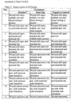
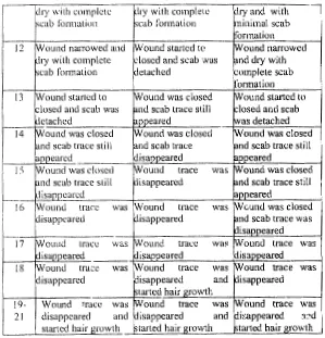
Wound hc::ling process (daily ob5ervatio) of all groups was tabulated in Tabel 2 below.
Table 2. Gross Lesion of all Groups.
セMMM - - - · - · · · · - · - - - · - - · - · - · · · - · · · - - - - · . · · · · ·
-Dav Treatment
• b・エ。、ゥョ・GセG@ Stem Sap Neestive Control
I
I Wotmd still open, セvッオョ、@ still open, rwound still open,eddish, wet and eddish, wet and eddish, wet and
blood doting is セャッッイャ@ doting ゥセ[@ セャッッ、@ doting is
present. present. present.
2 Wound still open. red !Wound still open,
iW
ound still open,o yellowish and wet eddish, wet and eddish and wet
white granule
3 Wound still open, Wound still open,
iW
ouad still openyellowish and ninimal dry anc セョ、@ wet.
ninimal d!.Y_ eddish black granule
4 Wound sti!l open and Wound still open and rwound still open
n!nimal d1y minimal drv セョ、@ minimal drv
5 Wound still open, Wound still open but Wound still open
ninimal dry and narrowed and minimal but narrowed and
exudated dry ninimal dry
6 Wound still open, Wound narrowed with Wound still open
ninimal dry and ninimal scab and minimal dry
exudated formation
7 Wound still open and Wound narrowed 'Nith Wound still open
ninimal dry ninimal scab and minimal dry
formation
--g·
Wound narrowed, dry Wound narrowed with Wound narrowedand with minimal minimal scab and minimal dry
セ」。「@ fromation
--
ヲァAセョ。エゥッョ@Wound narrowed
9 Wound narrowed, dry Wound narrowed, dry
and with minimal and with moderate and minimal dry
scab fonnation scab fonnation
10 Wound narrowed and Wound narrowed and Wound narrowed,
py
with moderate dry with complete dry tl.nd withscab fonnation scab fonnation minimal scab
fonnation
II :Wound narrowed and
iW
ound narrowed andiW
ound narrowed, [image:5.841.481.784.92.520.2]lry with complete ·cab ronnatiun
lry with complete ·'cab formation
l!y and with
ninimal scab formation
!2 !Wound nmTowed and セwッオョ、@ started tc
hy with complete ·loscd and scab was
ound narrowed . nd dry with
:omplcte scab !formation
-cab formation letached
13 ound started to
!4
!)
16
17 /Wound trace was /Wound trace キ。セ@ !Wound trace was
18 /Wound tra;:e was !Wound trace was !Wound trace was
!9-
I
W mmd trace was セ@ ound trace was round trace was21 disanneared and isaooeared and isaooeared Zャセ、@
40
b
Figure I. Gross lesion at day-7th post incision. a) Negative control; b)
Banana stem
sap
mtd c). Betadint."'In the stem s3p group, scab formation was started at day 7 post
incision (PI), while in the Bet!ldine"' scab was formed at day 8 PI and in
the negative control groups at day I 0 PI. (Table 2). Scab is the m3llifestation of the granulation tissue, with earlier scab formation implicated that formation of granulation tissue was started earlier (Kalangi 2004). Detachment of scab and wmmd closed were appeared
more early in the scab group, this condition implies that the skin was
going back normally and the wound healing process entered the final stage. Disappering of the wound trace indicated that WO\IDd healing
process was completed acheived.
Growth of the hair indicated that wounded skin were
morphologically a.'ld functionally back to normal (Pinkus
&
Mehregan1982). Growth of the hair was earlier detected on the セQ・ュ@ sap group
(at day 18 PI) this implies that treatment with stem sap caused WO\IDd healing process faster and better (Table 2).
Neutrophiles
Neutrophiies is the one of blood component that play an important role in the earlier response to inflamatory, phagocytosis, killing the microbes and (Lever 1986).
The 。ーーセZ。イ。ョ」・@ of neutrophiles is acted as the first
leucocytes response to the acute intlamat')ry in order to clean up the
wound from contaminant microbes by phagocytic activity (Kalangi
2004). The number of ncutrophiles on day 3rd PI in the stem sap group
was significantly higher compared to the Betadine" and negative control
groups (P<0.05) as shown in Table 3. The high number ofneutrophiles
is indicated that the clean up and phagocytic activity was earlier
occured in the stem sap group compared to other groups.
[image:6.841.87.386.44.355.2]bl
--
....--
-· セ@ ᄋMセMᄋ@ ber ofN - ---- - -hi! - - --...-Day Treatment
Betadinc<!O Stem Sap Negative Control
3 233.33 ± 47.93B 440.00 ± 46.94A 203.33 ± 30.92B
5 266.67 ± 118.09" 2)6.67± 8.02A 171.00 ± 40.26A
7 !46.00 ± 65.82AB 122.67 ± 45.94B 232.00 ± 32.92A
14 88.67 ± J4. 74A 60.00 ± 11.27/\ 66.33 ± l7.67A
21 94.33 ± 16.072/\ 59.()0 ± 28.58AB 37.67±5.18
MMMᄋMMセ@
Note: The same alphabet (superscript) indicated no significant difference (P>0.05).
G[セ@
·-.cJ
o•
セ@
. ,,, ..
.
·
...
--..,1
jゥャᄋセᄋᄋZ@
. . .セ@
;j' -:··,co· .. •· ')4'
"
,\;
:,.
MセM セ@
! .. :· ..
セセIiヲ@
t
Mセセᄋ@
....
'\.•.
;o, ...
:';
,, .• -· !'f'
·:> •
セN@
セ@
.,
...
BセN@
ᄋᄋセ@
.•.
',.0 , ..
·-·.,
...
·
セL@
it. ·. セ@ ·..•
セ@
-···· |Dセ@ "セN@
« ...
,fi,,,
' !·. .· ..
Figure 2. Number of neutrophiles day 3rd
PI.
a). Betadine"'; b). Bananas stem sap and c).negative. Control. HE Staining. 1200X.Kalangi (2004) stated that the present of neutrophiles in the wound is the tirst response of the body defense by fagocitic activity and will be decreased in line with cleaness of the wound tissue. The high number of neutrophiles in the stem sap group at day 3rd and followed by gradually decreasing up to day 21st (Table 3) indicated that the wound is clean enough from contaminant microbes therfore the requirement of neutrophiles was also decreased. The presence of neutrophiles and r.1acrophages on the wound area is sinergistic effect in order to clean up the wound (Nadesul 2003). The function of neutrophiles as phagocytic cells for clean up the wound tissue
was
very optimal in the earlier stage and then was gradually replaced by macrophages in the end stage of wound healing process.42
Macrophages
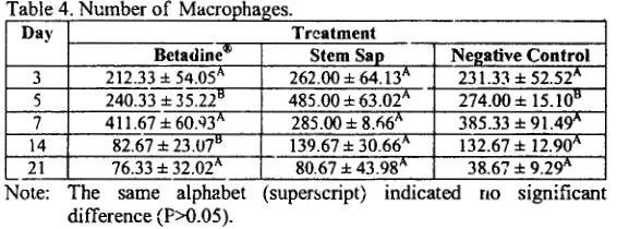
Macrophages !s ;me of the bigger size of white blood cells with ability to digest the microbes, antigens and others substances which nonnally not circulated but present on the blood vessel associated -tissue (Yahya 2005). The function ofmacrophages is for fagositize and elimination (clean up) of tissue debris, killing of microbes and (Yahya 2005). The number ofmacrophages at day 5th PI on stem sap group was significantly high (P<O 05) compared to other groups (Table 4). The condition mentioned above indicated that in the stem sap groups the fagocytic activity was high compared to the other two, and this implies the faster clean up of the wounded tis5ue by the sap.
---
·-Number of M ---- ---h MセMDay Treatment
Betadine"" Stem Sap Negative Control
3 212.33 ± 54.05A 262.00 ± 64.13" 231.33 ± 52.52"
5 240.33 ± SUNRRセ@ 485.00 ± 63.02A 274.00 ± 15.10"
7 411.67 ± 6Q.'l3A 285.00 ± 8.fl6A 385.33 ± 91.49"
14 82.67 ± 23.07" 139.67 ± 30.66A 132.67 ± 12.9QA
21 76.33 ± 32.02" 80.67 ± 43.98" 38.67 ± 9.29A
Note: The same alphabet (superscript) indicated no significant difference (P>0.05).
Biologically, macrophages released the active substances such as vasoactive mediators, chemotactic, grmvth factors and enzymes including proteases (Kalangi 2004 ). In the wound healing process, macrophages formed a granulation tissue together with neo-capillary and connective tissue. The number of macrophages on the stem sap group was highP.r compared to Betadine® and negative control groups in every observation day (Table 4). The high number of macrophages will produce a lot of growth factors which will stimulate the growth of new cells (cell proliferation) and faster formation of granulation tissue that affected to the acceleratton of wound healing process.
Lymphocytes
In the immune systems, beside phagositoses, elimination of infectious or toxical agents is aiso by formation of antibody. The function of lymphocytes is as natural killer which could destroy alien substances or produce specific antibody (Guyton & Halll997).
[image:7.841.495.778.179.284.2]MMセMM -· . ᄋセMMMMMM -- --- --·--
-Treatment Day b・エャャ、ゥョ・セG@
Stem Sap Negative Control 3 13.67 ± 4.736 33.33 ± 9.87A 14.00 ± 5.578 5 15.67 ± Jj3H 27.67 ± 4.73A 15.67 ± 1.158 7 9.33 ± 3.51A 10.00 ± 6.08A I 0.33 ± 3.051\: 14 17.67 ± 9.50A 14.00 ± 2.65A 18.67 ± 14.50"' 21 24.00 ± 9.85A 6.67 ± 4.1608 14.67 ±
1.53AB-Note: The same alphll.bet (superscript) indicated no significant difference (P>0.05).
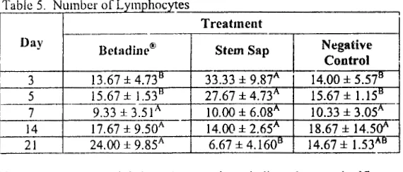
When specific lymphocyteswas activated by antigen, lymphocytes will proliferated and produce antibody (Guyton & Hall 1997). The numbt·r of lymphocytes at day 3rd and 5th PI were significantly higher (P<O.OS) compared to the Betadine<&· and negative control groups (Tabe!S). The high presence of lymphocytes 011 the stem
sap group indicated that lymphocytes act as body immune system together with neutrophiles and macrophages.
In the immune system mechanism, the presence of lymphocytes could be influenced by the presence of macrophages. Pathogenic microbes or substances will be phagocytize earlier by macrophages then their antigenic substances will be released into the cytosol. These antigens will be in contact with lymphocytes which stimulate the proliferation of lymphocytes (Guyton & Hall 1997). In this study, stem sap seem could stimulated the presence of macrophages which trigern .. xllymphocytes to proliferate for producing antibody.
N eo-capillaries Formation
Nco-capillary formation is one of a multistage mechanism in the wound healing process which a step of connective tissue re-modelling (Vegad 1996 ). In this study, there was a non-significant difference (P>0.05) on all ァセッオーウ@ in the nco-capillary fonnation.
44
Table 6. Numb - - ---- --
----Day
セ・エ。、ゥョ・セG@
TreatmPnt Stem Sap Ne2ative Control3 39.6i ± !9.7il'- 66.33 ± 26.03A 65.67 ± 18.01A
5 I 08.67 ± 30.14A 163.67 ± 71.35A 76.00 ± 14.53A 7 155.00 ± 77.3'57\ 132.33 ± 58.20}\ 213.67
±
64.08A 14 27.00-J: 14.5¥ 44.33 ± 17.10 ... 35.00±
1.73A 21 40.33 ± 17.797. 41.00±
29.51}\ 20.67 ± 12.9if Note: The same alphahet (superscript) indic:-tted no significantdifference ( P>0.05).
According to Vegad (1996) nco-capillary was fonned in the process of granulation tissue formation which started 24 hours PI and will be in the maxi!Pum at day 5th PI in order to fullfiled the nutrient intake for cells repair.
Increasing number of nco-capillary was early appeared on the stem sap group at clay 5th PI (Table 6). This phenomenon indicated that stem sap stimulate the fonnation of nco-capillary therefore the nutrient intake is fulfilled sufficiently for the necessity of cell proliferation and healing process. At day 7th PI the decreasing requirement of nutrient in
the affected tissue will also decreasing the number of vasculary until! the oedema process was disappeared (V egad 1996). In the stem sap group, the decreasing number of nco-capillary was detected at day 5th PI while on the other groups was nottced a:. day 7th PI (Table 6), this condition indicated that decreasing of oed<!tna reaction was quickly developed in the stem sap group.
Re-epithelization
Based on Stadelman in Kalangi (2004 ), re-epithelization is one of the multisi:age mechanisms on wound healing process that include mobilization, migration, mitoses, and epithelial cell differentiation. These stages will re - conditioned the skin integrity. Mitoses and epithelial cell migration is functioned for re-conC:itioned of skin integrity. In our present study, there was no significantly difference (?>0.05) on the re-epithelization between groups (Tabel 7), this condition it seem due to no 5timulation effect of stem sap on the process of re-epithelization (Pigure 3. ).
[image:8.841.106.391.51.173.2]Table 7. P . . . fR - - . he!" ----セ@_ ... セ@...
Treatment
MMMMセANG⦅IG@ ... - Betadinr'"; Stem Sap . Negative Control
3
-· ---o±OA _______
--o±o·;;--
o± o ...
5 34 ± 15" 45 ±4" 31 ± 20"
7 56:!: 4.711 63 ± 32-" 64 ± 33"
14 100 ±
o"
100±
OA 100 ± 0!\21 100 :l: OA 100 ±
o ...
100 ±o ...
:Note: The same alphabet (superscript) indicated no significant dif!erence (P>0,05 ).
Re - epithelization is a process of repairing the skin epithelial
cells to facilitate the closing of wound on healing process. If re-epithelization develops quickly, the structure fonnation of epidennis layer will also quickly, therefore the repairing of the skin to become
nonnal is also stimulated (Pinkus & Mehregan 1982). At day 5th
PI,
thepercentage of re-epithelization in the stem sap group was higher compared to others, even statil:.ticaly there was no significantly difference (P>0.05), this implies that re-epithelization process was quickly developed in the stem sap group than the two others. At the day 14th and 21st there were a similar percentage on the re-epithelization process iu all groups; this figure indicated that the body response to the repairing process of the wounded tissue has been maximum ancl optimally achieved.
Connective Tissue
Co1mective tissue is a main component on wound healing process in order to increase and repair the skin/ tissue integrity (Kalangi 2004). In the stem sap group, the score ot connectiv;: tissue density was high compared to the Betadine(l(o and negative control groups (Table 8), this figure indicated that stem sap has an effect on stimulating .tle development of connective tissue which influences the strength of the
repaired tissue (Figure 3).
46
Table 8. Connective T" --- D
---·
Day Treatment
nctadinc"'
-
Stem San Nc!!ativc Control3 + + + + ++ + + + +
-·
5 + ++ ++ f !-+ ++ ++ ++ +
7 ++ ++ ++ ++ ++ ++ ++ ++ ++
+
14 ++ ++ ++ ++ ++ ++ +++ +++ +++
1- + + + + + t
2i ++ ++ ++ I·+ ++ ++ ++++ ++++ +++
·1-+ + + t·+ + +· + +·
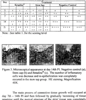
[image:9.841.473.771.50.398.2]Note : Sec table
1.
for エセ・@ scoring levelFigure 3. Microscopical appearance at day 14th Pl. Negative control (a);
Stem sap (b) and Betadine@ (c). The number ofintlamatory cells was decrease andre-epithelization was completely
occured in the stem sap group. HE staining. Magnification
40X.
The main ーイッ」・セウ@ of connective tissue growth will occured at
day 7th - 14th PI and then followed by gradually increasing of tissue
repairing until the normal structure of the skin! tissue was completely achieved (Kalangi 2004). At day 14th one of the replicant on the stem sap group has achieved the maximum level (+ + + +) while the other groups achieved this level at day 21st PI (Table 8), this condition
showed that the stem sap 。」」セォイ。エ・、@ the skin/ tissue nonnalization.
Conclusion
I. Banana stem sap accelerated the wound healing process.
2. Banana stem sap accelerated the detachment ofwouud scab
[image:9.841.102.383.50.164.2]..
)4. Oanana stem sap did not increased fom1ation of neo-capilary
and re-epithelisation
5. Banana stem sap increased the growth of connective tissue
6. Banana stem sap is seem could be use as a medicinal substance
for treated the wound healing
7. Further study for clarification of the mechanism of action as
well as toxicological effect of banana stem sap on the wound healing process is required.
References
Anonym. 2002. Healing and Repair
hllp://medweh.hham.ac.uk/hltp/depts/path/Teaching/toundat/re pair/healing.html. [ 15 Juni 2006]
.2005 Pisang, Man.faat gizi Hingga Obat Luka
http://www.republika.co.id/suplemen/cetak_detail.asp?mid=2
&id= 19l805&kat_id=l 05&kat_id
I
=150&kat_id2=187.[I 0
.Tuli 2006]
DiPietro LA eta/. 2001. Wound Healing
in
MIP-Hi·i· and mcpセャᄋGᆳMicc.
American Journal of Pathology.
Bum and ShockTrauma
Institute, Loyola University
MedicalCenter.
Maywood, Illine>is; 159:457-463.
http://a.ip.amjpathol.org/_cgi/content/fulll 159/2/457
/Fl.
[26Mei 2006].
Fishman TD. 1995. Phases of wound Healing
ィエエーZOOキキキNュ・、ゥ」セャ・、オN」ッュOーィ。ウ・ウNィエュ@ . [ 10 Juli 2006]
Guyton AC, JE Hall. 1997. Buku Ajar Fisiologi Kedokteran. Edisi 9.
Editor; Irawati SctiawPn. Penerbit buku Kedokteran, Jakarta.
Kalangi SJR. 2004. Peran Kolagen pada Persembuhan Luka.
http://www.dexa-medica.com/test/htdocs/dexamedica/article_.tiles/kolagen.pdf. [15 Juni 2006)
Kardono LBS, N Artanti, lD Dcwiyanti, T Basuki, K Padmawinata.
2003. Selected indonesian Medicinal Plants: Monographs and
Descriptions. PT Gramcdia Widiasarana Indonesia. Jakarta.
Lever WF, SL Gundula. 1986. Histopathology ofThe skin; Histology of
the Skin. Him. 8-36, Morphology of the Cells in the Dermal Iniiltrale. 1-llm. 47-55. J.B. Lippincott Company. Philadelphia.
Malole MBM, CSU Pramono. 1989. Pt:nggunaan Hewan
Laboratorium. Departement of Pendidikan dan Kebudayaan,
Bogor.
Mayasari NLPI. 2003. Kaiian Patologi Khasiat Tanaman Lidah Buaya
(Aloe vera) dalam Proses p・イウ・ュ「オィ。セ@ Luka Kulit pada
Mencit (Mus musculus albinus). FKH IPB. Bogor.
Munadjim. I 983. Teknologi Pengolahan Pisang. Gramedia, Jakarta.
Nadesul H. 1003. Biologi Sistem Kekebalan.
http://www.medicastore.com/med/detail. [1 5 Juni 2006]
Pinkus I-1, AH Mehregan. 198:!. A Guide to Dermatohistopathology
3'ct
Edition; Nonnal Structure; of Skin.
Him
5-38, Ulcers.Him
193-200. Appleton Century Crofts, New York.
Priosoeryanto BP. 2003. Penyembuh Luka di Sekitar Kita.
ィエエーZOOキキキNォッュー。セN」ッュOォッュー。ウM
cetak/0307 /24/inspira'ii/451512.htm. [15 Juni 2006]
Satuhu S, A Supriyadi. I 999. Pisang Budidaya, Pengolahan dan
Prospek Pasar. Cet. ke 10. Penebar Swadaya, Jakarta.
Soegiri J. 2006. Tartaman Berkhasiat Indonesia. Volume I. IPB Press.
Bogor .
Vegad JL. 1996. A Teet book of Veterinary General Pathology: Healing
and Repair. Him. 134-153. Vikas Publi!;hing House Put Ltd,
Nc;v Delhi.
Yahya H. 2005. Kastil yang Terkepung: Tuhuh Manusia.
www.harunyahya.com/indo/buku/tubuh 003.htm [ 15 Juni