ZALEHAMUSTAFA AND K. ELIZABETHTANNER University of Glasgow,
Glasgow, UK
INTRODUCTION
The materials requirements for composites for biomedi-cal applications include all the usual mechanibiomedi-cal property requirements for general engineering composites of stiff-ness, strength, toughstiff-ness, and so on. However, there are additional biological requirements, the materials have to be biocompatible, that is, not produce a disadvantageous biological response in the required application [1,2] includ-ing in the case of degradable materials the degradation products must also be biocompatible. An additional desir-able property is that the material should be bioactive, that is, it should produce a beneficial response in the appropri-ate application [1,2]. Bioactive means that the body’s cells react beneficially with the material, and in the case of hard tissue applications, the bone cells, instead of producing a layer of fibrous tissue between the patient’s own bone and the implant, produce new bone directly on the implant. This direct bone–implant contact leads to a mechanically stronger interface and thus a stronger and more durable bone–implant structure.
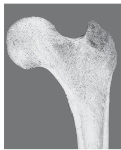
From a mechanical point of view, bone has two major functions, to provide a stiff skeleton against which the muscles can contract to provide locomotion and secondly to protect the major organs such as the brain, protected by the skull, and the heart and lungs, protected by the rib cage (Fig. 1). Biologically, bone provides a store of cal-cium and phosphate ions that are essential for biological function, while the marrow is the source for red and white blood cells [3]. At the macroscopic scale, there are two types of bone. The first type of bone is known ascortical, which is a nearly solid material, making up the outer surfaces of all bones, with a porosity of<3% in normal people [3]. The second type of bone is porous, is called eithercancellousortrabecular bone, being a foam of bone made up of small struts calledtrabeculae. Cancellous bone is found throughout the middle of bones such as the ribs and the skull and at the end of long bones below the joint surface. The porosity ranges from 30% to 90%, depending on the position in the body, age, and activity level of the person. Both cortical and cancellous bones become more porous with age and conditions, such as osteoporosis that particularly affects postmenopausal women. At the struc-tural and microstrucstruc-tural levels, cortical and cancellous bones have different structures in the form of osteons in cortical bone and trabeculae in cancellous bone. At the nanostructural level, all bone material is a composite of collagen, a natural polymer, filled with a calcium phos-phate mineral, thus a particulate-reinforced composite. There are three major types of bone cells, osteoblasts that
Wiley Encyclopedia of Composites, Second Edition. Edited by Luigi Nicolais and Assunta Borzacchiello. 2012 John Wiley & Sons, Inc. Published 2012 by John Wiley & Sons, Inc.
mature bone, and osteoclasts that resorb old and damaged bone. The properties of bone as a composite material are discussed in greater detail in the article titledComposite
Scaffolds for Bone Tissue Regenerationelsewhere in
this handbook. Hard tissue implants could either be going into or on to the shaft of bones and, thus, can contact cortical or cancellous bone or both. Young’s modulus of cortical bone is 7–25 GPa, while that for cancellous bone is 100–1000 MPa and their strengths are 50–150 and<50 MPa, respectively; all these mechanical properties depend on age, gender, activity level, and pathology in addition to the usual composite mechanical testing parameters of orientation and test velocity [4,5].
The major uses of hard tissue implants are as joint replacement to treat problems such as osteoarthritis, in fracture fixation where the bone ends need to be held sta-tionary relative to each other until the fracture has healed, filling of bony defects, such as after a tumor has been removed, and, the newest application, tissue engineering. For applications such as joint replacement, the material needs to retain its properties for the rest of the patient’s life, while for fracture fixation or tissue engineering scaf-folds, the material should degrade once the function of the implant has been taken over by newly formed tissue [1,2]. In tissue engineering, cells are removed, usually from the patient themselves, grown up in the laboratory to increase the number of biologically active cells and then seeded into a scaffold that is then implanted back into the patient [6]. The scaffold material needs to be an open-celled foam of a degradable material so that the cells can be dis-tributed throughout the scaffold material, body fluids can flow through to the cells to provide nutrients and remove waste products, and finally the scaffold material needs to degrade at the same rate as new material is produced by the body. The earliest composite implants used in the 1970s and 1980s were nondegradable, but in the last 30 years, degradable materials have been investigated and been used clinically.
Considering composites that have been developed specifically for biomedical applications, a range of matrix materials and fillers have been tested individually and been shown to be biocompatible. However, virtually no coupling agents have undergone biocompatibility testing and, therefore, most biomedical composites avoid the use of coupling agents and rely solely on mechanical interlocking to bond the filler and the matrix. Currently, most composites used in biomedical applications are either particulate-filled polymers or drawn polymer fibers in a polymer matrix; a few more recent applications use bioactive glass (BG) fibers. Most polymers used for the matrix phase are biocompatible, but only a few are bioactive and being polymers all are lower modulus than cortical bone. However, many calcium phosphate ceramic, glass, or glass–ceramic fillers are bioactive, particularly in bony applications, as well as having higher moduli; thus, their use should increase stiffness and bioactivity of the composite and can increase the strength.
Figure 1. Section through a proximal femur (thigh bone) show-ing cortical (lower walls) and cancellous bone (center of the femoral head).
The testing of biomedical composites includes the usual mechanical tests to assess the mechanical behavior of the material including fatigue tested at a frequency and in an environment that models the human body. Addition-ally, there are four groups of biological testing required with increasing levels of complexity and cost, two types of in vitrotests that are performed in the laboratory,in vivo tests performed in animals, and finally clinical trials. The first of the in vitro tests is soaking in a solution that mimics the appropriate body fluid to assess both how the material will break down after implantation and how the material’s properties change with time. While accelerated tests can be performed at raised temperature, most bioma-terials testing is performed at body temperature (37◦C), owing to the sensitivity of polymers to temperature effects, particularly in terms of their degradation behavior. In many applications, saline is the appropriate solution, but for potentially bone-bioactive materials, a simulated body fluid (SBF), such as that developed by Kokubo et al. [7] that is chemically very close to blood plasma, will be used. If a bone-bioactive material is placed in one of these solu-tions, a layer of apatite will be deposited on the surface, as would happen on implantation in the body, whereas a bioinert material does not produce this response. The second type ofin vitrotest uses cells and either the cells are grown in an elutant of the material, produced by soak-ing the material in a cell culture medium, or the cells are grown on the material surface. The elutant tests are to check for cytotoxicity, while the culturing of cells on the material is to assess the direct interaction of cells with the material, including how the cells attach to the material and whether their grown rate is increased or decreased by
this contact with the material. The type of cell grown will depend on the potential application of the material and for bone applications is commonly either a line of bone cancer cells or bone cells taken from human hip joints at total hip replacement. Assuming that the new material has passed these tests as being biocompatible and even bioactive the last set of tests before human use is implantation in an animal, thus is know asin vivotesting. Finally, a clinical trial will be performed before the new material or device is released for general use. The costs of bothin vivo test-ing and clinical trials are high and are highly controlled by national regulations. In the United States, approval is given by the Food and Drug Administration (FDA), while in Europe, approval is shown by a CE mark [8].
PRODUCTION OF BIOACTIVE COMPOSITES
All the techniques used for conventional composite mate-rials production and polymerization are applicable to biomaterials production, although the number of potential matrix and filler materials are constrained by biocom-patibility requirements. Biomedical composites based on thermoplastics and thermosets can be produced by conven-tional composite production methods such as compound-ing, extrusion, and injection molding and the application of preimpregation to produce ‘‘prepregs.’’ In the production of fiber-reinforced composites, techniques such as filament winding have been used to produce devices including arti-ficial intervertebral discs [9,10]. Selective laser sintering of both powders of composite materials and of the individ-ual phases has been performed to produce custom built porous materials [11–13].
Nondegradable Bioactive Composites
carbon-fiber epoxy composites is that the implants cannot be bent to fit the patient in the operating theater, unlike the equivalent metal plates where plastic deformation is possible, thus restricting the use of these composite plates to the human forearm where the bones are close to straight or to either preformed or low modulus implants as were used in the skull.
A different approach was taken Bonfield and colleagues, who proposed the use of high density polyethylene (HDPE) reinforced with hydroxyapatite (HA). Their philosophy was the ‘‘bone-analog’’ concept, that is, to produce a mate-rial with similar mechanical and biological properties to bone, consider the natural material, and try to repro-duce it. In HA–polyethylene (PE), the bone mineral was modeled by the HA and PE replaced the collagen, the HA providing stiffness and bioactivity, while the PE was responsible for ductility and toughness; at 40 vol% HA, the stiffness had increased from 0.94 to 4.29 GPa [19,20]. The first clinical application of this composite was as orbital floor implants and showed good biological responses [21,22]. Thereafter, the 40 vol% HA composite, HAPEX, was used as the shaft of middle ear implants with the pos-sibility of the supporting bone bonding with the HAPEX shaft, generating long-term stability of the implant; the high density of the composite was expected to improve the sound transfer through the implant shaft and the shaft could easily be trimmed intraoperatively to fit the indi-vidual patient. Goldenberg and Driver [23] reviewed the implant in 233 patients and found that the success rate was 56.8% defined by good hearing with only 5.3% implant extrusion occurred and 7.7% slippage. Meijer et al. [24], when reviewing some of the implants removed owing to reoccurrence of the original clinical problem, found good tissue cover, indicating better tissue acceptance than that occurred with previous implants of the same design, but manufactured using either Proplast or Plastipore.
Various attempts have been made to increase the mechanical properties of HAPEX. HAPEX was manu-factured by compounding extrusion of spray-dried HA particles in HDPE. Josephet al. [25,26] varied the molecu-lar weight of the PE to produce an injection molding grade of HAPEX and found that increasing both the filler con-tent of the composite and the molecular weight of the PE increased the viscosity of the melted composite. A similar effect was seen when using the nonsintered HA used in HAPEX rather than a sintered HA powder with the same mean particle size, but a specific surface area one-tenth of the nonsintered grade.
Ward and Bonfield and their colleagues attempted to produce HAPEX with higher and anisotropic mechan-ical properties by using hydrostatic extrusion [27,28]. Optimization of the extrusion ratio leads to a maximum flexural stiffness of 9 GPa and strength of 91 MPa in the longitudinal direction, although some slight reductions in the transverse properties were seen at the highest extrusion ratios.
A different approach to increase the mechanical properties was using very stiff drawn HDPE fibers as an additional filler in HA/HDPE composites [29,30]. They reported increases in stiffness and strength values to 17 GPa and 113 MPa respectively, suggesting that the
mechanical performance of the composite can be further increased. Alternatively, Reis and colleagues [31–33] used the shear-controlled orientation injection molding (SCORIM) method by applying a macroscopic shear stress field at the melt/solid interface of the polymer during the molding cycle. This molding technique produced anisotropy in the HDPE and HDPE/HA composite and yielded stiffness values between 5 and 7 GPa. None of these higher modulus and anisotropic composites have progressed through to clinical applications, probably because of the complexity of applying the processing methods in a ‘‘clean’’ materials production environment as would be needed for clinical applications. This problem of ensuring that no contaminants enter during the pro-duction of medical grade composites has been a problem for many investigators.
Since these studies other authors have produced mineral-reinforced polymer composites, using either HA or to give a degradable mineral filler using tricalcium phosphate (TCP). HA (Ca10(PO4)6(OH)2) is stoichiometric version of bone mineral and, depending of the degree of crystallinity, either does not degrade or only degrades slowly, while TCP (Ca3(PO4)2) degrades more quickly in the body and is commonly used as a filler with the aim of increasing bone repair due to the supply of calcium and phosphate ions. The degradation rate of TCP depends on whether it has the α- orβ-crystallographic structure and increasing the crystallinity decreases the degradation rate. Polyetheretherketone (PEEK) is a high performance engineering polymer with good biocompatibility in vivo. Bioactive PEEK composites are produced usually by compounding of the polymer with calcium phosphates including HA or β-TCP. Petrovic et al. [34] studied bioactive PEEK filled with 5–40% β-TCP and reported proliferation normal human osteoblasts onto the compos-ite. The finding leads to conclusion that the pure PEEK was nontoxic and has good biological interactions. Abu Bakaret al. [13] studied the use of HA/PEEK composites for bone substitution implants. The PEEK was reinforced with smooth thermal sprayed spherical HA particles via compounding followed by injection molding. The stiffness increased with the reinforcement volume up to the maxi-mum tested of 40 vol% HA particles from 2.8 to 16.0 GPa; however, the ductility decreased and the strength reduced from 69 to 45.4 MPa. These composite were also laser sintered to produce porous implants that were testedin vivoand showed good biological responses. Wenget al. [35] successfully produced nanoscale composites of PEEK–HA with 5 and 15 vol% of nanosized HA. The results showed the tensile strength increased with additional of the filler up to 15 vol%. The maximum strength was 93 MPa at 5 vol% and microscopic study showed that the HA particles are well bonded to the matrix.
(DMA) up to 560 MPa at 37◦C. Cell seeding with an osteoblast cell line showed the material to be bioactive, with mineralization occurring after 28 days in culture without the addition of dexamethazone, which is normally added to tissue culture medium to get cultured osteoblasts to mineralize.
Degradable Bioactive Composite Production
Biodegradable polymers and composites are used where the implant is no longer needed once the bone has healed, such as fracture healing and tissue engineering scaf-folds. The most widely studied synthetic biodegradable polymers for hard tissue applications are the polyesters including poly(lactic acid) (PLA), poly(glycolic acid) (PGA), poly(hydroxybutyrate) (PHB), poly(lactic-co-glycolic acid) (PLGA), and poly(ε-caprolactone) (PCL). Degradation mechanisms of these polymers include surface and bulk erosion and the polymers have a range of degradation rates in the body. In the case of PLA, the polymer has two stereotactic forms known asLandD. TheLform produces a highly crystalline bulk polymer with slow degradation rates, whereas the D form is less organized and thus has a faster degradation rate [40]. PHB copolymerizes with polyhydroxyvalerate (PHV) and increasing the PHV content decreases the stiffness, but increases the ductility of the bulk polymer. The processing route can be tailored to enhance the mechanical properties of the specimens. Poly-L-lactic acid (PLLA) usually has tensile strength and modulus of 50–70 MPa and 3–5 GPa, respectively, while PGA has a tensile modulus of 6–7 GPa and tensile strength of 60–100 MPa. To make degradable composites bioactive, inorganic components such as glasses, glass ceramics, and calcium phosphates are often used to pro-duce composites; the fillers act to enhance the mechanical stiffness and strength as well as bioactivity.
The earliest biodegradable polymer composite was PHB reinforced with HA. Doyleet al. [41] studied bioactive PHB composite filled with HA; the modulus reached 11 GPa at 40 vol% filler. In vivostudies showed that the stiffness of the composite reduced to 5 GPa after four months of immersion in SBF and the degradation rate was con-trolled by the composition and processing condition. There was also no chronic inflammatory response reported after 12 months implantation, indicating that PHB has good compatibility properties.
Solvent casting is a simple method to produce degrad-able composites usually at room temperature and to produce for simple shapes such as flat sheets. The polymer is dissolved in an appropriate solvent at a concentration to give a suitable viscosity. The solution is cast onto a nonstick surface, such as glass, and allowed to dry slowly. The film can be removed once the solvent has evaporated. Composites are produced by mechanically mixing a filler into the solution to produce good dispersion of the filler and to prevent particle agglomeration. Misra et al. [42] adopted this method to produce PHB reinforced with 5 or 20 wt% Bioglass45 S5. The addition of the Bioglass powder reduced the tensile modulus; however, DMA test-ing showed increases in the storage and loss moduli. It was suggested that difference was due to dewetting of
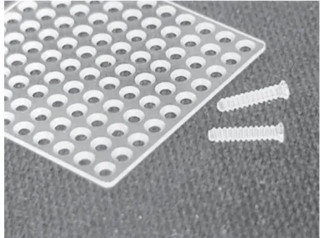
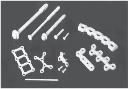
Figure 2. Screws, washers, a pin, and plates made of forged and then machined u-HA/PLLA composites.Source: From Shikinami and Okuno [43].
the polymer–glass interface or agglomeration of the filler particles, leading to premature failure of the composite interface at higher stress levels than those used in DMA. The composite also show bioactivity when soaked in SBF solution.
Shikinami and Okuno [43] successfully produced PLA/HA composites with strengths above that of cortical bone. They synthesized their own HA particles and mixed them into a PLLA/dichloromethane solution followed by precipitation. The composite was processed via extrusion and forging before it was finally machined into fracture fixation devices (Fig. 2). Increasing the HA content increases the modulus up to the maximum filler level of 50 wt% HA with the bending strength reported of 270 MPa exceeding that of cortical bone and modulus up to 12 GPa. The bending strength remained above 200 MPa for the first 25 weeks in phosphate-buffered saline (PBS) solution; thereafter, it was gradually decreased to 120 MPa after 52 weeks (Fig. 3).
Wright-Charlesworthet al. [44,45] produced 0–40 wt% HA in PLLA using compounding extrusion and injection molding. The addition of HA, as expected, increased the stiffness, but at the same time, decreased the strength of the PLLA with less drawing of the polymer matrix between the particles during the failure process. Soaking in PBS solution at 37◦
C showed that increasing the HA content decreased the degradation rate. As with most degradable composites, the strength seems to decrease faster than stiffness for all compositions. The ideal fracture fixation implant stiffness should reduce faster than the strength to allow gradual load transfer from the implant to the healing bone, while retaining strength to prevent refacture.
m
f
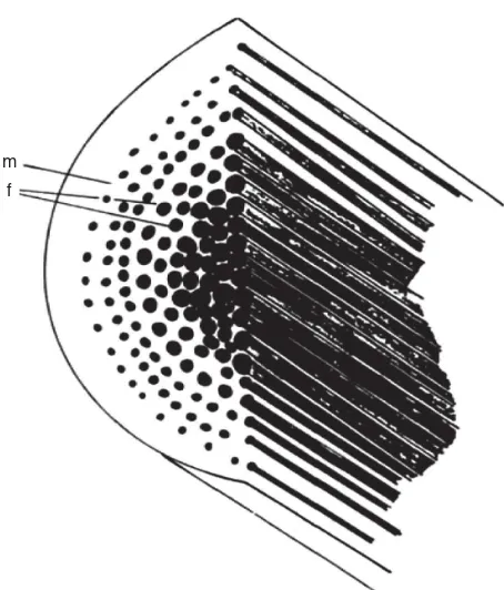
Figure 3. A schematic illustration of self-reinforced composite consisting of matrix (m) reinforced with parallel reinforcing lements (f).Source: From T¨orm ¨al ¨aet al. [49].
to produce a composite with reinforcing elements and binding matrix with the same chemical composition. T¨orm ¨al ¨a and colleagues [49–51] manufactured sintered self-reinforced, absorbable polyglycolide (SR-PGA), and injection molding PGA rods with 1.5–3.2 mm diameter presented bending moduli of 10–12 and 7 GPa, respec-tively. The bending strengths were 260 and 218 MPa with shear strengths of 192 and 95 MPa, respectively. Degradation studies also showed that the strength of the specimens could be retained more than eight weeks. These self-reinforced implants were developed through to clinical application. However, the company developed to exploit the fiber-reinforced composite technology has subsequently concentrated on different PLA, PGA, and trimethylene carbonate (TMC) copolymers.
Triphasic composites of PLA–PLA–TCP were produced with flexural strength of 65–80 MPa and flexural modulus more than 7 GPa [52,53]. The composites were produced by producing a prepreg of PLA96, that is, 96% PLLA, fibers in a PLA70(70% PLLA and 30% PLDLA) matrix that was reinforced with HA or TCP. The strength was maintained up to 12 weeks soaking in buffered saline and, thereafter, lost stiffness and strength slowly. The presence of the calcium phosphates also increased the bioactivity of the materials when assessed by soaking in PBS solution and also slowed the degradation, compared to PLLA/PLDLLA composite prepared in the same manner, but without any calcium phosphate. The pH also remained near neutral for longer as the TCP degradation chemically buffered the lactic acid produced as the PLA degraded and furthermore slowed the degradation.
Bone Tissue Engineering Scaffolds
Until recently, materials were implanted into the body and the body was required to provide cells to the new implant surface. However, tissue engineering has been developed where cells are taken, commonly from the patient’s own body, seeded into a porous scaffold and allowed to grow before being reimplanted back into the body [6]. The space forin vitrocell growth allows faster cell division than in the body and thus an increase in cell number. One of the earliest applications was for cartilage defects where cells are taken from a nonlead bearing area of joint cartilage and expanded up and then replaced in a damaged load bearing area of cartilage. Scaffolds are designed to boost tissue repair rate by providing a large surface area to sup-port revascularization, that is, restoration of the vascular system to allow the supply of nutrients and removal of waste products from the new tissue, and by integration of the regenerating tissue within the system. A scaffold needs to be open-celled porous with mechanical properties such that the implant will not collapse either on implantation or during use, until the newly formed tissue has taken over the mechanical function of the implant. In the case of bone tissue engineering, the scaffolds also need to be bioactive in addition to the usual biocompatible requirement. There are a range of methods to produce porous scaffolds such as thermally induced phase separation (TIPS), solvent casting with particulate leaching, sintering, microsphere templating, and others.
TIPS can be used to produce scaffolds with very high porosities up to 97% and very good pore interconnectivity that are usually used for tissues such as nerve, muscle, tendon, and ligament [54,55]. In this method, the poly-mer is dissolved and glass or ceramic powder then added into the polymer solution. For example, to produce high porosity PLDLLA/Bioglass composite scaffolds, the mix-ture was transferred into a flask and sonicated, followed by quenching in liquid nitrogen for 2 h at −196◦
C. The frozen mixture was later held at−10◦C; the solvent was sublimated at −10◦C and then at 0◦C for 48 h, followed by drying at room temperature in a vacuum oven until the weight is constant. Maquet and colleagues [54–58] using the TIPS method successfully produced structure with tubular macropores of 100µm, interconnected with micropores 10–50µm in diameter. After adding 40 wt% Bioglass, the pore volume decreased from 9.5 to 5.7 cm3/g, with no apparent change observed in the overall pore morphology. Both the PLDLLA/Bioglass composites and the unfilled PLDLLA foams maintained their structural integrity up to 16 weeks.
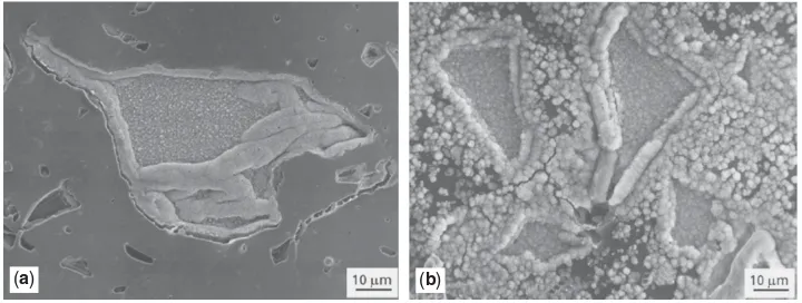
Figure 4. Typical morphologies of porous polymer foams produced by different techniques and structure of cancellous bone: (a) Thermal induced phase separation, (b) sol-vent casting and particle leach-ing, (c) cancellous bone, and (d) microsphere—sintering.Source: Part (a), from Maquetet al. [56]; part (b), from Wenget al. [35]; part (c), from Gibson [64]; part (d), from Luet al. [62].
(a) (b)
(c) (d)
Dunnet al. [59] used salt leaching to manufactured scaf-folds of 10 wt% HA in a blend of PCL and poly(DL-lactic acid-co-glycolic acid) at 10:90 or 40:60 PCL: Degrada-tion testing was performed in PBS and serum-containing media, showing the pH decrease despite the present of HA in the composite. In a study by Li and Chang [60], porous scaffold of poly(β-hydroxybutyrate-co-β-hydroxyvalerate) (PHBV) reinforced with 0–40 wt% Wollastonite has been produced with porosity between 73% and 80% using the porogen method. The composite was found to be bioac-tive when soaked in SBF and the contact angle decreased with increasing amount of Wollastonite, making the mate-rials to be more hydrophilic and thus increasing cell attachment. Wenget al. [35] used sugar leaching to man-ufacture porous scaffolds of PLLA reinforced with 20 wt% plasma-sprayed calcium phosphates. The spherical parti-cles of CaP had smooth amorphous surfaces with crys-talline cores. The surface layer dissolved easily when soaked in SBF or after ultrasonication in distilled water. The composite scaffold was expected to be highly bioactive as the amorphous calcium phosphate dissolved and then reprecipitated on the internal and external surfaces of the scaffold, giving a large number of potential nucleation sites on the scaffold surface.
Polymeric scaffolds also can be produced using a micro-sphere sintering process. Micromicro-spheres of a ceramic and polymer composite are synthesized using emulsion/solvent evaporation technique, followed by sintering the com-posite microspheres to produce 3D porous scaffolds. Lu et al. [61–63] successfully produced 3D composites of degradable polymers and BG using this method. First, PLA–PGA copolymer (polylactide-co-glycolide, PLAGA) Bioglass composite microspheres were produced through
a water–oil–water emulsion technique and then com-pressed and heated to sinter the spheres together. This resulted in an interconnected porous structure with an average porosity of 40% with 90µm pore diameter. The mechanical properties reported were close to those of can-cellous bone. Furthermore, cell culture studies show a good response to the materials. This method was further developed by Yao and colleagues [63] to produce porous 3D scaffolds of PLGA/BG. They showed the materials have a very good bioactivity response and promoting osteogenesis of marrow stromal cells. Figure 4a,b and d shows vari-ous typical microsphere-sintered 3D structure produced by different methods in comparison to cancellous bone (Fig. 4c).
MECHANICAL PROPERTY TESTING
Friedrich and Karsch [65] where debonding of the poly-mer matrix from the filler surface is followed by drawing of polymer fibrils between the filler particles. While their studies were applied to tensile testing, the same failure mode is also seen after impact, fatigue, and creep loading. Dynamic mechanical analysis (DMA) including temper-ature effects (DMTA) has been popular as small specimens can be used and, with the costs and difficulties of produc-ing biomedical composites, such studies have benefits. A typical flexion DMA test sample is 1×2×23 mm3, while compression samples are small cubes or cylinders a few millimeters in height. Nazhatet al. [40,67–69] in a series of studies used DMA to investigate the mechanical prop-erties of different composites. In their first study [66], they compared the storage modulus (E′) measured using DMA
with Young’s modulus (E) of HA/PE composites and found a linear corrolation given by
E′
=0.98E+0.95 (R2=0.996). (1)
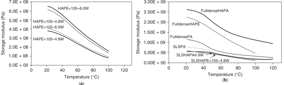
In subsequent studies, Nazhat with various coauthors investigated various composites developed by Bleachet al. [53], Blakeret al. [69], and Misraet al. [42]. Zhanget al. [70] used DMA to optimize the processing parameters for selective laser-sintered HA in polyamide and showed that for a given scan speed optimizing the laser power could increase the modulus by a factor of 1.5 (Fig. 5a). In all these DMTA studies, the transition temperatures seen for the nonreinforced polymer are also seen in the composite (Fig. 5b).
Younesi and Bahrololoom [71] studied the effect of molecular weight, particle size, and Ringer’s solu-tion on the impact properties of surface treated of polypropylene-filled HA composite. Their samples were notched Izod specimens of 64×10.2×6.4 mm3. About 6.4 mm was used instead of the standard specimen thickness of 3.2 mm to give higher bending resistance. Samples were tested both dry and after soaking in Ringer’s solution for 30 days. Impact tests were carried out using drop weight impact testing that was performed by Zhang and Tanner [72,73] using 4-mm-thick 60 mm
square samples, and they showed that increasing the molecular weight from 50,000 to 250,000 increased the total energy absorbed by a factor of 18 while adding 40 vol% spray-dried HA reduced the total energy absorbed by a factor of 12. They further found that replacing the spray-dried HA with a sintered HA with the same particle size but one-tenth the specific surface area increased the total energy absorbed by a factor between 1.5 and 2.0 depending on the volume fraction of filler.
The classic biomaterial to undergo fatigue testing is bone cement, given that the major failure of bone cement is due to the one million or so load cycles applied per year [74]. Most bone cements are polymethylmethacrylate with various amounts of fillers in the form of radiopacifiers and antibiotics, and these are considered too low filler concentrations to be described as a composite material.
Ton Thatet al. [75,76] studied the fatigue behavior of HA-reinforced PE in fully reversed axial tension compres-sion and fully reversed torcompres-sion using cylindrical dumbbell, which were machined according to the ASTM E466 stan-dard. Fatigue tests were then carried out using sinusoidal loading at a frequency of 2 Hz at various loading levels, in saline solution at 37◦
C.S–Ncurves were established, showing that in tension–compression fatigue, the cycles to failure range from 1000 cycles at ±13 MPa to more than one million cycles at±4.4 MPa. In torsion mode, the fatigue cycles range from 100 cycles at 75% of the ultimate shear stress to more than one million cycles for the 25% of the ultimate shear stress loading level.
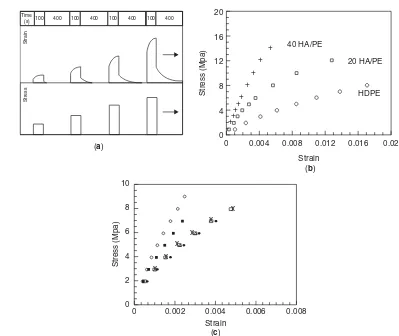
In general, polymers have poor creep resistance which results in poor service life. Some of the early studies by Suwanprateebet al. [77–79] are on HA-reinforced HDPE. While conventional creep testing is extremely time con-suming, isochronous testing allows a large amount of initial data to be collected [81]. Here, a small stress is applied for a short time, say 100 s, the strain at the end of this time is measured and then the stress is removed for four times the original loading time (400 s); a higher stress is applied for 100 s and again the strain is measured at the end of the loading time. This load unload process is applied for gradually increasing stress levels (Fig. 6a)
0.0E + 00
0 20 40 60 80 Temperature (°C)
100 120 1.0E + 08
2.0E + 08 3.0E + 08 4.0E + 08 5.0E + 08 6.0E + 08 7.0E + 08
Storage modulus (Pa)
0.00E + 00 1.00E + 09 1.50E + 09 2.00E + 09 2.50E + 09 3.00E + 09
5.00E + 08
0 20 40 60 80 Temperature (°C)
100 120
Storage modulus (Pa)
HAPE<105–6.0W
HAPE<105–4.8W
HAPE<105–4.8W HAPE<105–6.0W
FulldenseHAPA
FulldenseHAPE
FulldensePA
SLSPA
SLSHAPA4.9W
SLSHAPE<105–4.8W
(a) (b)
Time
(s) 100 400 100 400 100 400 100 400
Strain
Stress
0
0 0.004 0.008 0.012 0.016 0.02 4
8 12 16 20
Stress (Mpa)
Strain
Stress (Mpa)
Strain (c)
(b) (a)
40 HA/PE
20 HA/PE
HDPE
++ ++
+ +
+ +
+
0
0 0.002 0.004 0.006 0.008 2
4 6 8 10
x x
x x
x x
Figure 6. Isochronous creep testing of HA PE composites showing (a) strain and stress variations with time in isochronous testing, (b) the effect of adding 20 and 40 vol% HA to HDPE, and (c) the effect of soaking 40 vol% HAPE for◦nonimmersed;•7 days; * 30 days;×90 days; and150 days. Source: From Suwanprateebet al. [77,78].
and finally an isochronous stress–strain curve is plotted. While allowing a large amount of data to be collected in a relatively short time, isochronous testing also allows the choice of the most appropriate stress levels for long-term creep testing. Isochronous creep testing can also allow various factors to be compared. They found that adding 20 and then 40 vol% HA halved the creep strain for a given stress level (Fig. 6b). They showed that 40 vol% HA in PE can sustain static tensile loads of up to 6 MPa in saline solution. The importance of soaking samples before testing were considered by Suwanprateebet al. [78] who showed that despite the nondegradability of PE, soaking in saline at 37◦C caused the absorption of 0.1 wt% of liquid, which had minimal effects on the creep behavior, whereas with 40 vol% HA 1 wt% liquid was absorbed that nearly doubled the isochronous creep strain for a given stress (Fig. 6c).
Younesi and Bahrololoom [80] studied the creep behav-ior of the surface treated of polypropylene-filled HA com-posited in Ringer’s solution. The samples were tested at dry and wet conditions, which was soaking time for 30 days in Ringer’s solution. The test was carried out at 35% of their corresponding ultimate tensile strength (UTS) in physiological environment. They found that the creep
resistance increased by decreasing the HA particle size, as this increased the contact surface between particles and the matrix. The decrease in friction reduces the motion and slippage between the polymers chains and thus reduces the deformation between the matrices, resulting in stronger composite. These findings are supported by Starkovaet al. [81], who reported that adding nanoparticles filler reduces the strain in the primary as well as secondary creep stage by restricting the movement of polymer chains, thus producing higher creep resistance properties.
saline solution, bending strength of pure CPLA was con-stant, but declines rapidly thereafter. Niemel ¨a [83] used shear testing to characterize self-reinforced poly-L-lactide (SRPLA) and TCP/SRPLA composites afterin vitro hydrol-ysis. Results showed that all SRPLA rods retained their initial shear strength virtually unchanged up to 30–36 weeks, but thereafter the composite strength decreased rapidly. After 52 weeks of degradation testing, the com-posite appeared to have 15% of initial shear strength in comparison to TCP/SRPLA that managed to retain about 60–70% of the initial value. Kobayashi and Yamadi [84] used conventional tensile testing to study the strain rate dependency of mechanical properties of β-TCP/(PLLA). They used rectangular specimens produced using injec-tion molding technique and then immersed in SBF for 8, 16, and 24 weeks before testing. They found that the modulus and strength increased with strain rate, but that soaking reduced these increases.
Suwanprateebet al. [85] used flexural testing to study the influence of three-dimensional printing fabrication technique for bioactive HA/bis-GMA (bisphenol A-glycidyl methacrylate) based composite.
In the field of tissue engineering, among other param-eters that need to be considered in scaffold fabrications is that they should have appropriate and controllable mechanical properties, for example, elastic constants and compressive strength to provide sufficient temporary sup-port for cells to enable tissue regeneration. Lu et al. [86] developed a three-dimensional porous composite of PLAGA and 45 S5 BG (PLAGA–BG composite) scaffold for bone tissue. Compression testing showed that adding the BG granules to the composite yielded higher compressive modulus than PLAGA alone. Blakeret al. [87] performed similar compression testing to study the mechanical prop-erties of highly porous PDLLA/Bioglass produced by TIPS technique. Compression tests were performed axially and transversely from 2 to 300 kPa. They also used DMA to establish the storage modulus (E′
), loss modulus (E′′
), and mechanical loss tangent (tan δ). Results indicated that mechanical anisotropy was controlled by the direction of the macropores.
Microhardness studies, using a Vickers diamond indenter, were carried out by Sousa and colleagues [32] to investigate the structure development and interfacial interaction of preferred orientation in HDPE/HA compos-ite. In the latter study, they investigated the influence
of the processing on the mechanical performance of HA-reinforced biodegradable starch-based blends and HDPE [33].
IN VITRO BIOACTIVITY AND BIOCOMPATIBILTY TESTING
The biocompatibility of materials is an essential prop-erty for their use inside or in contact with the human body. Materials must to be nontoxic but should act as a substrate to promote cell spreading and growth in cer-tain applications, while in other applications, the surface should prevent cell attachment. The second requirement is bioactivity.In vitrobioactivity testing was initiated by Kokuboet al. [7] who showed that materials that are bone bioactivein vivowill develop a hydroxy-carbonate apatite (HCA) layer when placed in a SBF, which has similar ion levels as those of blood plasma. This work was extended by Hench [88,89] who defined a bioactivity index based on the inverse of the time to get the entire surface of the biomaterial covered with an HCA layer (Fig. 7). Hench compared various formulations of Bioglass particles to find the highest bioactivity formulation. In composites work, Huanget al. [90] used this technique to compare 20 and 40 vol% Bioglass in PE and showed that after seven days the HCA layer had covered almost all the surface of the composite, while with only 20 vol% Bioglass very little of HCA layer has progressed beyond from the particles onto the PE (Fig. 5). Maquetet al. [56] manufactured foams of PDLLA reinforced with Bioglass particles as bone tissue scaffolds. Thein vitrobioactivity results showed the rate of apatite deposition both on the surface and throughout the foams increased with increasing Bioglass content. At the same time, they also found some apatite deposition on the nonfilled PDLLA foams.
In vitroevaluation of the biocompatibility usually pro-vides initial screening often using cell lines to minimize the variability in terms of metabolism, distribution, absorp-tion and to maximize the cell response to possible toxicity reaction that may have. The results of in vitro testing of bioactive materials can indicate whether the material increases the response of the cells to the material, that is, it is bioactive as well as whether the material is biocom-patible. Two types of cells are used: immortalized cell lines are obtained from either malignant tumors or where a cell line has been transformed and primary cells are obtained
(a) (b)
from people or animals and then grown up. The immor-talized cells can be grown up and divided and grown up again an infinite number of times (each step being called a passage) so that they are convenient to use, but they are pathological cells. In contrast, primary bone cells are commonly harvested from discarded femoral heads after hip replacement, and so are considered normal cells but suffer from variability depending on age, gender, etc. of the individual from which they are taken. Furthermore, they can only be used for a limited number of passages as they differentiate from their cell type after a number of passages. A wide range of materials have been inves-tigated both for bioactivity and biocompatibility; some of these studies are described below.
Brauer and coworkers [91,92] produced a porous mate-rial using a methacrylate-modified oligolactide polymer network and phosphate-based glasses in the system P2O5–CaO–MgO–Na2O–TiO2. These glasses were used to reinforce 10 wt% methacrylic acid 2-hydroxyethylester (pHEMA) copolymer at ∼1:3.0 polymer-to-glass weight ratio. The porous structure was produced through the salt leaching method. In vitro cell culture testing was carried out using the MC3T3-E1 osteoblast cell line. They reported that the materials was nontoxic and biocompatible.
The foams produced by Maquet et al. [56] were later tested by Blaker et al. [93]in vitro cell culture using the osteosarcoma (bone cancer) line MG-63 for up to eight days. In cell culture studies, the osteosarcoma cells were deposited on the outer layer of the foam. They reported that the highest cell count was from 5 wt% Bioglass foams, but overall good attachment to the materials was found.
In vitro testing using three different cell lines, macrophages, osteoblasts, and osteoclasts, were used to study the biocompatibility of the PHBV composite filled with 40–50 wt% HA or TCP [94]. The three cell lines used cover those cells that are involved in the inflammatory response to materials, bone deposition, and bone resorp-tion, respectively. The results showed that the addition of bioactive calcium phosphates enhances the cell response. In the macrophage study, they found low inflammatory responses with the reinforced materials with a slightly higher response with the nonfilled PHBV. The osteoblast study, that is, using cells involved in bone deposition, showed that plain HA or PHBV filled with HA produced more calcium and more mineralization than unfilled PHBV or when filled with TCP. In the osteoclast-like cells study, the cells attached to the composite were seen to be active but unable to resorpt the materials. In contrast, resorption pits were observed on samples of bone.
IN VIVO BIOCOMPATIBILITY TESTING
Full in vivo biocompatibility testing involves tests such as irritation, intracutaneous reactivity, systemic toxicity (acute toxicity), subchronic toxicity, genotoxicity (affects later generations), hemocompatibility (i.e., blood compati-bility), chronic toxicity, carcinogeniticity (cancer causing), biodegradation, and immune responses [2]. If an entirely new material is developed, it will need to undergo the full set of tests; however, for composite biomaterials, usu-ally the individual component materials have been tested.
Therefore, for a composite that has undergone in vitro testing, thein vivotest is usually to assess the response to the material when in a body and under mechanical loading. Thein vivostudy allows long-term investigation of the biofunctionality in the complex biological environ-ment before proceeding with clinical testing. As it involves animal implantation testing, it has to comply all aspects of legal rules and ethics on animal experimentation of the country concerned.
Shikinmari and Okuno [43] successfully developed high modulus HA/PLLA rods, which was later used by Furukawa and colleagues [95,96] for in vivo testing in rabbits both subcutaneously and in the femoral medullary cavity. The HA was either calcined, to increase the cystallinity, or noncalcined. The subcutaneous implan-tation was used to observe the material’s degradation behavior. After 52 weeks, the reduction of the molecular weight was reported to be less than 10%; however, the bending strength remained high enough to provide sufficient mechanical support to the system. In their second study, initial intermedullary implantation was studied between 2 and 25 weeks. They reported a fibrous layer surrounding the implants of nonreinforced PLLA, while the reinforced composite exhibited bone contact by two weeks and the amount of contact gradually increased. More bone contact was observed in 40 wt% than that in 30 wt% HA composites and those composites filled with the uncalcined HA. In the third paper [97], they reported that after four yearsin vivo testing the diameter of the rods has reduced to 77% of their original cross section. While after six or seven years, in some sections there was no material remaining; it was also observed that bone had replaced the lost composite. In vivo studies very rarely run beyond even one year because of the short life span of rabbits; thus, over seven years in these studies are probably unique.
N ¨arhi et al. [98] evaluated biological reaction to the bioactive composite and the biologic behavior of a poly(ε-caprolactone-co-DL-lactide) composite filled with 40 or 60 wt% BG implanted in the long bones of rabbits for 8 and 16 weeks. In eight weeks, the implants were covered with fibrous capsule and no direct contact with bone was observed. After 16 weeks, the capsule had thinned in all samples with bone ingrowth observed, indicating that the materials is compatible with the bone tissue. Similar findings were later reported by Ranne and coworkers [99] when implanted in rat subcutaneous tissue.
(a)
(d)
(h) (i) (j)
(e) (f)
[image:11.720.163.551.102.281.2](b) (c)
Figure 8. Comparison of healing after 12 weeks of radial bone defect: (a–c) with no implant in defect, (d–f) with gelatin/nHAP/fibrin scaffold without rhBMP-2 in the defect, and (h–j) gelatin/nHAP/fibrin scaffold with rhBMP-2 repaired the defect.Source: From Liuet al. [101].
Liu et al. [101] developed a hybrid scaffold of gelatin/nanohydroxypatite (nHAP) by incorporating human BMP-2 (rhBMP-2) to enhance bone regeneration. Microscopy showed the scaffold to have 3D porous structure and DNA assay test revealed that the structure was noncytotoxic and could promote cell proliferation.In vivotesting carried out in a rabbit model indicated good healing of the skull defect after 12 weeks of implantation (Fig. 8).
CLINICAL USES
As discussed earlier, the first clinical use of composites was in the form of carbon-fiber-reinforced epoxy resin as facture fixation plates [15–17]. While Downeset al. [21,22] used HA/HDPE composite for orbital floor implants and this material then progressed to extensive use in the shaft of middle ear implants as HAPEX [23,24]. Since 1990, the numbers of publications on fracture fixation in the human hand using bioabsorbable devices have increased. SR-PLLA pins (diameter 1.5 or 2.0 mm) [104] were used in clinical studies for the fixation of small fragment frac-tures and osteotomies. It was reported that in 27 patients, uneventful recovery of the function was accomplished in four fractures of the metacarpal and three of the proximal phalanx [103]. Arataet al. [102] reported that in 26 cases of digital replantation using intramedullary SR-PLLA rods, no cases of nonunion or infection occurred. How-ever, in one patient transient bone resorption occurred. They summarized that the intramedullary bioabsorbable rods gave stable, simple, and effective osteosynthesis in digital replantation. Bioabsorbable pins and screws can be used to stabilize fusions. Rokkanenet al. [106] wrote that in rheumatoid arthritis patients, bioabsorbable poly-mers (such as PLLA) with slow degradation are preferred. SR-PLLA pins with cross section of 1.5–2.0 mm were observed to provide stable fixation for 18 interphalangeal, metacarpophalangeal, and carpometacarpal arthrodeses when tested in patients with rheumatoid arthritis [104]. No complications were observed and the authors concluded
Figure 9. Macropore bioabsorbable sheet and screw of 70:30 PLLDLA polymer.Source: From Parket al. [104].
that the results were comparable with earlier studies on use of metal implants. Furthermore, in the same study, 18 wrist joints were fused uneventfully with 3.2 mm SR-PLLA pins and only one patient reported a superficial infection postoperatively.
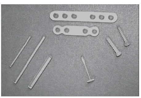
[image:11.720.372.600.334.503.2]Figure 10. Self-reinforced poly-L-lactide (SR-PLLA) and self-reinforced poly-L/DL-lactide (SR-P(L/DL)LA) miniplates, pins (diameter 1.5, 2.0, and 3.2 mm), tacks, and screws (diameter 2.0 and 1.5 mm) for bone fixation in the hand.Source: From Waris et al. [107].
no soft tissue inflammatory or clinical complications were recorded.
Composites have been developed that have been used in patients since the 1970s; the early composites, such as carbon-reinforced epoxy [15–17], were used in a few patients and then were either too complex to use or had insufficient advantages over similar devices manufactured of more conventional metal implants. One composite mate-rial, HAPEX, did enter extensive clinical use but owing to low mechanical properties could only be used in the orbital floor or middle ear implants. However, it did show that the use of composites allowed bioactivity to be combined with the ductility of polymeric phases.
The conventional composite production techniques such as the development of prepregs and then compression molding have been applied to biomaterials and have allowed the production of composites with higher mechan-ical properties [49–53]. Self-reinforced PLA composites have entered extensive clinical use (Fig. 10).
The latest developments in composites are as bioactive scaffolds for bone tissue engineering. There are a series of new composites under development and in various stages of testing fromin vitrothrough to clinical trials. It is hoped that in the next few years some of these will come into general use.
Composites based on the application of conventional materials production techniques to biologically compat-ible materials have been used since the 1970s. Their application is gradually increasing because of the bene-fits of modulus compatibility with the materials they are replacing and the bioactivity of the some of the individ-ual materials used in the composite manufacture. Both degradable and nondegradable materials can be manufac-tured depending on the starter materials. Optimization of the filler content and processing regime should be used to optimize both the mechanical and biological proper-ties. The applications can run from low modulus porous degradable scaffolds containing bioactive molecules used for tissue engineering to high modulus (for a composite) nondegradable implants allowing to permanently replace
a lightly load bearing bone. Currently, no composite mate-rial has been used successfully to replace a major load bearing bone, but that is only a matter of time.
REFERENCES
1. Williams DF. Williams dictionary of biomaterials. Liverpool: Liverpool University Press; 1999.
2. Anderson J. An introduction to materials in medicine. San Diego (CA): Elsevier Press; 2004. pp. 296–304.
3. Martini FH. Fundamentals of anatomy and physiology. Singapore: Prentice Hall; 2007.
4. Bonfield W, Grynpas MD. Nature 1977;270:453–454. 5. Currey JD. Proc Inst Mech Eng [H] J Eng Med 1998;
212-H:399–412.
6. El Haj AJ, Cartmell SH. Proc Inst Mech Eng [H] J Eng Med 2010;224-H:1523–1532.
7. Kokubo T, Kushitani H, Sakka S,et al. J Biomed Mater Res 1990;24:721–734.
8. Alcock B. In: Ambrosio L, Tanner KE, editor. Biomaterials for spinal applications. Cambridge: CRC Woodhead; 2012. Chapter 2. In press.
9. Gloria A, Causa F, De Santis R,et al. J Mater Sci Mater Med 2007;18:2159–2165.
10. Gloria A, De Santis R, Ambrosio L,et al. J Biomater Appl 2011;25:795–810.
11. Savalani MM, Hao L, Zhang Y,et al. Proc Inst Mech Eng [H] J Eng Med 2007;221-H:873–886.
12. Tan KH, Chua CK, Leong KF, et al. Biomaterials 2003; 24:3115–3123.
13. Abu Bakar MS, Cheang P, Khor KA. Mat Sci Eng A 2003;34 S:55–63.
14. Saringer W, Nobauer-Huhmann I, Knosp E. Acta Neurochir 2002;144:1193–1203.
15. Hastings GW. Composites 1978;9:193–197.
16. Bradley JS, Hasting GW, Johnson-Nurse C. Biomaterials 1980;1:38–40.
17. Ali MS, French TA, Hasting GW,et al. J Bone Joint Surg Br 1990;72-B:586–591.
18. Al-Shawi AK, Smith SP, Anderson GH. J Arthroplasty 2002;17:320–324.
19. Bonfield W, Grynpas M, Tully AE, et al. Biomaterials 1981;2:185–186.
20. Wang M, Porter D, Bonfield W. Br Ceram Trans 1994;93:91–95.
21. Downes RN, Vardy S, Tanner KE,et al. In: Bonfield W, Hast-ings GW, Tanner KE, editors. Bioceramics 4 - proceedHast-ings of the fourth international symposium on ceramics in medicine. Oxford: Butterworth-Heinnemann Ltd; 1991. pp. 239–246. 22. Tanner KE, Downes RN, Bonfield W. Br Ceram Trans
1994;93:104–107.
23. Goldenberg RA, Driver M. Otolaryngol Head Neck Surg 2000;122:635–642.
24. Meijer AGW, Segenhout HM, Albers FWJ, et al. ORL 2002;64:173–179.
25. Joseph R, McGregor WJ, Martyn MT, et al. Biomaterials 2002;23:4295–4302.
26. Joseph R, McGregor WJ, Martyn MT,et al. Polym Eng Sci 2002;42:326–335.
28. Ward IM, Bonfield W, Ladizesky NH. Polym Int 1997;43:333–337.
29. Bonner M, Saunders LS, Ward IM, et al. J Mater Sci 2002;37:325–334.
30. Ladizesky NH, Pirhonen EM, Appleyard DB,et al. Compos Sci Technol 1998;58:419–434.
31. Reis RL, Cunha AM, Oliveira MJ,et al. Mater Res Innov 2001;4:263–272.
32. Sousa RA, Reis RL, Cunha AM, et al. J Appl Polym Sci 2002;86:2866–2872.
33. Sousa RA, Reis RL, Cunha AM,et al. J Compos Sci Technol 2003;86:389–402.
34. Petrovic L, Pohle D, Munstedt H, et al. J Biomed Sci 2006;13:41–46.
35. Weng J, Wang M, Chen J. Biomaterials 2002;23:2623–2629. 36. Hao L, Savalani MM, Zhang Y,et al. Proc I Mech Eng [H] J
Eng Med 2006;220-H:521–531.
37. Hao L, Savalani MM, Zhang Y,et al. Proc Roy Soc London A 2007;463:1857–1869.
38. Zhang Y, Hao L, Savalani MM,et al. J Biomed Mater Res A 2008;86A:606–616.
39. Zhang Y, Hao L, Savalani MM,et al. J Biomed Mater Res A 2009;91A:1018–1027.
40. Chu CC. In: Park JB, Bronzino JD, editors. Biomaterials: principles and applications. Boca Raton (FL): Pub, CRC Press; 2003. pp. 95–115.
41. Doyle C, Tanner E, Bonfield W. Biomaterials 1991;12: 841–847.
42. Misra SK, Nazhat SN, Valappil SP,et al. Biomacromolecules 2007;8:2112–2119.
43. Shikinami Y, Okuno M. Biomaterials 1999;20:859–877. 44. Wright-Charlesworth DD, Miller DM, Miskioglu I,et al. J
Biomed Mater Res A 2005;74A:388–396.
45. Wright-Charlesworth DD, King JA, Miller DM, et al. J Biomed Mater Res A 2006;78A:541–549.
46. Fambri L, Pegoretti A, Fenner R, et al. Polymer 1997;38: 79–85.
47. Eling B, Gogolewski S, Pennings AJ. Polymer 1982;23:1587–1593.
48. Leenslag JW, Gogolewski S, Pennings AJ. J Appl Polym Sci 1984;29:2829–2842.
49. T¨orm ¨al ¨a P, Vasenius J, Vainionp ¨a ¨a S,et al. J Biomed Mater Res 1991;25:1–22.
50. Majola A, Vainionp ¨a ¨a S, Rokkanen P, et al. J Mater Sci Mater Med 1992;3:43–47.
51. Pihlajam ¨aki H, B¨ostman O, Hirvensalo E,et al. J Bone Joint Surg 1992;74-B:853–857.
52. Bleach NC, Tanner KE, Kellom ¨aki M, et al. J Mater Sci Mater Med 2001;12:911–915.
53. Bleach NC, Nazhat SN, Tanner KE, et al. Biomaterials 2002;23:1579–1585.
54. Boccaccini AR, Maquet V. Compos Sci Technol 2003;63:2417–2429.
55. Maquet V, Martin D, Scholtes F, et al. Biomaterials 2001;22:1137–1146.
56. Maquet V, Boccaccini AR, Pravata L,et al. J Biomed Mater Res A 2003;66A:335–346.
57. Maquet V, Boccaccini AR, Pravata L, et al. Biomaterials 2004;25:4185–4194.
58. Boccaccini AR, Blaker JJ. Expert Opin Med Dev 2005;2:303–317.
59. Dunn AS, Campbell PG, Marra KG. J Mater Sci Mater Med 2001;12:673–677.
60. Li HY, Chang J. Polym Degrad Stab 2005;87:301–307.
61. Lu HH, Tang A, Oh SC, et al. Biomaterials 2005;26: 6323–6334.
62. Lu HH, El-Amin SF, Scott KD,et al. J Biomed Mater Res A 2003;64A:465–474.
63. Yao J, Radin S, Leboy PS, et al. Biomaterials 2005;26:1935–1943.
64. Gibson LJ. J Biomech 1985;18:317–328.
65. Friedrich K, Karch UA. J Mater Sci 1981;16:2167–2175. 66. Nazhat SN, Joseph R, Wang M, et al. J Mater Sci Mater
Med 2000;11:621–628.
67. Blaker JJ, Nazhat SN, Boccaccini AR. Biomaterials 2004;25:1319–1329.
68. Nazhat SN, Kellom ¨aki M, T¨orm ¨al ¨a P,et al. J Biomed Mater Res Appl Biomater 2001;58:335–343.
69. Blaker JJ, Maquet V, J´erˆome R, et al. Acta Biomater 2005;1:643–652.
70. Zhang Y, Tanner KE. J Mater Sci Mater Med 2008;19: 761–766.
71. Younesi M, Bahrololoom ME. J Compos Mater 2010;44: 2785–2799.
72. Zhang Y, Tanner KE. J Mater Sci Mater Med 2003;14: 63–68.
73. Zhang Y, Hao L, Savalani MM,et al. J Mater Sci Mater Med 2009;91A(4):1018–1027.
74. Wallbridge N, Dowson D. Eng Med 1982;11:95–96. 75. Ton That PT, Tanner KE, Bonfield W. J Biomed Mat Res
2000;51A:453–460.
76. Ton That PT, Tanner KE, Bonfield W. J Biomed Mater Res 2000;51A:461–468.
77. Suwanprateeb J, Tanner KE, Turner S,et al. J Mater Sci Mater Med 1995;6:804–807.
78. Suwanprateeb J, Tanner KE, Turner S,et al. J Mater Sci Mater Med 1997;8:469–472.
79. Suwanprateeb J, Tanner KE, Turner S, et al. J Biomed Mater Res 1998;39:16–22.
80. Younesi M, Bahrololoom ME. J Compos Mater 2011;45:513–523.
81. Starkova O, Jing-Lei Y, Zhong Z. Compos Sci Technol 2007;67:2691–2698.
82. Kikuchi M, Koyama Y, Takakuda K,et al. J Biomed Mater Res 2002;62:265–272.
83. Niemel ¨a T. Polym Degrad Stab 2005;89:492–500.
84. Kobayashi S, Yamadi S. Compos Sci Technol 2010;70:1820–1825.
85. Suwanprateeb J, Sanngam R, Suwanpreuk W. J Mater Sci Mater Med 2008;19:2637–2645.
86. Lu HH, El-Amin SF, Scott KD,et al. J Biomed Mater Res 2003;64A:465–474.
87. Blaker JJ, Bismarck A, Boccaccini AR,et al. Acta Biomate-rialia 2010;6:756–762.
88. Hench LL. J Am Ceram Soc 1991;74:1487–1510.
89. Thompson ID, Hench LL. Proc Inst Mech Eng [H] J Eng Med 1998;212(H):127–136.
90. Huang J, Di Silvio L, Wang M,et al. J Mater Sci Mater Med 1997;8:809–813.
92. Brauer DS, R ¨ussel C, Vogt S,et al. J Mater Sci Mater Med 2008;19:121–127.
93. Blaker JJ, Gough JE, Maquet V,et al. J Biomed Mater Res A 2003;67A:1401–1411.
94. Cool SM, Kenny B, Wu A, et al. J Biomed Mater Res A 2007;82A:599–610.
95. Furukawa T, Matsusue Y, Yasunaga T, et al. J Biomed Mater Res 2000;50:410–419.
96. Furukawa T, Matsusue Y, Yasunaga T,et al. Biomaterials 2000;21:889–898.
97. Hasegawa S, Ishii S, Tamura J, et al. Biomaterials 2006;27:1327–1332.
98. Narhi TO, Jansen JA, Jaakkola T, et al. Biomaterials 2003;24:1697–1704.
99. Ranne T, Tirri T, Yli-Urpo A,et al. J Bioact Compat Polym 2007;22:249–264.
100. van der Pol U, Mathieu L, Zeiter S,et al. Acta Biomaterialia 2010;6:3755–3762.
101. Liu Y, Lu Y, Tian X,et al. Biomaterials 2009;30:6276–6285. 102. Arata J, Ishikawa K, Sawabe K, et al. Ann Plast Surg
2003;50:350–353.
103. Rokkanen PU, B¨ostman O, Hirvensalo E,et al. Biomaterials 2000;21:2607–2613.
104. Juutilainen T, Patiala H. Scand J Rheumatol 1995; 24:228–233.
105. Park MS, Aryan HE, Ozgur BM, et al. Neurosurgery 2004;54:631–635.
106. Vaccaro AR, Sahni D, Pahl MA, et al. Spine 2006; 31:2091–2094.
107. Waris E, Ashammakhi N, Kaarela O,et al. J Hand Surg 2004;29B:(6):590–598.
FURTHER READING
Johnson AJW, Herschler BA. Acta Biomate 2011;7:16–30. DOI: 10.1016/j.actbio.2010.07.012.
Tanner KE. Proc Inst Mech Eng [H] 2010;224-H:1359–1372. DOI: 10.1243/09544119JEim823.
Tanner KE. In: Wuisman PIJM, Smit TH, editors. Degradable polymers for skeletal implants. Cambridge: Pub Nova; 2009. pp. 73–93.
![Figure 4. TypicalGibson [64]; part (d), from Lufrom Weng(a), from Maquetmorphologiesofporous polymer foams produced bydifferent techniques and structureofcancellousbone:(a)Thermalinduced phase separation, (b) sol-ventcastingandparticleleach-ing,(c)cancello](https://thumb-ap.123doks.com/thumbv2/123dok/568400.67209/6.720.251.609.101.376/typicalgibson-maquetmorphologiesofporous-bydifferent-techniques-structureofcancellousbone-thermalinduced-separation-ventcastingandparticleleach.webp)