Assessment
of
reactivities of
$phoid
fever
sera
s3-2
against outer membrane
protein
preparations
from
strains of Salmonella typhi
in
Jakarta
Lucky H. Moehario,
E.S. Firdaus,
P. SudarmonoAbstrak
Suatu upaya penilaian reaktivitas serum pasien demam tifuid di Jakarta terhadap protein membran luar (Outer Membrane Proteùt, OMP) Salmonella typhi telah dilakukan. OMP dari
l0
isolat klùùk S. typhi telah diisolasi dengan soniknsi dan menggunakan buffer yang berisi triton X-100 2 7o. Elektroforesis dengan gel Polyacrylamide Sodium Dodecyl Sulfate memperlihatkan protein major Omp C, Omp F dan Omp A dengan berat molekul 38, 37 clan 34 kDa dalam hampir semua sampel. Akan tetapi 3 dari sampeL Omp tidak memperli-hatkan protein 34 kDa. Meskipun tidak menonjol, polipeptida 20,5 kDa ditemukan pada semua sampel Omp, polipeptirta 25 kDa dite-mukan pada 4 sampel dan po[ipeptida 29 kDa ditemukan pada 6 sampel. Selanjutnya polipepticta minor dengan berat molekttl yang berkisar antara 20,5 kDa - 139,9 kDa iuga ditemukan pada semua sampel. Immunoblotting Omp dengan serum kelinci yang diimunisasi memperlihatkan reaktivitas yang kuat terhadap porin, polipeptida 37, 38 dan 29 kDa. Serum pasien demam tifuid tampafuya'berisi IgMyong tidttk memperlihatkan reaktivitas silang dengan protein majory tetapi memperlihatkan reaktivitas terhadap polipepticla minor
Abstract
An attempt was caruied out to assess reactivity of sera from patients with typhoid fever against outer membrane protein (OMp) preparations tlerived from local strain of Salmonella typhi. OMPs from
l0
clinical isoLates of S. typhi were isolatetl by sonication and through the use of buffer containing Triton X-100 2Vo. SDS-PAGE of these OMPs showed sixdffirent
electrophoretic profiles in whiclt major proteins with molecular weight (MW) 38, 37 and 34 kDa corresportding to OmpC, OmpF ancl OmpA respectit,eLy were present itt most of the OMPs. Three isolates of S. typhi, however, ditL not show the 34 kDa protein. Although txot as prominent, polypeptitle with MW 20.5 kDa was observed inall
OMPs while polypeptide with MW 25 and 29 kDa in 4 ancl 6 OMP preparations respectively. Int-munoblotting of the OMPs with immunisecl rabbils\era showed strong reactivity against the porins, the 37, 38 kDa, antl the 29 kDA polypeptide. Sera from patients with typhoitl fever contained IgM antibodies which did not react with tlrc porirzs and the OmpA, howetter, they showed reactivity to the minor poLypeptides.INTRODUCTION
Typhoid
fever
is
one
of
infectious
diseases
which
gave rise
to
animportant
health
problem
especially in
developing countries.
In
Indonesia
the
diseaseis
en-demic and remains
a major
public
health problem
with number
of
casesper year 360-900 per
100,000inhabitants
in
which
urban
areas
have higher
inci-dence
than rural
areasl.
Furlher,
casefatality
rate in
urban
areasis higher
compared
to
other
countries2,3Salmonella
typhi is an etiologic
agent and has been
shown
by many investigators
to
contain
outer
mem-brane
proteins (OMPs) which stimulated immune
re-sponsesin
acute and
convalescence
stagesof
the
dis-ease4-7.
The proteins induced production
of
antibo-Departme,xt of Microbiology, FacuLty of Medicine University
of
Indonesia, Jakarta I 0320, Inclonesndies
in
significant level
sothat they were
considered
of
diagnostic
values-I3.
However, those studies
sug-gested
the
presence
of
more than one polypeptides
that
were specific to
the microbe.
Analysis
of
strains
of S. typhi
associated
with
spo-radic
cases
of
typhoid fever and occasional out
breaks
suggested
the existence
of
genetic diversity
among
S.
typhi
s112inst4'15.The study indicated
that
some strains
with
sharedgenomic
DNA pattern
were
distributed within
SoutheastAsia
regions
i.e.
Malay-sia, Indonesia
and Thailandl-s.In
this
study we examined characteristics
of
OMP
preparations derived
from
local S.
typhi
strains
andtheir
ability
in stimulating immune
responses
in
pa-tients
with typhoid fever in
Jakarla.
If
specific
OMPs
were
present,
it
might be
used
for diagnostic
of
the diseasein Indonesia
as
well
as
in
other
areas
in
theSoutheast
Asia
since
related
strains
might
present
in
Suppl
I -
1998MATERIALS AND METHODS
Bacterial
strains
Clinical
isolates
of S. typhi O9,Vi:d
were
derived
from ten patients
with
typhoid fever
in
Jakarta. The
microbes were cultured
and serotyped
in
our
labora-tory
using standard procedures. S.
typhi TyZ
were
used as a standard strain.
Isolation of outer
membrane proteins
S. typhi isolates were cultured ovemight
in
nutrient
broth
which
also contained yeast extract
0.2Vo and
glucose l.25Vo
at37oC. Overnight
cultures were
sub-cultured
in 10-3 dilution
at37oC
for
6 hours
to
reachgrowth
at
mid
logarithmic phase (OD 0.35
aI660nm).
OMPs isolation were
carried
out as
de-scribed previouslyll'16.
Cell
suspensions
in
10 mM
Na2POa
pH
7.2 were disrupted
by
sonication over ice
using
Branson ultrasonic
sonifier
(Branson
Ultra-sonic Corporation).
Sonicated
materials were
centri-fuged
at 7000xg
for
10min to precipitate cell
debris.
Supernatants
were collected
and centrifuged
at200,000xg
for
30min.
Membrane fractions recovered
and
resuspended
in
ZVaTriton-X
100
and 10 mM
Na2HPOa,
incubated at3loC
for 15
min
andrecentri-fuged. Pellets
obtained
were
washed in
10
mM
Na2HPOa and
resuspended
in
sample
buffer
for
SDSPAGE. Protein concentration
was measuredby
stand-ard procedurelT.
SDS-PAGE and
Immunoblotting
OMPs
were solubilized
in
sample
buffer containing
2Vo
SDS
and 0.05VoB-mercapto
ethanol
andelectro-phoresed on
SDS-PAGE containing
I2Vo acrylamide.Electrophoresis
wasperfomed
at 30 mAfor
I
h usingMiniprotean
II
apparatus(BioRad).
OMPs were
elec-:.::::::::i:j::LDl
,:i ::,:li: l+t l.rûf . :. :.. i 1l-O ;;:
::,:r , Flrt .,:,:,.. -T:
::,:::,,,,, :r r::,: r,t:i+;ûjF
tj.ii :::: : 4!rù:j;
,ii ::i tlhtrlÈ
Diagnostic
45trophoretic transferred
to
nitrocellulose
membranesat 30 mA for 2.5 h
using
Mini
trans-blot
electro-phoretic
transfer
cell (BioRad);
the
blocked
nitrocel-lulose
membranes were
cut into strips
and incubated
in
immunised rabbits'sera
with
1:100
dilution
or
pa-tients'
sera
in
1:200
dilution.
After
washing with
PBS-Tween
0.05Vo,the antibody
treated membranes
were incubated
for
t
h at room
temperature with
horseradish peroxidase-conjugated
goat anti
rabbit
IgG
(1:2500) (BioRad)
or anti
human
IgG
(1:500)
(BioRad)
or IgM
(1:800) (Sigma Immuno
Chemi-cals). Immunoreactive
bands
were visualised
by
ad-dition of 2mM 4-chloro-2-naphtol
and H2O2.
Rabbit Immunisation
Two isolates
of
S.
typhi
in
the
form
of
whole
cells
were used as antigens. Preparation
of the
antigens
andimrnunisation
procedures were
carried out
asde-scribed
previouslyl8.
Human
sera
Pooled
serafrom
ten patients
with culture confirmed
typhoid fever,
in
acute stage
within first
week
of
fe-ver, were analysed
for IgM
and
IgG
antibody
re-sponsesto
OMPs.
Control
sera
were obtained from
ten
healthy blood
donors.
RESUITS AND
DISCUSSIONS
SDS-PAGE showed
major
proteins
with
MW
38kDa,
37
kDa
and 34
kDA
corresponded
to
OmpC,
OmpF
and
OmpA respectively
in
OMP
preparations
derived
from
strandard strain,
S.typhiTy2,
aswell
asin
most
of
the OMPs
from
10
clinical
isolates
of
S.typhi. Three
isolates,
however,
did
not show the
34kDa polypeptide.
Minor proteins
were observed
with
molecular sizes ranging
from
less than
20 kDa
to
ffi r7jtl
T1 Qr.o i..{:t{.rE!
[image:2.595.80.536.554.690.2].+lÉ-t :-- !4'S:
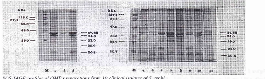
Figure 1. SDS-PAGE profiles of OMP preparations from
I0
clinical isolates of S. typhi.Major proteins (38, 37 and 34 kDtt) were present in most of tlrc S. lyphi OMP preparations (l.ane 2, 3, 5, 7, B, I0 and
l1) as
weLL as ittaround
140kDa. Polypetide with
MW 20.5
kDa
wasobserved
in all
OMPs, while polypeptides
with MV/
29
and25 kDa were
observed
in
6
and 4
OMps
re-spectively. One OMPs showed
a
polypeptide with
MW
less than 20kDa (Figure
1).Electrophoretic
pro-files
of
major proteins were in
agreement
with
thosepreviously
reported6,1l.
In
this
study,
however,
we
demonstrated that
eachisolates
of
S.typhi might
con-tain outer
membrane
proteins which similar
in
their
major
proteins
but differ in minor
ones.We
found
the
presenceof
six different OMPs'profiles
in
our
^S.ry-phi isolates
in
comparison
to .S. typhi
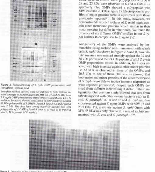
Ty2.Antigenicity
of
the
OMPs were
analysed
by
im-munoblot using rabbits'
sera
immunised
with
whole
cells
S.typhi. As
shown
in Figure
2
Aand B, two
rab-bits' immune
serareacted
strongly
against the 37
and 38kDa
porins
and the29
kDaprorein of all
5 S.typhi
OMP
preparations tested.
In
addition, both
sera
re-actedwith high intensity
against other
minor
proteins
i.e. 63
kDa as observed
in
three
of
the OMps,
and20.5
kDa
in
one
of
them. The results showed that
both
major
andminor
proteins
of
the outer membrane
of
S.typhi were
able
to induce immune
responses
aswere reported previously6, despite each
OMps
de-rived from different
isolates
might
differ
in their
an-tigenicity. Our previous study
showed
that
serafrom
rabbits
injected with
other enteric bacteria such
as E.coli,
S.
paratyphi
A, B
and
C
and
S. typhimurium
cross-reacted
against S.typhi
OMPs
with
MW
37
and
23.5
kDa. Yet, reactivity
against S.
typhi Omps with
MW
55kDa
was
only
observed
in
seraof rabbits
[image:3.595.49.287.90.390.2] [image:3.595.47.556.110.677.2]im-munised
with
E
coli
and S.paratyphi
Ct8. Figure2.
Immunoblotting of S. typhi OMP preparations withtwo rabbits' immune sera,
Sera from rabbits injected with two
dffirent
S. typhi isolates re-29 kDafrom lane l-5). In tivity againstand PaneL B
Lane 2,3,4). Also they hclve strong reactivity against 20.5 kDa polypeptide of
I
OMPs (Panel A lane 4) as well as in panel BIane 3. M is protein MW marken
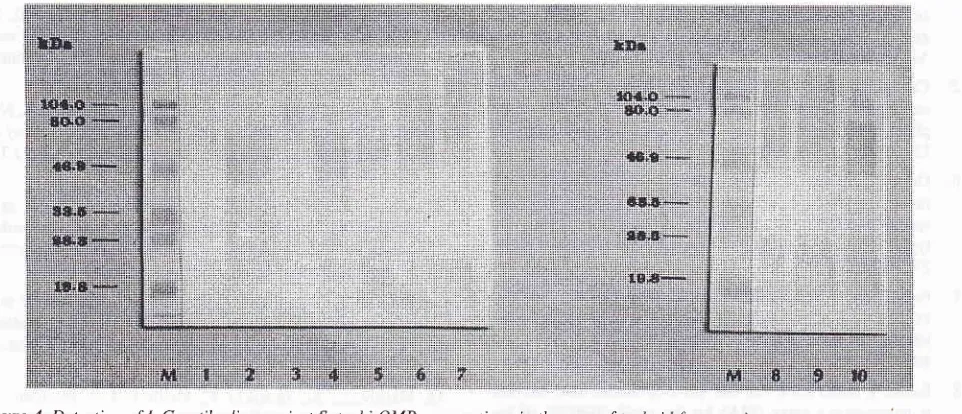
Figure 3. Detection of IgM antibodies against S. typhi OMP preparations in the sera of typhoidfever patients.
Acute sera from
10
cuhures were pooled and dilwted 1:80A" Thes
nst major proteins (3g, 3ZOMPs were
observe
x. 30 kDa seemed. to showSuppl
I
-
1998 Diagnostic 47Figure 4. Detection of IgG antibodies against S. typhi OMP preparations in the sera of typhoid fever patients.
Pooled patients' sera were tliluted I:200 and anti-human IgG HRP-conjugate was diluted I :500. No reactivity against the S. typhi OMP
preparations was observed.
M
is protein MW marker Lane I -10 are OMP preparations froml0
clinical isoLates of S. typhi.To determine the
antigenicity
of
the S.typhi
OMPs
in
human,
IgM
and
IgG
antibodies against
these
pro-teins
in
the
seraof typhoid fever
patients were
exam-ined
by immunoblotting.
The results were
asfollows:
pooled patients'
sera
did
not
react
to
major
proteins
of
all
S.
typhi
Omp preparations
used
in
the
study,suggesting
neither
IgM
nor
IgG
immunoglobulins
were present
in
responseto OmpC, OmpF
andOmpA
(Figure
3
and4).
Some degree
of
reactivity
of IgM
antibodies
against
minor polypeptides
present
in
theOMP
preparations
were
observed
(Figure
3). Protein
band
with
MW
approx. 30
kDa
in
one
of
the OMP
preparations
seemedto
show stronger reaction
to
the sera (seeFigure
3).
Moreover,
when anti-human
IgG
HRP-conjugate was used
as second
antibody
in The
immunoblotting
analysis,
no distinctive
reactive
pro-tein
bandswere observed (Figure 4).
Immunoblotting
using
pooled control
serashowed
no
IgM
or IgG
an-tibody
responses
against
all of
the S.
typhi
OMP
preparations (data
not shown). Earlier
investigations
reporled the
presenceof
IgM
andIgG
immunoglobu-lin
responses
directed against S. typhi porins
in
thetyphoid fever patienls' se1n5,6,12'19'20.
pufthsr, minor
polypeptides
of
the OMPs
with
MW
50, 53 and
56kDa
were
found
to
be immunogenic
in
human8'10,12.Our
results, however,
did
not
confirm
theirs,
this
might indicate the possibility
of
the presence
of
dif-ferent antigenic determinants
in
our
OMP
prepara-tions,
or
else
the antigenicity
of
S. typhi OMPs
hassome degree
of
variability
within
strains. Moreover,
the possible
existence
of
natural variation
in
the
re-sponse
to
S. typhi OMPs
among
human
populations
should
also be considered.
In
the present study, we
demonstrated
that S. typhi
OMPs
areimmunogenic in rabbits
administered
with
the bacteria,
in
that
IgG
antibodies recognised
theporins and other polypeptides
of
the OMP
prepara-tions.
In typhoid
fever patients, however,
primary
andsecondary
immune
responses against
the porins
arenot
detected,
although we
observed
that thè
patients
have
IgM
antibodies against minor polypeptides
of
rheS.typhi OMPs.
In
briei
the presenceof
antibodies
against S.
ryphi OMPs might be an indication
of
in-fection
of the microbe. However,
the use
of
any
spe-cific
immunodominant polypeptides
of
the S. typhi
OMPs
for
diagnostic
of
typhoid fever
is in
need
for
further
study.Acknowledgment
This
work was
supported
by
Competitive
ResearchGrant
(RUT)
No:
1OO4/SP-KD|PPIT|M95 from
Ministry
of
Research
andTechnology, Indonesia.
REFERENCES
l.
Simanjuntak CH, Hoffman SL, Punjabi NH et al,Epidemiol-ogy of typhoid fever in Paseh, rural area in West Java.
Cer-min Dunia Kedokteran 1987;
45
\6-8.2.
Simanjuntak CH. Development typhoid fever oral vaccine.Medika 1990;3:213-8.
3.
Sudarmono P, Raji M. Features of typhoid fever in Indonesia.In Pang
!
Koh CL, Puthucheary SD (eds). Typhoid Fever, Strategies for the 90's Kuala Lumpur, World Scientific 1991;I 1-5.
4.
Chou PY Wan KC and Tsang RSW. Crossed5
of typhoid patients and carriers: Demonstration of the pres-ence of typhoid-specific antibodies to a non-O, non-H,
non-Vi antigens. Infect Immunity 1984; 43: 1110-3.
Calderon I, Lobos SR, Rojas HA, Palomino C, et al. Antibod-ies to porin antigens of Salmonella typhi induced during
ty-phoid infection
in
humans. Infect Immunity 1986; 52:209-12.
Ortiz
V
IsibasiA,
Garcia-OrtigozaE,
Kumante J.Im-munoblot detectionof
class-specific humoral immune re-sponse to outer membrane protein isolated from Salmonella typhi in humans with typhoid fever. J Clin Microbiol 1989;27: I64Q-5.
Fernandez-Beros ME, Gonzales C, Mclntosh
MA,
CabelloFC. Immune response to the iron-deprivation-induced
pro-teins
of
Salmonella typhiin
typhoid fever. Infect Immunity 1989; 57:I27l-5.
Ismail A, Kader ZSA, Hai OK. Development of a dot enzyme
immunosorbent assay
(EIA) for
the rapid diagnosisof
ty-phoid fever. In PangI
Koh CL, Puthucheary SD (eds). Ty-phoid Fever, Strategies for the 90's (Kuala Lumpur, World Scientificl99I:
201-6.Hai OK, Ismail A, Choo KE, Kader ZK. Clinical field trials on the dot enzyme immunosorbent assy (EIA) for diagnosis
of typhoid fever. In
P*g
T, Koh CL, Puthucheary SD (eds).Ïlphoid
Fever, Strategies for the 90's Kuala Lumpur, World Scientific 199 1 : 207 -13.10. Gam LH, Devi S, Puthucheary Sd, Koh CL et al. Immune
re-activity
of
typhoid sera with OMPs extracted from Salmo-nelLa typhi. In Pang T, Koh CL, Puthucheary SD (eds). Ty-phoid Fever, Strategiesfor
the 90's Kuala Lumpur, World Scientific 1991; 183-7.11. Verdugo-Rodriquez A, Lopez-Vidal
I
PuenteJL,
Ruiz-Palacios GM et al. Early diagnosis of typhoid fever by an
en-zyme immunoassay using Salmonella typhi membrane pro-tein preperations. Eur J Clin Microbiol Infect Dis 1993; l2:
248-54.
12. Verdugo-Rodriquez A, Gam LH, Devi S, Koh CL et
al.
De-tectionof
antibodies against Salmonella typhi outer mem-brane protein (OMP) preparationin
typhoid fever patients.Asian Pacific J Allergy Immunol 1993;
ll
45-52,13. Jackson AA, Ismail A, Tuan Ibrahim TA, Kader ZSA, Nawi
NM. Retrospective review of dot enzyme immunoâssay test
for typhoid fever in an endemic area. Southeast Asian J Trop Med Public Health 1995; 26:625-30.
14. Thong
KL,
CheongYM,
Puthucheary S,Koh CL, et
al.Epidemiologic analysis of sporadic Salmonella typhi isolates and those from outbreaks by pulsed-field gel electrophoresis.
J Clin Microbiol 1994; 32:
ll35-41.
15. Thong
KL,
Puthucheary S, Yassin RM Sudarmono P, et al.l995Analysis
of SalmonelLa
4rplrl isolatesfrom
SoutheastAsia
by
pulsed-field gel electrophoresis.J Clin
Microbiol1994;33: 1938-41.
16. Matsuyama
SI,
InokuchiK,
MizushimaS.
Promotorex-change between ompF and ompC, genes for osmbregulated major outer membrane proteins
of
Esherichiacoli
K-12. JBacteriol 1984; 158:
l04l-7.
17. Bradford MM. A rapid and sensitive method for the quanti-tation of microgram quantities of protein utilizing the
princi-ple
of
protein-dye binding. Analytical Biochem 1976;72:248-54.
18. Moehario
I
H, BelaB
and Sudarmono P. Studyof
specific proteinof
Salmonella typhifor
diagnosisof
typhoid fever. Southeast Asia J Trop Med Public Health 1995; 26:230-2.19. Mora
GG
Barnier RA, Toro CS, Asenjo CA.Regulation ofSalmonella typhi porins. In Pang
I
Koh CL, Puthucheary SD (eds). Typhoid Fever, Strategies for the 90's Kuala Lumpur, World Scientificl99l;
64.20. Paniagua J, Isibasi A, Garcia JA, Gonzalea CR, et al. Porins
as protective immunogens against typhoid fever. In Pang T,
Kob CL, Puthucheary SD (eds). Typhoid Fever, Strategies
for the 90's, Kuala Lumpur World Scientific 1991; 65-70
8