Partial Characterisation of Salmonella gallinarum Clinical Isolate and Expression of Its
Antigenic Outer Membrane Protein C (OmpC) Gene In Planta
Ee Leen Pang1
, Hwei-San Loh1,2*
1
School of Biosciences, Faculty of Science, The University of Nottingham Malaysia Campus, Jalan Broga, 43500 Semenyih, Selangor, Malaysia.
2Biotechnology Research Centre, The University of Nottingham Malaysia Campus, Jalan Broga,
43500 Semenyih, Selangor, Malaysia.
ABSTRACT
Fowl typhoid’s epidemiology and disease intervention have been extensively studied since 1950's owing to its high mortality and morbidity rates. Even up-to-date, outbreaks are incessantly haunting poultry industries of major continents. Salmonella gallinarum, the etiologic agent of fowl typhoid, was used to develop a series of vacci-nation regime. However, treatments are gradually losing effectiveness due to residual virulence from mutated strains and rapid evolution of multidrug resistance isolates. Hence, in planta subunit vaccine production is pro -posed to surpass current limitations. The homotrimeric osmoporin (outer membrane protein C) is a potent can-didate antigen that confers momentous stimulation of humoral and cell-mediated immune responses in broilers. This research signified the potential development of a plantexpressed OmpC immunogen. The project scope em -barked on the identification of S. gallinarum clinical isolate, construction of expression cassette and delivery of constructs into Nicotiana benthamiana via agroinfiltration. The OmpC transcripts and proteins were detected suc-cessfully at the molecular weights of ~1.002 kbp and ~35 kDa, respectively. These preliminary findings pave the feasibility of biomanufacturing a safe and cost-effective fowl typhoid vaccine that would confer multi-protection against other significant Salmonella infections attributed to the high sequence homology of the OmpC gene. Speed improvement is demonstrated and transient expression appears to outperform conventional platforms in expediting vaccine production for an emerging pandemic strain.
Keywords: OmpC gene, KDEL signal, Salmonella gallinarum, subunit vaccine
Fuelled by the trend of increasing urbanisation, meat demands are progressively elevating where 29% increment is specifically pressured upon poultry meat production [1]. Accounting for almost half of world meat trading, figures of 9,700,000 and 10,500,000 tonnes were recorded in years 2004 and 2008, respec-tively [2,3]. Poultry products which serve as cheap source of protein are now widely known to be threat-ened by Salmonella diseases [4]. Therefore, in the course of demanding increment for international trad-ing, the risk of global dissemination of poultry infec-tions is concurrently exacerbated. In fact, huge
invest-ments have already been laid down by European countries in salmonellosis disease control. Hitherto, se-ries of molecular diagnostics tools have been deployed for rapid serotyping of clinical isolates such as real-time multiplex polymerase chain reaction (qPCR) assay [5]. Nevertheless, more constructive approaches are an-ticipated; of which extensive researches are currently on-going to pave a way for efficient subunit vaccines production that takes consumer safety concerns into account. It is because the issue is previously neglected in terms of antibiotics and live-attenuated strain vac-cines application [6]. Pathogenicity of host-adapted Salmonella gallinarum is primarily noted through the epidemic spread of fowl typhoid (FT) in chickens and turkeys, often instigating high morbidity and mortality rates. The first outbreak of the infectious septicaemia disease was reported back in England during 1888. Blood poisoning usually leads to enlargement of liver and spleen, sometimes with signs of white necrotic foci INTRODUCTION
*Corresponding author: Hwei-San Loh
School of Biosciences, Faculty of Science, The University of Nottingham Malaysia Campus, Jalan Broga, 43500 Semenyih, Selangor, Malaysia
in other organs of the infected poultry [7,8]. Albeit eradication programme had been successful in Europe and other Western countries via vaccination and sur-veillance programmes, but severe losses were suffered by pandemic outbreaks in Asia, Latin America and Africa [9,10]. The severity of FT was highlighted when a total of 227 disease outbreaks were detected in Haryana alone simply within a 4-year period, affecting 9 out of 20 districts [11]. Death rate of acute FT can range from 10-93% [12]. Without underestimating the risk, 100% mortality rate had been reported in one-day-old broiler chicks after infected by 108
colony-forming unit/ml of S. gallinarum [13].
The upsurge of FT prevalence had pioneered the development of 9R live-attenuated vaccine with semi-rough lipopolysaccharide [14] that would not interfere with egg production and blood testing as compared to dead vaccines of no value. Dejectedly, minimal protec-tion against highly virulent field strains and egg trans-mission of 9R strain were reported [15]. Because of natural variation, this is further complicated with the emergence of new strains that could hijack existing im-mune responses due to poor cross protection from the conventional vaccine. Albeit drugs application (i.e. chloramphenicol, tetracyclines and sulphonamides) are employed to control disease outspread, constant evolu-tion of multi-drug resistant field strains has gradually hampered the effectiveness of eradication programmes. Dealing with residual virulence induced by whole cell bacteria and live strain vaccines, subunit vaccines de-velopment from immunodominant protein components were extensively researched for new vaccine formula-tion. Bacterial outer membrane protein is a potent in-ducer of both humoral and cell-mediated immune re-sponses [16]. A remarkable breakthrough was achieved when administration of partially purified OmpC from S. gallinarum conferred superior protection as com-pared to whole cell-based and 9R strain live vaccines [17]. Selection of immunogen on the basis of long-last-ing protection is vital. The trimeric β-barrel protein had satisfactorily fulfilled the criteria as 53-70% protec-tion was passed on by immunised breeders to newly-hatched chicks after challenged with 20-500 half-maxi-mum lethal dose of the pathogen [18].
With the popularised trend of exploiting plant sys-tems as biofactories for the production of pharmaceuti-cal products, this context aims to present a framework for rapid development of in planta vaccine candidate based on agroinfiltration strategy [19]. Hitherto, the transient expression platform has gained increasing adoption as a robust and efficient gene delivery
methodology for numerous biopharmaceutical proteins production. It is to serve as a prompt remedy to tackle conventional regime, which commonly entangles with more tedious works in the course of harvesting a safe and efficient vaccine for poultry at infested fields. Prac-tically, plants stand out as a better alternative to dis-place prokaryotic and eukaryotic expression systems with improved efficiency, biocontainment warranty, higher safety and convenient storage. As plants are im-plicated not to harbour mammalian pathogens, this would then eliminate concerns of protein contamina-tion with viruses, endotoxins or oncogenic sequences; correspondingly reduce the cost associated with screen-ing and purification [20].
It is noteworthy to mention, the OmpC candidate antigen has never been isolated for heterologous ex-pression in tobacco as described in this novel study. As the “old” vaccine is inadequate in combating new vari-ants, continuous vaccine development is ultimately es-sential without being jeopardised by expensive and long lead time approach. Thereby, this paper reports the feasibility of a rapid vaccine development upon de-tection of an emerging outbreak. A clinical isolate of Salmonella disease was used here to demonstrate an in-dustrial-like application in a safer and speedy produc-tion platform.
Identification of Etiologic Agent
A clinical isolate of S. gallinarum obtained from an undisclosed local farm (Peninsular of Malaysia) was firstly subjected to morphological and biochemical identification. The colonial growth response was tested on several media such as Nutrient agar, MacConkey agar and Xylose-Lysine-Deoxycholate agar, using closely-related enterobacteria (Escherichia coli) for comparison. Differential staining methodology was used to confirm the presence of gram-negative rods un-der microscopic examination. The identity of the FT etiologic agent was reinforced through biochemical analyses on the utilisation of sugars and amino acids, motility observation and Analytical Profile Index (API) test.
Bacterial Genomic DNA Extraction and OmpC Gene Isolation
maximise gDNA template harvest. The extraction was conducted as follow: (i) lysis of 10 ml bacterial culture in buffer [40 mM Tris acetate (pH 7.8), 20 mM sodium acetate, 1 mM ethylenediaminetetraacetic acid (EDTA) and 1% sodium dodecyl sulphate (SDS)]; (ii) removal of cellular debris by adding 5M sodium chloride (NaCl); (iii) separate protein precipitates via chloro-form treatment; (iv) ethanol precipitation and RNase A incubation; and (v) purification prior re-suspension in 1X TE buffer. Alongside, the primers targeted for am-plification of full-length OmpC gene [0.969 kilobase pair (kbp)] were designed according to published se-quence (Accession Number: NC_011274). For cloning purpose, NruI and XmaI restriction sites were intro-duced into 5’ and 3’ ends of the construct. Eukaryotic KDEL consensus sequence was also incorporated to the C-terminus of OmpC as product localisation signal. Detailed design of forward (PF1ompC) and reverse (PR1ompC) primers was specified in Figure 1a. A PCR cocktail was prepared by mixing regents (1X PCR buf-fer, 1.5 mM MgCl2, 0.3 mM dNTPs, 0.5 µM of each primer and 1.5 units/µl of Taq polymerase) with 100.0 ng/µl of extracted DNA template to isolate the anti-genic gene. Optimised OmpC gene amplification was programmed as follow: (i) pre-denaturation step at 94ºC; (ii) 30 amplification cycles with 94ºC for denatu-ration, 67ºC for annealing and 72ºC for elongation; and lastly (iii) the final extension of amplicons at 72ºC.
Recombinant Vector Construction
Amplicons collected from PCR were purified using Expin™ Combo GP (GeneAll®, Korea). As both pEAQ-HT vector (Accession Number: GQ497234) and OmpC insert had similar cutting site for NruI and XmaI, a two-step restriction endonuclease (RE) diges-tion was performed (Figure 1b). Subsequent ligadiges-tion of digested fragments was performed via T4 DNA Ligase (New England Biolabs, UK) according to manufac-turer’s instructions. Heat-shock approach was then em-ployed to introduce the recombinant vector, pEAQ::OmpC-KDEL into competent XL-1 E. coli cells. Putative recombinants were subjected to PCR-based screening to confirm the successful incorporation of the OmpC insert. Verification of positive clone was re-inforced by extracting plasmids of transformed bacteria via Exprep™ Plasmid Quick (GeneAll®, Korea) for RE and PCR analyses. To facilitate rapid screening, XhoI was used instead of XmaI since it shared common Buf-fer R as NruI. Upon confirmation, the extracted pEAQ::OmpC-KDEL was sent for nucleotide sequenc-ing (Bioneer, Korea).
Plant Transformation via Agroinfiltration
Nicotiana benthamiana seeds were planted and grown up to 10-week old under controlled environ-ment of 24ºC. The recombinant vector, pEAQ::OmpC-KDEL was incorporated into competent Agrobac-terium tumefaciens strain LBA4404 to establish stable transformant for downstream agroinfiltration. Broth cultures harbouring constructs of pEAQ::OmpC-KDEL, pEAQ::GFP and pEAQ-HT were grown for 48 hours at 28ºC and 200 rpm continuous shaking condi-tion. These agrobacterial cultures were then harvested at OD600= 0.4 by 4ºC centrifugation (6,000 rpm for 10 minutes) and re-suspended in MMA solution [10 mM MES (2-[N-morpholino]ethanesulfonic acid) at pH 5.6, 10 mM magnesium chloride and 100 µM acetosy-ringone] [22]. Abaxial sides of tobacco expanded leaves were pricked before infiltrated with above mentioned suspensions using a needless syringe. Three plant repli-cates were prepared for each construct along with a healthy tobacco as mock-infiltration control. Trans-formed plants were then allowed to resume growth and harvested at 3, 6 and 9 days post inoculation (dpi). Ap-parent morphological distortion of infected leaves was also recorded.
RNA Assessment of Infiltrated Leaves
To monitor expression and stability of foreign OmpC gene post-infiltration, TRIZOL® reagent (In-vitrogen, USA) was used to extract the total RNA from 100 mg of grounded tobacco leaves [23] with liquid ni-trogen. Phase separation was then achieved by 20% (v/v) chloroform treatment. Next, the extracted aque-ous layer was subjected to precipitation by isopropanol and ethanol followed by elution of RNA in 100 mM Tris-HCl (pH 8.5). Two-step reverse-transcription PCR (RT-PCR) was performed to generate cDNA samples using Omniscript RT Kit (Qiagen, Netherlands) ac-cording to manufacturer’s instructions. Initial denatu-ration of template RNA was done at 65ºC for 5 min-utes before added into the master mix [1X Buffer RT, 0.5 mM dNTPs, 1.0 µM of each primer, 500.0 ng/µl RNA and 4.0 units/µl Omniscript Reverse Transcrip-tase]. RT cocktail was then allowed to react at 37ºC for 2 hours before proceeding to amplification of OmpC cDNA. Lastly, targeted transcripts were analysed via 1% (w/v) agarose gel electrophoresis, along with non-RT controls to trace for any potential DNA contamina-tion.
Evaluation of Protein Expression
was done as previously described [22]. Following grinding of 200 mg leaf tissues, samples were sus-pended in 1X volume of Extraction Buffer [50 mM Tris-HCl at pH 7.25, 150 mM NaCl, 2 mM EDTA and 0.1% (v/v) Triton X-100]. After high-speed centrifuga-tion (14,000 rpm) at 4ºC for 10 minutes, the super-natant was collected for quantification using Pierce® 660 nm Protein Assay Reagent (Thermo Scientific, USA) prior storage at -80ºC. SDS-polyacrylamide gel electrophoresis (SDS-PAGE) was performed at 120 V for 90 minutes to check the state of protein integrity. The 12% separating gel was stained with Coomassie blue and then analysed under GS-800™ calibrated den-sitometer (Bio-Rad, USA). For Western blot analysis, 20 µg/ml of protein extract was loaded in each well and subsequently electroblotted onto methanol-treated Polyvinylidene Difluoride (PVDF) membrane (MILI-PORE, USA). Blocking step was done by incubating the membrane with 1X PBS-Tween (0.05 M Sodium Dihydrogen Phosphate at pH 7.4, 0.15 M NaCl and 0.05% Tween-20) supplemented with 4% (w/v) milk. Primary antibody used for detection was rabbit anti-His polyclonal antibody (Abcam, UK) (1:5,000 dilu-tion). The mouse anti-glyderaldehy3-phosphate de-hydrogenase (GAPDH) monoclonal antibody (Thermo Scientific, USA) (1:1,000 dilution) was used as a load-ing control. After 2-hour shakload-ing at 50 rpm, the mem-brane was washed and followed sequentially by 1-hour incubation of rabbit HRP-linked secondary anti-body (Abcam, UK) (1:10,000 dilution). Another wash-ing step was repeated five times at 5-minute interval before 1 ml of 3, 3’, 5, 5’-tetramethylbenzidine (TMB) substrate was added. Finally, the colorimetric response of the immunoblot was recorded using the same cali-brated densitometer.
Salmonella gallinarum Characterisation
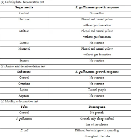
Comparatively slower growth was observed from the smooth, domed and glistening salmonellae colonies. In addition, selective conditions of Mac-Conkey and Xylose-Lysine-Deoxycholate agar plates had recorded the bacterial response as a non-lactose fermenter and hydrogen sulphide-negative enteric col-iform. From the microscopic observation, the pinkish non-sporulating microbes were seen to be arranged as a single entity that conformed to the measured 1.0-2.5 µm length and 0.3-1.5 µm width [8]. Examination of S. gallinarum biochemical properties as summarised in Table 1 revealed catabolic breakdown of dextrose, mal-tose and mannitol which was indicated by phenol red
without any gas evolvement (Figure 2a). Next, the colour change of bromocresol purple had denoted posi-tive reaction in decarboxylase tube containing lysine only (Figure 2b). Evidence of bacterial non-locomotive feature was recorded based on the confined growth from the line of inoculation (Figure 2c). Nevertheless, a 7-digit code of 4004102 was generated based on the sheet attached with API kit.
Isolation of Antigenic Gene
Extraction of intact bacterial gDNA (data not shown) was successful, yielding two bands of ~2.5 kbp and >10 kbp in size on the agarose gel. Consecutively, amplification of the OmpC gene from the genomic template was achieved using designated primers. With the annealing temperature set at 67ºC, ~1.002 kbp am-plicons were obtained and thereafter named as OmpC-KDEL.
Recombinant Vector Construction
Recombinant vector, namely pEAQ::OmpC-KDEL was generated as illustrated in Figure 1b. The presence of the transgene in E. coli clones was verified by PCR (Figure 3). XmaI- and NruI- digested amplicons and pEAQ-HT with molecular sizes of ∼0.988 kbp and
∼9.973 kbp, were ligated through complementary
se-quences to yield the expression cassette (Figure 1b). Se-quence alignment results had scored 99.8% of data
RESULTS AND DISCUSSION
similarity when BLAST analysis was performed against the Accession Number NC_011274.
Morphological Observation of Plants
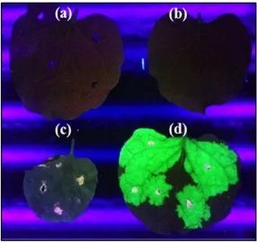
Following agroinfiltration, a systemic spread of plant virus was noted; with abnormal morphologies like leaf chlorosis, crinkle and/or necrosis of tobacco host. Emission of green fluorescence was detected from pEAQ::GFP-infected leaves upon sample collection (Figure 4).
Heterologous Expression of OmpC Gene at RNA and Protein Levels
The RT-PCR profiles as depicted in Figure 5 con-firmed successful expression of the antigenic gene.
Bands with a molecular size of ∼1.002 kbp were
ob-tained for samples at 3, 6 and 9 dpi, conformed to the positive controls (lanes 13 and 28). Healthy and pEAQ-HT-infiltrated leaves did not show amplification
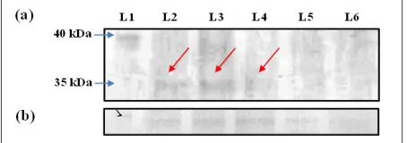
as expected. For preliminary testing, a positive signal was detected by dot blot assay of pEAQ::OmpC-KDEL-infiltrated samples (data not shown). Compatible hybridization was further verified via Western blot analysis (Figure 6a). The presence of OmpC protein at ∼35 kDa (UniProtKB Database: B5RC93) was detected in all plant samples infected with pEAQ::OmpC-KDEL. Contrariwise, none was detected for pEAQ-HT-infected and healthy ones.
Correspondingly, the presence of ∼34 kDa
housekeeping GAPDH protein was identified from all plant samples (Figure 6b).
In agreement to the general features of salmonellae, the avian-adapted S. gallinarum was identified as gram-negative non-sporogenic tiny rods that grew slowly on nutritive and selective media. In view of biochemical deficiency, an appreciable growth of the serotype could be limited by the auxotrophic requirement for thiamin, which was not usually formulated into customary
Figure 1. Schematic diagrams of (a) PF1ompC and PR1ompC primers designed for OmpC gene isolation, with additional sequences added onto 5’ or 3’ end; and (b) construction of gene expression cassette pEAQ::OmpC-KDEL, as adopted from Sainsbury et al. [35]).
bacteriological media [24]. Based on the carbohydrate fermentation test, catabolic breakdown of dextrose, maltose and mannitol into acids were displayed by S. gallinarum. Interestingly, no formation of gas bubbles was detected as compared to E. coli. Despite that, results obtained did conform to other findings [25, 26]. Vast biochemical similarities between the two avian-specific serovar Gallinarum and serovar Pullorum had been ascribed to their monophyletic divergence. Hence, ornithine decarboxylation test was incorporated to vali-date the identity of S. gallinarum due to its naturally mutated speC gene that blocks putrescine biosynthesis [25,27]. The credibility of data on ornithine and lysine decarboxylation was in fact strengthened by previous report [28]. However, a discrepancy was noted for argi-nine decarboxylase activity as the pathway had been discovered previously [27]. Repetitive tests were done and it could be narrowed down to two factors: (i) pH shift induced by arginase, arginine dihydrolase and di-amine oxidase or (ii) widespread interchange of genetic elements between compatible strains given the diversity among isolates [29-31]. Besides, a locomotive test was crucial to distinguish salmonellae into serotypes of paratyphoid and avian specific. The absence of peritrichous flagella in the latter group could due to
the faulty assemblage of flagellar subunits or dysfunctional motor proteins [30].
Guided by concrete evidences of OmpC as im-munogen [32], a pioneering study was established here to present the reproducibility of plant-based transient expression as a time- and cost-effective platform in de-veloping a vaccine candidate against fowl typhoid. The binary vector pEAQ-HT has built-in CaMV35S pro-moter (P-35S) and Nos terminator (NosT) flanking be-tween the T-DNA borders to mediate high OmpC-KDEL expression. The presence of p19 silencing suppressor is expected to promote expression through sequestration of small-interfering RNAs generated by Dicer [33]. Considering the plant protein glycosyla-tion pattern, KDEL inserglycosyla-tion was projected to min-imise protein conformational changes that would im-pact neutralising antibodies production. Accumulation of ER-resident proteins could benefit the yield harvest attributed to 20-100 folds of improved expression, greater stability and minimised exo-proteinase degrada-tion in cytoplasm [34].
BLASTn had recorded 99.8% of maximum identity and an E-value of 0, indicating that the similarity score was significant to prove that sequences were homologs. In other words, convincing evidence of successful
re-Figure 4. Detection of green fluorescence under an ultraviolet box. The expression of GFP protein was compared between (a) pEAQ-HT-infected (mock control), (b) healthy tobacco, (c) pEAQ::OmpC-KDEL-infiltrated and (d) pEAQ::GFP-infected (positive control) leaves as a checkpoint for successful agroinfiltration.
combinant DNA construction was achieved. A tran-sient expression assay was opted here to assess the ca-pacity of plant system to express the foreign antigenic sequence preceding stable transgenesis [35]. Based on the RT-PCR profiles, amplicons of OmpC-KDEL (∼1.002 kbp) were obtained at all days tested, signify-ing positive expression at the transcriptional level. The possibility of OmpC-KDEL expression being affected by individual host’s physiological condition could not be excluded as low transcript abundance was also noted (Figure 5, lanes 7 and 8). Only samples of 6 dpi were selected for subsequent immunoblotting since OmpC transcripts at this time point were consistently detected. Compatible hybridization between His-tagged OmpC and anti-His antibody through western blotting assay had further confirmed successful expression of recombinant antigen where an expected band size of ~35 kDa was obtained only in pEAQ::OmpC-KDEL-infiltrated samples. Besides, protein bands of 50 kDa
were postulated to be background proteins of tobacco host that reacted with the anti-His antibody (data not shown). As the Ribulose-1, 5- bisphosphate carboxy-lase/oxygenase (Rubisco) constitutes at least 50% of the total soluble protein in C3 plants [36], it is likely that the 50 kDa product represents the degrading frag-ment of Rubisco large subunit. Turnover could be prompted by oxidative stress suffered during CPMV-based infection. Nevertheless, solid detection of OmpC up to protein stage was achieved. It is also interesting to note that satisfactory OmpC expression could be achieved even without codon optimisation of the native bacterial sequence.
Nevertheless, His-tag detection could not serve as a definite resemblance of antibody hybridization directed specifically against S. gallinarum OmpC. Also, because a polyclonal anti-His was used, it could not confer high stringency like monoclonal antibody that only binds to single epitopes. Thus, this would limit the detection of
Figure 6. Immunoblotting profiles of protein samples extracted from infiltrated N. benthamiana leaves at 6 dpi reacted with (a) anti-His and (b) anti-GAPDH antibodies. L1: Spectra™ Multicolor Broad Range Protein Ladder; L2-L4: Replicates of pEAQ::OmpC-KDEL-infected plant samples; L5: Healthy tobacco; L6: pEAQ-HT infiltration control. The OmpC protein was successfully detected at ~35 kDa region for all samples infiltrated with pEAQ::OmpC-KDEL (red arrows).
low OmpC expression by which merely fade bands were obtained along with non-specific interference by most probably Rubisco. Albeit His-tagged fusion was valued for downstream purification capacity, the im-practicality of the assay to detect poorly expressed pro-tein, however was reported [37], attributed to interfer-ing background and length of the procedure. Enrich-ment procedure could be employed to pool eluents of His-tagged protein from nickel affinity column prior purifying via high-performance liquid chromatography to enhance the performance of anti-OmpC-based de-tection [38].
In this study, rapid biochemical identification of S. gallinarum clinical isolate along with successful expres-sion of OmpC-KDEL in N. benthamiana signify its momentous value as a potential plant-based vaccine de-velopment against emerging fowl typhoid. The high degree of sequence homology between Salmonella porins further enhances the practicality of recombinant OmpC in conferring protection against other infectious serotypes of significant importance in poultry indus-tries. Nevertheless, confirmatory hybridization of anti-OmpC against the immunogen is proposed to achieve more comprehensive study for functional validation in future. Once the immunogenic protection is proven to be promising, this research could then be exemplified as a platform for the transformation of palatable plant species such as alfalfa and corn to function as an edible vaccine.
Comprehensive scope which includes clinical strain identification, antigenic protein extraction and OmpC expression studies had highlighted the robustness of the transient technology in delivering pandemic vac-cines within the shortest time frame. Considering from the production standpoint, the advantages of speed and cost would vastly benefit from the instantaneous sup-ply of potent vaccines to global market prior waves of any further epidemic loss.
Deepest gratitude is expressed for the insightful comments and technical assistance from Mr. Eu Sheng Wang and Ms. Xiao Ying Chan (University of Notting-ham Malaysia Campus). We would also like to thank Professor George Lomonossoff (John Innes Centre, Norwich, UK) for supplying the pEAQ-HT vectors.
1. Wattagnet (2012) Next decade to bring change, challenge
for poultry industry. http://www.wattagnet.com/Next_ decade_to_bring_change,_challenge_for_poultry_in-dustry.html.
2. Windhorst H (2006) Changes in poultry production and trade worldwide. World's Poultry Science Journal. 62: 585-602.
3. Economic and Social Development Department, FAO Corporate Document Repository (2009) Meat and meat products. http://www.fao.org/docrep/011/ai482e/ai482e08. htm.
4. Cogan T, Humphrey T (2003) The rise and fall of Salmo-nella enteritidis in the UK. Journal of Applied Microbiol-ogy. 94: 114S–119S.
5. O'Regan E, McCabe E, Burgess C, McGuinness S, Barry T, Duffy G, et al (2008) Development of a real-time multi-plex PCR assay for the detection of multiple Salmonella serotypes in chicken samples. BMC Microbiology. 8: DOI: 10.1186/1471-2180-8-156.
6. Barrow P, Wallis T (2000) Vaccination against Salmonella infections in food animals: Rationale, theoretical basis and practical application. In Wray C, Wray A, eds. Salmonella in domestic animals. CABI Publishing. Oxon. 323-340. 7. Shivaprasad H, Barrow P. (2008). Pullorum disease and
fowl typhoid. In Saif Y, Fadly A, Glisson J, McDougald L, Nolan L, Swayne D, eds. Diseases of poultry. 12th edi -tion. Oxford: Blackwell Publishing. 2008: 620-636. 8. World Organization for Animal Health (OIE) (2012)
Fowl typhoid and Pullorum disease, chapter 2.3.11. In: Manual of diagnostic tests and vaccines for terrestrial ani-mals, ed. OIE, 7th ed., pp. 538–548. Paris.
9. Jones M, Wigley P, Page K, Hulme S, Barrow P (2001) Salmonella enterica serovar Gallinarum requires the Sal-monella pathogenicity island 2 type III secretion system but not the Salmonella pathogenicity island 1 type III se-cretion system for virulence in chickens. Infection and Im-munity. 69: 5471-5476.
10. Paiva J, Penha Filho R, Arguello Y, Silva M, Gardin Y, Resende F, et al (2009) Efficacy of several Salmonella vac-cination programs against experimental challenge with Salmonella gallinarum in commercial brown layer and broiler breeder hens. Brazilian Journal of Poultry Science. 11: 65-72.
11. Kumar T, Mahajan N, Rakha N (2010) Epidemiology of fowl typhoid in Haryana, India. World's Poultry Science Journal. 66: 503-510.
12. Cobb S (2011) The spread of pathogens through trade in poultry meat: overview and recent developments. Scien-tific and Technical Review - International Office of Epi-zootics. 30: 149-164.
13. Wong R, Tellez G, Valladares J, Hargis B (1996) Pathogenicity of Salmonella gallinarum on an experimen-ACKNOWLEDGMENT
tal infection of one-day-old broiler chicks. Poultry Science. 75:44.
14. Smith H (1956) The use of live vaccines in experimental Salmonella gallinarum infection in chickens with observa-tions on their interference effect. Journal of Hygiene. 54: 419-432.
15. Silva E, Snoeyenbos G, Weinack O, Smyser C (1981) Studies on the use of 9R strain of Salmonella gallinarum as a vaccine in chickens. Avian Diseases. 25: 38-52. 16. Bouzoubaa K., Nagaraja K, Newman J, Pomeroy B (1987)
Use of membrane proteins from Salmonella gallinarum for prevention of fowl typhoid infection in chickens. Avian Diseases. 31: 699-704.
17. Lee H, Kim S, Kim K, Mo I, Woo Y, Kwon Y, et al (1997) Immunogenicity of outer membrane protein extracted from Salmonella gallinarum in chickens. Korean Journal of Veterinary Research. 37: 555-568.
18. Gómez-Verduzco G, Téllez G, Quintana A, Isibasi A, Or-tiz-Navarrete V (2010) Humoral immune response in breeding hens and protective immunity provided by ad-ministration of purified Salmonella Gallinarum porins. Poultry Science. 89: 495-500.
19. Loh HS, Wayah SB (2014) Optimizations of laboratory-scale vacuum-assisted agroinfiltration for delivery of a transgene in Nicotiana benthamiana. Asian Journal of Biotechnology. 6: 1-14.
20. Fischer R, Emans N (2000) Molecular farming of pharma-ceutical proteins. Transgenic Research. 9: 279-299. 21. Chen W, Kuo T (1993) A simple and rapid method for
the preparation of gram-negative bacterial genomic DNA. Nucleic Acids Research. 21: 2260.
22. Pua TL, Loh HS, Massawe F, Tan CS, Omar AR (2012) Expression of insoluble influenza neuraminidase type 1 (NA1) protein in tobacco. Journal of Tropical Life Science. 2: 62-71.
23. MacRae E (2007) Extraction of plant RNA. In Hilario E, Mackay J, eds. Protocols for nucleic acid analysis by nonradioactive probes. 2nd edition. Humana Press Inc. New Jersey. 15-24.
24. Stokes J, Bayne H (1958) Growth-factor-dependent strains of Salmonellae. Journal of Bacteriology. 76: 417-421. 25. Shivaprasad H (2003) Pullorum disease and fowl typhoid.
In Saif Y, ed. Diseases of poultry. 11th edition. Iowa State Press. Iowa. 568-582.
26. Willshaw G, Cheasty T, Smith H (2000) Escherichia coli. In Lund B, Baird-Parker T, Gould G, eds. The microbiological safety and quality of food (Vol. II). Aspen Publishers, Inc. Gaithersburg. 1136-1177.
27. Thomson N, Clayton D, Windhorst D, Vernikos G, Davidson S, Churcher C, et al. (2008) Comparative genome analysis of Salmonella Enteritidis PT4 and
Salmo-nella Gallinarum 287/91 provides insights into evolution-ary and host adaptation pathways. Genome Research. 18: 1624-1637.
28. Kwon YK., Kim A, Kang MS, Her M, Jung BY, Lee KM, et al. (2010) Prevalence and characterization of Salmo-nella Gallinarum in the chicken in Korea during 2000 to 2008. Poultry Science. 89: 236-242.
29. Kersters K, De Ley J (1971) Enzymatic tests with resting cells and cell-free extracts. In Norris J, Ribbons D, eds. Methods in microbiology (Vol. 6A). Academic Press Inc. Ltd. London. 33-52.
30. D' Aoust J (2000) Salmonella. In Lund B, Baird-Parker T, & Gould G, eds. The microbiological safety and quality of food (Vol. II). Aspen Publishers. Gaithersburg. 1233-1299. 31. Lee YJ, Kim KS, Kwon YK, Tak RB (2003) Biochemical characteristics and antimicrobials susceptibility of Salmo-nella gallinarum isolated in Korea. Journal of Veterinary Science. 4: 161-166.
32. Lee YS, Choi KS, Kim MC, Han JC, Park JS, Shah DH, et al. (2005) Purification of outer membrane protein C (OmpC) from Salmonella gallinarum for characterisation of its immunogenicity. Indian Journal of Animal Sciences. 75: 164-167.
33. Szittya G, Dalmay T, Burgyan J (2010) Plant antiviral defense: Gene silencing pathway. In Mahy B, van Regenmortel M, eds. Desk encyclopedia of plant and fungal virology. Academic Press. Oxford. 141-149. 34. Bischoff F (2004) A top-down view of molecular farming
from the pharmaceutical industry: Requirements and expectations. In Fischer R, Schillberg S, eds. Molecular farming: Plant-made pharmaceuticals and technical proteins. Wiley-VCH Verlag GmbH & Co. Weinheim. 267-290.
35. Sainsbury F, Liu L, Lomonossoff G (2009) Cowpea mosaic virus-based systems for the expression of antigens and antibodies in plants. In Faye L, Gomord V, eds. Methods in molecular biology, recombinant proteins from plants (Vol. 483). Humana Press. New York. 25-39.
36. Makino A, Mae T, Ohira K (1983) Photosynthesis and ribulose-1, 5-bisphosphate carboxylase/oxygenase in rice leaves: Changes in photosynthesis and enzymes involved in carbon assimilation from leaf development through senescence. Plant Physiology. 178: 147-155.
37. Astatke M (2008) Comparison of HisDetectorTM Nickel-NTA conjugates with a single-step antibody method for the detection of His-tagged proteins. Nature Methods / Application Notes. DOI: 10.1038/an5492.