MICR
I
N
D
0
N
Accredited at level "A" until November 2013 No.64a/Dikti/Kep/2010
Patron
Koesnandar, 2013 Chief Editor
Debbie S Retnoningrum, 2016 Erutorlal Board Members
Antonius Suwanto, 2016 I utinj Kramadibrata, 2013 K -esnandar, 2013 Bambang Ponco Priosoecyanto, 2013
Brett Neilan, 2016 Damayanti Bucbori, 2013
Maggy Thenawijaya Subartono, 2016 Maria Inge Lusida, 2016
Dessy Natalia, 2016
Ernawati Ari.fin Giri-Racbman, 2016 Fricdbelm Meinhardt, 2012 1 Wayan Tegub Wibawan, 2013 John Acton, 20 J 6
Managing Editor ls Hclianti, 2013 Electronic Editor lman Rusmana, 2016
Neung Tiaamroeng, 2016 Norio Kurosawa, 2016 Novik Nurbidayat, 2012 Prati'WiSudarmono,2012 Raija Laiho, 2016 Niknik Nurhayati, 2016 ls Hclianti, 2013 Biuiness Manager
Diana Nurani, 2016 Netty Widyastuti Si git, 2013 Editorial Office
Indonesian Society for Microbiology (Sekretariat PERM1)
1s•Ftoor, BPPT 2" Building, Jalan MH Tbamrin 8, Jakarta 10340, Indonesia Phone: +62-21-7560536 ext 124
Fax: +62-21-3169538
E-mail: [email protected] [email protected] URL: http://jumal.pcrmi.or.id/index.pbplmionJine Publisher
Indonesian Society for Microbiology. Published in March, June, September, and December Subscription Prices for One Year, not including shipping and handling
Individual rate
Institutional rate (institution or library) Bank
Indonesian (IDR) 150 (IDR) 240
000,-Overseas 200000,- 400000,-Bank Mandiri Cabang Menara Thamrin, Jakarta, Ace PERM1; Ace No I 03-0002080774 Printed by: CV. Sinar Jaya
ISSN 1978-3477, eISSN 2087-8575
Volume 6, Number 4, December 2012
Rasti Saraswati, 2013 Ratih Dewanti, 2016 Sri Hendrastuti Hidayat, 201 2
Sukma Nuswantara, 2013 Tani Wiryono Darmono, 2013 Tresnawati Purwadaria, 2012 Wcllyzar Sjamsuridzal, 2013 Yuan Kun Lee, 2016 Yulin Lcstari, 2012
GENERAL EXECUTIVE BOARD OF INDONESIAN SOCIETY FOR MICROBIOLOGY 2009-2013
Advisory Board: Prof Dr Haryanto Dbanutirto; Prof Dr Wahono Summyono; Prof dr Pratiwi Sudannono, PhD, SpMK; Prof Dr Ir Betty Sri Laksmi Jcnie, MS; Prof Dr IrTien Muchtadi; Prof dr Amin Soe\>&Ddri<>, PhD, SpMK; Dr Ir Witono Basuki, MSc; President: Dr Ir Kocsnandar, MEog; Vice Praldent.Cert111catlon: Prof Dr Ir Endang Slllriswati !Ubayu, MS; Vice Preslde.nt-lllteraatlonal Relations: Prof Dr Ir Antonius Suwanto, MSc; Vice President-Science and Tecbnolol)': ProfFedikAJlantam, PhD; Vice Pruldent· AppUcatlon oCTechAolo&Y: DnNuld B Nuaroho, MSi; General Secretaries: Ora Diana Nurani, MSi; Dr Ir Nur Hidayat, MSc; Trtuurer: Dr Niknik Nurhayati; dr Purwati, Sp PD; President oflodonesian Soclety for Lactic Acid Bacteria: Dr 1 Nengah Sujaya, MAgrSc; Pruldent of Communication Forum for lndoaeslan Culture CoUectlon Cur1tors: Dr. Puspita Lisdiyanli; President of lndooetl• Blologlcal Safety Association: Sri Harjati Suhardi, PhD; Mana&fn1 Direetor of Microbiology Indonesia: Dr Is Helianti, MSc; Certtlkatlon Committee: Dr Mi.rawati Sudiro; Ora Sumaria Sudian, Apt; Ir Dwi Kusuma lndriaoi, MP; InternadoDll RelatlonJ Committee: Ora Hannastini Sulriman, MAgr;
V111.0y Narita, PhD; Jimmy Hariantooo, PhD; Sciences 1ndTechD01o&Y Committee: Dr Debbie SoeficRetnoningrum; Dr Ir Wahyu Purbowasito Setyo Waskito, MSc; cir Abu Thohb
MICR®bialagy
I N O O N E $ 1 4 ISSN 1978-3477, elSSN 2087-8587 Vol 6, No 4. December 2012Abdul Hadi, Unjversitas Lambung Mangkurat, Indonesia
Abdul RaufShakoori, University of the Punjab, India
Agus lrianto, Universitas JenderaJ Soedirman, Indonesia Amarila Malik, Universitas
Indonesia, Indonesia
Angela Sessitsch, Austrian Institute of Technology, Austria Anne Nurbaity, Universitas
Padjadjaran, Indonesia Antonius Suwanto, lnstitut
Pertanian Bogor, Indonesia Arbakariya Ariff, Universiti
Putra Malaysia, Malaysia Astutiati Nurhasanah, Badan
Pengkajian dan Penerapan Teknologi, Indonesia Ayda T. Yusuf, InstitutTeknologi
Bandung, Indonesia
Begoiia Bartolome Sualdea, Instituto de Investigaci6n en Ciencias de La Alimentaci6n, Spain
Bernward Bisping, University of Hamburg, Germany Brett A. Neilan, University of
New South Wales, Australia Budiasih Wahyuntari, Badan
Pengkajian dan Penerapan Teknologi, Indonesia
Corale L. Brierley, Brierley Consultancy LLC, United States of America
ChWui Ong, Multimedia University,
Malaysia
Debbie Retnoningrum, Institut Teknologi Bandung, Indonesia David Miller, Carleton University,
Canada
Dessy Natalia, lnstitut Teknologi Bandung, Indonesia
Diley Mudhakir, InstitutTeknologi Bandung, Indonesia
Din Syafruddin, Eijkman Institute for Molecular Biology, Indonesia
Douglas E. Rawlings, University of Stellenbosch. South Africa Dwi Suryanto, Universitas
·Sumatera Utara, Indonesia Elena Kiseleva, Siberian Branch
of the Russian Academy of Sciences, Russia
Elyzana Dewi Putrianti, Max Planck Institute for Infection Biology, Germany
ACKNOWLEDGMENT
VOLUME6
Ernawati Arifin Giri Rahman, Institut Teknologi Bandung, Indonesia
Friedbelm Meinhardt, University of Muenster, Germany Hadiwiyono, Universitas Sebelas
Maret, Indonesia
Hiroaki Okamoto, Jichi Medical University, Japan
Hyung Kwoun Kim, The Catholic University of Korea, South Korea
Iman Rusmana, Institut Pertanian Bogor, Indonesia
I Nengab Sujaya, Universitas Udayana, Indonesia
lnggrid S. Surono, Universitas Indonesia, Indonesia
Is Helianti, Badan Pengkajian dan Penernpan Teknologi, Indonesia
Kartini Kramadibrata, Lembaga
Ilmu Pengetahuan Indonesia, Indonesia
Kieran Jordan, MooreparkFood Research Centre, Ireland Koesnandar Sastrowijono, Badan
Pengkajian dan Penerapan Teknologi, Indonesia
Liew Siew Ling, Universiti
Keba.npn Malaysia, Malaysia
Lili Rosana Mesak, University of British Columbia, Canada Luis Andres Yanabal, Universidad
de LosAndes, Venezuela
Maggy Tbenawidjaja Suhartono, Institut Pertanian Bogor, Indonesia
Mark Sumarab, Agriculture and Agri-F ood Canada Chercbeur Scientifique, Canada
Marla Tuffin, University of Western Cape, South Africa Masanori Matsuoka, National
Institute of Infectious Diseases, Japan
Michele T. Hoffman, University of Arizona, United States of America
Min-Tze Liong, Universiti Sains Malaysia, Malaysia
Mukhamad Syaifudin, Badan Tenaga Nuklir Nasional, Indonesia
MuJyanto, Uruversitas Matraman, Indonesia
Nakamichi Watanabe, Showa Women's University, Japan Neni Nuraeni, Biofarma. Indonesia
Available onbne at bnp://jumal.permi.or.idlindex.pbp/mionlinc
Neung Tiaamroeng, Suranaree University of Technology, Thailand
Niknik Nurhayati, Badan Pengkajian dan Penerapan Teknologi, Indonesia Norio Kurosawa, Soka University,
Japan
Orazio Taglialatela-Scafati, Universita di Napoli " Federico Il", Italy
Oslan Jumadi, Universitas Negeri Makassar, Indonesia Ong Soon An, Univers iti
Malaysa Perlis, Malaysia Osmund Holm-Hansen, University
of San Diego, United States of America
Pradnya Pralhad Kanekar, Agharkar Research Institute, India
Ratna Wahyuni, Universitas Airlangga, Indonesia
R. Russell M . Paterson, University ofMinho, Portugal
Ra rah R. A. Maheswari, Ins ti tut Pertanian Bogor, Indonesia Rintis Novianti, Eijkman Institute
for Molecular Biology, Indonesia
Robert Djonggi Maruli Shnanung kalit, Badan Penelitian Bioteknologi dan Sumberdaya Genetik Pettmian, Indonesia Robert Nout, Wageningen
University, Netherlands Rofiq Sunaryanto, Badan
Pengkajian dan Penerapan Teknologi, Indonesia
Rosfarizan Mohamad, Universiti Putra Malaysia, Malaysia Sally Smith, University of
Adelaide, Australia
Sarjito, Universitas Diponcgoro, Indonesia
Scott O'Neill, Monash University, Australia
Siti Khodizab Chaerun, Peking University, China
Sri Hendrastuti IDdayat, Institut Pertanian Bogor, Indonesia Sri Sulandari, Universitas
Gadjah Mada, Indonesia Subhash J. Bhore, AIMST
· University, Malaysia
AcKNKOWLEDGMENT
Zentrum Miinchen, Germany
Toshio Sakamoto, Kanazawa University, Japan
Vanny Narita, Badan Pengkajian dan Penerapan Teknologi, Indonesia
Wardono Niloperbowo, Institut
Teknologi Bandung, Indonesia
Wibowo Mangunwardoyo,
Universitas Indonesia, InOOnesia
Yenni Bakbdar, Badan Pengkajian
dan Penerapan Teknologi,
Indonesia
Yuan Kun Lee, National
University of Singapore
Microbiol lodones
Yu-RyangPyun, Yonsei University, South
Korea
Zhiyong Li, Shanghai Jiao Tong
'
'
I
MICR®bialagy
Qmdohセsエa@ ISSN 1978-3477, e!SSN 2087-8587 Vol 6, No 4, December2012, p 165-172Available online at http://jurnal.permi.or.id/index.pbp/mionline DOI: 10.5454/mi.6.4.4
Genus Diversity of Actinomycetes in Cibinong Science Center,
West Java, Indonesia
YANTYATI WIDYASTUTI1*, PUSPITA LISDIYANTI1, SHANTI RATNAKOMALA1, GINA
KARTINA
1,
RONI RIDWAN
1,ROHMATUSSOLIHAT
1,NITA ROSALINDA PRAYITN0
1 ,
EVI
TRlANA
2,
NUNUK WIDHYASTUTr2, RASTI SARASWATI
3,RATIH DEWI HASTUTI
3 ,
YULIN LESTARI4, MISA OTOGUR0
5,
SHINil MIYADOH
5
,
HIDEKI YAMAMURA
5 ,
TOMOHIKO TAMURA\
ANDKATSUHIKO AND0
5'Research Center for Biotechnology, Indonesian Institute of Sciences (LJPI), Jalan Raya Bogor Km 46, Bogor 16911, Indonesian;
1
Research Center for Biology, Indonesian Institute of Sciences (LIP!), Jalan Raya Jakarta-Bogar Km 46, Bogor 16911, Indonesia;
J Indonesia Soil Research Institute, Departement of Agricultutre, Jalan Tentara Pe/ajar No 12, Bogor 16114, Indonesia;
'Faculty of Mathematics and Sciences, Institut Pertanian Bogor, Dramaga Campus, Bogor 16680, Indonesia;
1
NITE Biological Resource Center, National lnstituite of Technology and Evaluation,
Kazusakamatari, Kisarazu-shi, Chiba, Japan
Actinomycetes are microorganisms that play important role to support human health and known as soil microorganisms. The aim of the research was to describe genus diversity of actinomycetes in Cibinong Science Center (CSC), West Java. Samples for isolation were soil and plant litters. The samples were air dried and ground. We employed isolation methods: dry heat (DH), sodium dodecyl sulphates-yeast extract (SDS-YE), rehydration and centrifugation (RC), and oil separation (OS). A total of263 isolates of actinomycetes were isolated in CSC, in 2004-2006. Totally 58, 144, 50, and 11 isolates were isolated under each isolation methods, respectively. All isolates were identified using the 16S rRNA gene sequencing method. The results showed that the isolates were belonged to the family Kineosporiaceae, Micromonosporaceae, Nocardiaceae, Pseudonocardiaceae, Streptomycetaceae, Streptosporangiaceae, Mycobacteriaceae, Nocardioidaceae, Nocardiopsaceae, and
Thermomonosporaceae. There were 23 genera under those families. Homology value of the isolates based on BLAST search using 16S rRNA gene sequence data as queries showed that 136, 91, 30, and 6 isolates were セYYL@ 98, 97, and
s
96%, respectively, compared to the known sequence in data base. The later 6 isolates were interesting for further identification leading to new taxa. Recognized species of Streptomyces genera under the member of the Streptomycetaceae were dominant among other isolates.Keywords: l 6S rRNAgene sequencing, actinomycetes, diversity, Indonesia
Actinomycetes merupakan mikroorganisma tanab yang mem.punyai peran pada bidang kesehatan. Oleh karena itu, upaya pencarian species actinomycetes baru banyak dilakukan. Tujuan penelitian ini adalab melibat keanekargaman actinomycetes tingk.at genus di Cibinong Science Center (CSC), Jawa Barat. Sampel untuk isolasi actinomycetes adalab tanab dan serasab. Metode isolasi yang digunakan adalab dry heat (DH), sodium dodesil sulfat-yeast extract (SDS-YE), rehydration and centrifugation (RC), dan oil separation (OS). Sebanyak 263 isolat actinomycetes telab diperoleh dari CSC pada tahun 2004-2006. Dari jumlab tersebut 58 dari metode
DH, 144 isolat dari SDS-YE, 50 dari RC dan 11 dari OS isolat. ldentifikasi isolat dilakukan menggun.akan
metoda sekuen gen 168 rRNA. Hasilnya menunjukkan adanya famili Kineosporiaceae, Micromonosporaceae, Nocardiaceae, Pseudonocardiaceae, Streptomycetaceae, Streptosporangiaceae, Mycobacteriaceae, Nocardioidaceae, Nocardiopsaceae, dan Thermomonosporaceae. Dari famili tersebut diperoleh 23 genus. Nilai kesesuaian isolat berdasarkan analisis BLAST menggunakan sekuen l 6S gen rRNA sebagai queri dibandingkan dengan species yang terdaftar pada data base menunjukkan bahwa 131, 91, 30, dan 6 isolat masing-masing mempunyai kesamaan sebesar S99, 98, 97, dan S96%. Enam isolat dengan kesesuaian S96% sangat menarik untuk diungk.ap sebagai taxa baru. Pada penelitian ini, 、ゥー・イッセ・ィ@ isolat terbanyak dari genus Streptomyces.
Katakunci: actinomycetes, Indonesia, keragaman, sekuensing gen l 6S rRNA
Microorganisms play important role in our activities, although some have pathogenic properties but some have positive role to support human life.
*Corresponding author; Phone: +62-21-8754587, Fax.:+62-21-8754587, E-mail: yantyati. [email protected]
166 WIDYASTIJTI ET AL.
for new products of antibiotics are carried out in some parts of the world recently. Different ecological niches showed many interesting new taxa of actinomycetes and soil has been reported as the natural habitat of actinomycetes. Results of isolation of actinomycetes differed depend on condition of soil and vegetation above the soil (Li et al. 2006; Jiang et al. 2008) or mangrove ecosystem (Tamura et al. 2005; Tamura et al. 2006).
Indonesia is located in an equatorial area and most of the area is covered by its tropical rain forests. Tropical rain forests has been known to be the most biologically diverse ecosystems for plants, animals and microorganims on earth. Compared to that of plants and animals, information on diversity microorganisms is very limited. So far, there is no report on diversity of actinomycetes from Indonesia. During the collaborative research on ecology and taxonomy of actinomycetes in Indonesia between Indonesia and Japan, we have collected thousands of actinomycetes isolates from several sites in Indonesia. Some interesting new taxa has been reported, including
Streptomyces baliensis sp nov. from Bali (Otoguro et al. 2009), Dietzia timorensis from Timor (Yamamura
et al. 2010); Actinokineospora baliensis sp. nov.,
Actinokineospora cibodasensis sp. nov.,
Actinokineospora cianjurensis sp. nov. from Bali and Cibodas Botanic Garden (Lisdiyanti et al. 201 O); and
Actinophytocola timorensis sp. nov. and
Actinophytocola coralina sp. nov., from Kupang, East Nusa Tenggara (Otoguro et al. 2011). Cibinong Science Center (CSC), West Java, was selected as one site during the study. The aim of the research was to describe genus diversity of actinomycetes in CSC duringtheperiodof 2004-2006.
MATERIALS AND METHODS
Sample Collection and Preparation. Soil and plant litter samples were collected from different locations in CSC in the year of2004, 2005, and 2006. Soil samples were taken by digging about 15 cm from the top soil and and put into plastic bag to keep their humidity while plant litter samples were kept in a paper bag. All samples were air dried for 5-7 d, then ground and sieved through a 2 mm sieve.
Isolation of Actinomycetes. Four different isolation methods for actinomycetes were employed with different target of isolates first Sodium dodecyl sulphate-yeast extract (SDS-YE) (Hayakawa and Nonomura 1989) for general actinomycetes. One g of a
Microbiol lndones
dried soil sample was suspended in 10 mL of water· and stirred for 1 min using a thermo mixer. Then, 1
mL
of this suspension was transferred to SDS-YE solution (0.05% ofSDS and 6% of yeast extract were dissolved in 50 mM P-bufferpH 7.0). The suspension was heated at 40 °C for 20 min. SDS-YE solution was serially diluted and spread each 0.1mL
solutions to Humic acid-Vitamin (HV) medium (Hayakawa and Nonomura 1987). Second Rehydration-centrifugation(RC) (Hayakawa et al. 2000) for motile bearing arthrospore actinomycetes. Five hundred mg of dried soil sample were suspended in 10% of soil extract and P-buffer and kept at 28 °C for 1 h. to release the zoospores. Elimination of non-motile actinomyetes was carried out by centrifugation at 3000 g for 20 min. After centrifugation, the buffer was incubated at 28 °C for 30 min, to sediment the non-motile actinomycetes, while motile actinomycetes are swimming up in the supernatant. Supernatant was serially diluted and spread 0.1
mL
solutions to HV medium. The third dry heat (DH)(Nonomura and Ohara 1969) for heat resistant and rare actinomycetes. One gram of air dried soil samples were put in a glass petri dish and heatedin
oven at 100-120 °C for 1 h to eliminate filamentous bacteria and Streptomyces. The sample (0.1 mL solutionswas) serially diluted and spread to HV medium. Fourth, oil separation (OS) as modification of Ishigami's water-hexane distribution method use of olive-oil instead of hexane (Ishigami
et al.
2004) for lypolytic actinomycetes.This method is based on distribution with oil and water. Five hundred mg of soil sample were suspended in 5 mL of olive oil and mixed for 5min.
Then, 5 mL of water were added to oil and mixed again. Oil and water were distributed by centrifugation for 10 min. After centrifugation, 0.1 mL of oil was spread to HV medium with addition of Kabicidin (0.75 mg L'1) , Nalidixic acid (10 mg L' 1
) , and cィイッャエ・エイ。」ケ」ャゥョHUPュァlセ QIN@
HV Medium Preparation. HV agar plates supplemented with kabicidin (0.75 mg L'1
) and nalidixic acid (10 mg L'1) were prepared at least 4 d
before used, for optimum absorption of inoculated samples. Diluted samples were inoculated and spread until dry. Incubation was done at room temperature for 5-20 d, with occasionally observation after 5 d. Plates were kept in a plastic bag and put in paper box.
Isolation and Selection of Actinomycetes. Colonies appeared around 5 d of incubation represent fast growing and the rest were slow growing actinomycetes. Colonies of interest were picked up using sterile woody tooth pick and inoculated to yeast
I
Volume 6, 2012
extract-starch (YS) agar plates. Incubation was carried out at room temperature. Morphological observation of colonies grown in YS agar was done under a microscope. Detection of pigment produced by the isolates was observed from both upper and lower sides of the plates. Selection of the isolates was based on their different morphological appearance.
Identification of Isolates Based on 16S rRNA Gene Sequencing. Selected isolates were subjected for sequence analysis of l 6S rRNA gene. Genomic DNA of the isolates was isolated using DNA extraction
kit
(Promega, USA). The 16S rRNA gene of the isolates was amplified by PCR. PCR condition was as follows: pre denaturation 1.5 min at 96 °C; denaturation 10 sec at 96 °C, annealing 5 sec at 50 °C, elongation 4 min at 60 °C ( denaturation, annealing and elongation were run for 25 cycles) and final extension 5 min at 72 °C. The PCR product was sequenced using an ABI PRISM 3130 Genetic Analyzer (Applied Biosystem) according to the manufacturer's protocol. Cycle sequencing was performed using 6 primers, i.e. 9F GAGTITGATCCTGGCTCAG-3'), 515F (5'-GTGCCAGCAGCCGCGGT-3'), 1099F (5'-GCAAC GAGCGCAACCC-3 '), 536R (5'GTATTACCGCGG CTOCTG-3'), 1510R (5'-GGCTACCTIGTTACOA-3'), and 1541R (5'AAGGAGGTGATCCAGCC-3'). The 168 rRNA gene sequence data were aligned with published sequences of species of the related genus with validly published names available from EMBL/GenBank/DDBJ by using BLAST Search program (Altschul et al. 1990).Phylogenetic Analysis. The neighbour-joining (Saitou and Nei 1987), maximum-likelihood (Felsenstein 1981) and maximum- parsimony (Fitch 1971) algorithms of the Clustal_X 1.8 program (Thompson et al. 1997) and MEGA version 3.1 (Kumar et al. 2004) were used for constructing a phylogenetic tree. The robustness for individual branches were estimated by bootstrapping withh I 000 replicates (Felsenstein 1985). Phylogenetic tree was constructed based on several genera within a family.
RESULTS
Isolation and Selection of Actinomycetes. CSC is one campus area of the Indonesian Institute of Sciences (LIPI) of about 190 Ha, consisting of buildings, roads, ponds and gardens with several vegetations. Characteristic of the area including altitude 161-170 m, temperature 29.9-33.7 °C, humidity 46.9-63% and pH of soil 6-6.5. Collection of samples in CSC was carried
Microbiol lndones 167
out almost in the same season between June and September each year. Although there is global climate change, the period of sample collection may represents end of dry season and beginning of rainy season in Indonesia. Different number of soil and plant litter samples from several rhizospberes and vegetations were collected. We used several isolation methods for soil samples and isolated many isolates compare to that of plant litter samples. Nwnber of plant litter samples collected is also less than soil samples. Many colonies grew on HV medium from DH and SDS-YE isolation method and they appeared to be different each other so that we obtained many isolates from these isolation methods. Only RC method was used for isolation of actinomycetes from both soil and plant litter samples. There is no isolate obtained from both soil and plant litter by RC method in 2004, however we obtained many interested isolates by this method in 2005 and 2006. Actinomycetes isolated from plant litter by RC method did not grow well and difficult to purified, therefore no selected representative isolates in 2004 and 2006. We selected 6 isolates from plant litter by RC method in 2005. OS is a new modification method and was employed for soil samples (Table I).
Selection of interest isolates of actinomycetes was based on morphological observation and selected as representative of those isolates with similar appearance. We tried to select as diverse as possible
to
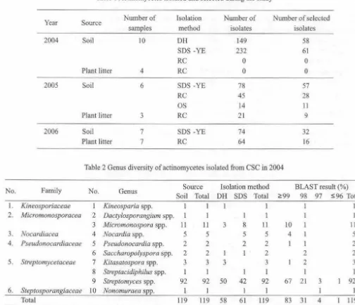
describe the genus diversity of actinomycetes in CSC during the study. Identification of Actinomycetes. We identified 119 isolates of actinomycetes and they belong to 10 genera and 6 families in 2004 (Table 2), there are more genera and families in 2005 (Table 3) and less genera and families in 2006 (Table 4). A total of 263 isolates represented of 10 genera and 10 families of actinomycetes were identified from this study (Table 5).Phylogenetic Tree of Interesting Isolates. Identification of the interesting isolates and their position in the phylogenetic tree showed that most of the isolates were separated from the known species of actinomycetes. Among the families of actinomycetes present in CSC, family
Kineosporiaceae
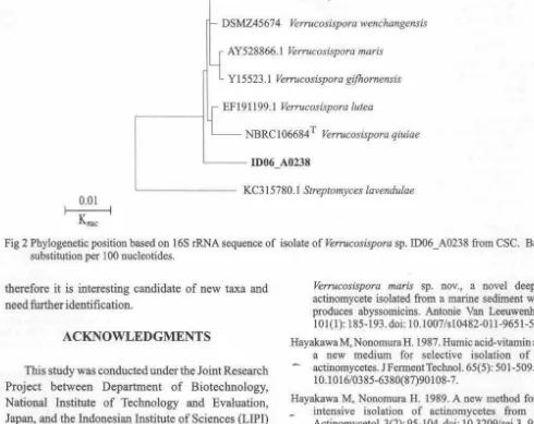
showed a cluster of interesting isolates as candidates of new genera (Fig I). We found a canditate of new taxa ofVerrucosispora
from CSC in 2005 (Fig 2).DISCUSSION
'
168 WIDYASTUTI ET Al. Microbiol lndones '
Table I Acrinomycctes isolated and selected during the study
Year Source Number of Isolation Number of Number of selected
samples method isolates isolates
2004 Soil 10 DH 149 58
SDS-YE 232 61
RC 0 0
Plant litter 4 RC 0 0
2005 Soil 6 SDS-YE 78 57
RC
45 28OS 14 11
Plant litter 3 RC 21 9
2006 Soil 7 SDS -YE 74 32
Plant litter 7 RC 64 16
Table 2 Genus diversity of actinomycetes isolated from CSC in 2004
No. Family No. Genus Source Isolation method BLAST result(%)
Soil
I. Kineosporiaceae Kineosparia spp. 2. Micromonosporacea 2 Dactylosporangium spp.
3 Micromonospora spp.
3. Nocardiacea 4 Nocardia spp. 4. Pseudonocardiaceae 5 Pseudonocardia spp.
6 Saccliaropolyspora spp. 5. Streptomycetaceae 7 Kitasatospora spp.
8 Streptacidiphi/11s spp. 9 Streptomyces spp. 6. Steptosporangiaceae 10 Nonomuraea spp.
Total
from other habitats or substrates such as marine sediments, mangrove mud, composted pig manure and animal dung (Kurtboke 2000). Based on the isolation method used, we collected a huge number of
Streptomyces from DH and SDS-YE methods. Actinomycetes are known to be heat resistant and able to stay in soil for a long period in dry soil. Heating of the soil at about 120 °C showed that Streptomyces are the most heat resistant actinomycetes. SDS-YE method based on SDS-YE solution containing 0.05% of SDS and 6% of yeast extract
in
50
mM P-buffer and RC method which was developed for isolation of motile actinomycetes (Hayakawa et al. 2000) showed that those procedure is suitable for isolation ofStreptomyces. Motile actinomycetes usually categorized as
rare
or non Streptomyces. However, we found many isolates of Streptomyces in the medium. tィセケ@ showed various morphological characteristics among others and interesting to be selected. Using OSl
11 5 2 2
3
I 92
l
119
Total DH SDS Total <?:99 98 97 S96 Total
l l l 1
I I
11 3 8 11 10 11
5 5 5 4 5
2 2 2 l 2
2 1 2 2 2
3 3 3 2 3
I l I
92 50 42 92 67 21 3 92
I 1
119 58 61 119 83 31 4 119
method we selected 11 isolates dominated by isolates from Streptomyces, followed by Mycobacterium and
Actinomadura genera. Naturally Streptomyces present abundantly in soil and can be isolated easily using all isolated methods used in this study. There are totally
93
from 186 isolates of selected Streptomyces and showed 99% or more BLAST similarity to the recognized species, this means the rest or a half number of the selected Streptomyces are not described yet. Under the
family Streptomycetaceae we also selected
3
isolates of genera Kitasatospora and 1 isolate of generaStreptacidiphilus, with 98% BLAST similarity to the recognized species.
[image:8.624.55.559.72.506.2]f'
Volume 6, 2012 Microbiol Indones 169
Table 3 Genus diversity of actinomycetes isolated from CSC in 2005
No Family No Genus Source Isolation method BLAST result (%)
Soil Litter Total SDS RC OSS Total 2!99 98 97 Total
Kineosporiaceae l Cryptosporangium spp. I 1
2 Kineosporia spp. 1 6 7 7 7 3 3 7 2 Micromonosporacea 3 Actinop/anes spp. 7 8 8 8 1 3 4 8
4 Dacty/osporangium spp. 4 4 2 2 4 4 4 5 Micromonospora spp. 6 6 3 3 6 4 2 6 3 Mycobacteriaceae 6 Mycobacterium spp. 3 3 3 3 3 3 4 Nocardiacea 7 Nocardia spp. 4 4 3 4 3 I 4
5 Nocardioidaceae 8 Kribbella 2 2 2 2 2 2 6 Pseudonocardiaceae 9 Amycolatopsis spp. l 1 l 1 1 7 Streptomycetaceae 10 Streptomyces spp. 60 61 41 13 7 61 19 31 11 61 8 Streptosporangiaceae 11 Microbispora spp. 1
12 Microtetraspora spp. 1 1 1 1
13 Nonomuraea spp. 2 2 2 2 2
9 Thermomonosporaceae 14 Actinomadura spp. 1 1 1
Total 94 8 102 55 36 11 102 41 44 17 102
Table 4 Genus diversity of actinomycetes isolated from CSC in 2006
No Family No Genus Source Isolation method BLAST result(%)
Soil 1 Micromonosporacea Actinoplanes spp. 2
2 Verrucosispora spp.
2 Nocardiacea 3 Nocardia spp. 1
4 Rhodococcus spp.
3 Nocardiopsaceae 5 Nocardiopsis spp.
4 Streptomycetaceae 6 Kitasatospora spp. 1
7 Streptomyces spp. 33
8 Planotetraspora spp.
5 Streptosporangiaceae 9 Streptosporangium spp. 1
Total 42
Nocardiopsaceae,
andThermomonosporaceae
eachappeared only one time during the course of the study. Good success of isolation is depend on variety of strategies to isolate rare and new taxa of actinomycetes. Four tsolation methods used in this study have different isolates target.
Isolates belong to the genera
Kineosporia
familyKineosporiaceae
are isolated from plant litter. It seemsthat their habitat is that kind of substrates and RC isolation method fit to get isolates of
Kineosporia.
Novel speciesKineosporia mesophila
was isolated from surface-sterilized stems of a ーィ。ョョ。」・オエゥ」セャ@ plantin
China (Liet al.
2009)andKineosporia babensis
wasSDS RC Total ;?! 99 98 97 S96 Total
2 2 1 2
1 I
1 1
I 1 1
1 1
23 10 33 7 13 9 4
33
1 1
1 1
28 14 42 12 16 9 5 42
isolated from plant litter in Vietnam (Sakiyama
et al.
2009). The position of isolates of the familyKineosporiaceae
selected in this study represented aline of descent distinct from previously described species of this family and further information are needed to describe the isolates (Fig 1 ).
There was only 1 species,
Verrucosispora
,
gifhornensis
under the genus Verrucosispora reportedpreviously (Rheims
et
al. 1998), however 5 other species were reported recently {Liao et al. 2009; Daiet
al.
2010; Goodfellowet al.
2012; Xiet al.
2012; Xie et170 WlDYASTIJTl ET AL. Microbiol Indones No 1. 2. 3.
4.
5. 6. 7. 89.
10. [image:10.621.73.533.86.769.2]I 0.01 I
Knuc
Table 5 Genus diversity of actinomycetes from CSC in 2004-2006
Family No
Kineo sporiaceae I
2
Micromonosporacea 3 4
5
6
Mycobacteriaceae 7
Nocardiacea 8 9
Nocardioidaceae IO
Nocardiospase 11
PseudonoCflrdiaceae 12
13
14
Streptomycetaceae 15 16 17 18
Streptosporangiaceae 19 20 21 22
Thermomonosporaceae 23
Total
Genus BLAST results(%)
セYY@
Kineosporia spp. 3
Cryptosporangium spp.
Dactylosporangium spp. 4
Micromonospora spp. 14
Actinoplanes spp. 2
Verrucosispora spp.
Mycobacterium spp. 3
Nocardia spp. 8
Rhodococcus spp.
Kribbella spp.
Nocardiopsis spp.
Pseudonocardia spp.
Saccharopolyspora spp.
Amycolatopsis spp.
Kitasatospora spp.
Streptacidiphilus spp.
Streptomyces spp. 93
Planotetraspora spp.
Nonomuraea Microbispora spp.
Microtetraspora spp.
Streptosporangium spp.
Actinomadura spp.
136
ID04-0565 - - ID05-A0745
ID05_A0902
98 97
4
1
3
4
42 2 l 2 3 I
65 23
91 30
S96
5
6
' - - - - AB003933 Kine,osporia rhizophila
086937 Kineosporia aurantiaca
A.9003931 Kineosporia aurantiaca
X871l0 Kineosporia aurantiaca
AF095336 Kineosporia aurantiaca
' - - - - ID05-A0908
'----lID05-A0909
ID05-A0907
セaPYPS@
ID05-A0443
AB003932 Kineosporia succinea
.__---i AB003935 Kineosporia rhamnosa
AB003934 Kineosporia rhamnosa
AF247813 Kineosporia radiotolerans
Tot.al 8 5 17 10 I 3 10 l 2 l 2 2 1 4 186 I 3 263
' - - - AB007420. l Kineosporia aurantiaci/.s
- - - - -- -- - - X92618 Kineosporia mikuniensis
085465 Cryptosporangium arvum
mos A01ss
1 セ MM M MMM PXUTVV@ Cryptosporangium japonicum
AB006168.l Cryptosporangium sp.
AB047490 Cryptosporangium aura
AB037007 Cryptosporangium minu
[image:10.621.48.543.108.775.2]'--- - - -085116 Streptomyces l<lllendulae
'
•
Volume 6, 2012 Microbiol lndones 171
- EU870859 .1 Verrucosispora sediminis
'-- DSMZ45674 Verrucosispora wenchangensis
[_J
AY528866. l Verrucosispora marisL
Y15523.1 Verrucosispora gifhornensisL
F 191199 .1 Verrucosispora luteaNBRCl 06684 T Verrucosispora qiuiae
,__ _ _ ID06 A0238
O.Ql L--- - - - KC315780.1 Streptomyces lavendulae
I I
[image:11.614.49.539.81.470.2]Knuc
Fig 2 Phylogenetic position based on 16$ rRNA sequence of isolate of Verrucosispora sp. ID06_A0238 from CSC. Bar, 1 substitUtion per 100 nucleotides.
therefore it is interesting candidate of new taxa and need further identification.
ACKNOWLEDGMENTS
This study was conducted under the Joint Research Project between Department of Biotechnology, National Institute of Technology and Evaluation, Japan, and the Indonesian Institute of Sciences (UPI) representing Indonesian Government Research Institutes. The authors would like to thank セッ「オケオォゥ@
Goto for providing the 16S rRNA gene sequences.
REFERENCES
Altschul SF, Gish W, Miller W, Myers EW, Lipman DJ. ' 1990. Basic local alignment search tool. J Mol Biol.
215(3):403-410.
Dai H-Q,. Wang J, Xin Y-H, Gang Pei.Shu-Kun Tang, Biao Ren, Alan Ward, Ji-Sheng Ruan, Wen-Jun Li and Li-Xin Zhang. 2010.Verrucosispora sediminis sp. nov., a cyclodipeptide-producing actinomycete from deep-sea sediment. Int J Syst Evol Microbiol. 60(8): 1807-1812. doi: 10.1099/ijs.0.017053-0.
Felsenstein J. 1981. Evolutionary trees from DNA _ sequences: a maximum likelihood approach. J Mo!
Evol.17(6): 368-376.doi: 10.1007/BF01734359.
Felsenstein J. 1985. Confidence limits on phylogenies: an approach using the bootstrap. Evolution 39(4): 783-- 791.doi: 10.2307/2408678.
Verrucosispora maris sp. nov., a novel deep-sea actinomycete isolated from a marine sediment which produces abyssomicins. Antonie Van Leeu_wenhoek,
101(1): 185-193.doi: l0.1007/sl0482-0ll-9651-5.
HayakawaM, Nonomura H. 1987. Burnie acid-vitamin agar,
a
new medium for selective isolation of soil - actinomycetes. JFermentTechnol. 65(5): 501.509. doi:10.1016/03 85-6380(87)90108-7.
Hayakawa M, Nonoroura H. 1989. A new method for the _ intensive isolation of actinomycetes from soil.
Actinomycetol. 3(2): 95-104. doi: 10.3209/saj.3_95.
Hayakawa M, Otoguro M, Takeuchi T, Yamazaki T, Iimura Y. 2000. Application of a method incorporating differential centrifugation for selective isolation of motile actinomycetes in soil and plant litter. Antonie van Leeuwenhoek 78(2): 171-185. doi:l0.1023/A:10265 79426265.
Ishigarni M, Nakagawa Y, Hayakawa M, limura Y. 2004. · FLO 11 is essential for flor formation caused by the
C-terminal deletion of NRG I in Saccharomyces
cerevisiae. FEMS Microbiol Lett. 237(2): 425-430. Jiang Y, Wiese J, Tang S-K, Xu L-H, Imhoff J F, Jiang C-L.
2008. Actinomycetospora chiangmaiensis gen nov., sp. nov, a new member of the family Pseudonocardiaceae.
Int J Syst Evol Microbiol. 58(2):408-413. doi: 10.1099/ijs.0.64976-0.
Kumar S, Tamura K, Nei M. 2004. MEGA3: integrated software for molecular evolutionary genetics analyses and sequence alignment. Brief Bioinform. 5(2): l 50-163. doi: l 0.1093/bib/5.2.150.
Kurtboke DI. 2000. Australian Actinomycetes: An
unexhausted source for biotechnological applications. Actinomycetol. 14(2): 43-53. doi: 10.3209/saj.14_ 43. Fitch WM. 1971. Toward defining the course of evolution:
minimum change for a specific tree topology. Syst - - Zool.20:406-416.doi: 10.2307/2412116.
Li W-J, Wang D, Zhang Y-Q, Xu L-H, Jiang C-L. 2006.
Kribbella yunnanensis sp. nov., Kribbella alba sp. nov., two novel species of genus Kribbel/a isolated from soils in Yunnan, China. Syst Appl Microbiol. 29(1): 29-35. doi: I0.1016/j.syapm.2005.06.005.
172 WIDYASTUTI ET AL
Li J, Zhao g-Z, Huang H-Y, Qin S, Zhu W-Y, Xu L-H, Li W-J. 2009. Kineosporia mesophila sp. nov., isolated from surface-sterilized stems of Tripterygium wilfordii. Int J Syst Evol Microbiol. 59(12):3150-3154. doi: 10. L099/ijs.0.012021-0.
Liao ZL, Tang SK, Guo L, Zhang YQ, Tian XP, Jiang CL, Xu LH, Li WJ.2009.Verrucosispora lutea sp. nov., isolated from a mangrove sediment sample. Int J Syst Evol Microbiol. 59(9): 2269-2273. doi: I 0.1099/ijs.0.008813-0.
Lisdiyanti P, Otoguro M, Ratnakomala S, Lestari Y, Hastuti RD, Triana E, Ando K, Widyastuti Y. 2010. Actinokineospora baliensis sp. nov., Actinokineospora cibodasensis sp. nov., Actinokineospora cianjurensis sp. nov., isolated from soil and plant litter. Int J Syst Evol Microbial. 60(1 ): 2331-2335. doi: I 0. 1099/ijs.0.013276-0.
Nonomura H, Ohara Y. 1969. Distribution of actinomycetes in soil. VIl. A culture method effective for both preferential isolation and enumeration ofMicrobispora
and Streptosporangium strains in soil. (Part 1) Classification of the isolates. J Fennent Technol. 47: 463-469.
Otoguro M, Ratnakomala S, Lestari Y, Hastuti RD, Triana E, Widyastuti Y, Ando K. 2009. Streptomyces baliensis sp. nov., isolated from Balinese soil. Int J Syst Evol Microbiol. 59(2): 2158-2161. doi: 10.l 099/ijs.0.007179--0.
Otoguro M, Yamamura H, Tamura T, lrzaldi R, Ratnakomala S, Ridwan R, Kartina G, Triana E, Nurkanto A, Lestari Y, Lisdiymiti P, Widyastuti Y, Katsuhiko A. 2011.
Actinophytocola timorensis sp. nov. and
Actinophytoco/a coralina sp. oov., isolated from soil. Intl J Syst Evol Microbiol. 61(4): 834-838. doi:
I 0.1099/ijs.0.023432-0.
Saitou N, Nei M. 1987. The neighbor-joining method: a new - method for reconstructing phylogenetic trees. Mol Biol
.
'"
Microbiol lndones
Evol. 4(4): 406-425.
Sakiyama Y, Thao NKN, Giang NM, Miyadoh S, Hop DV, Ando K. 2009. Kineosporia babensis sp. nov., isolated from plant litter in Vietnam. Int J Syst Evol Microbiol. 59 (3): 550-554. doi: I 0.1099/ijs.0.002907-0.
Rheims H, Schumann P, Rohde M, Stackebrandt E. 1998.
Venucosispora gijhomensis gen. nov., sp. nov., a new
- member of the actinobacterial fumi1y Micromonosporaceae.
IntJ Syst Evol Microbiol. 48: 1119-1127.
Tamura T, Sakane T. 2005. Asanoa iriomote11Sis sp. nov., isolated from mangrove soil. Int J Syst Evol Microbiol. 55(2): 725-727. doi: 10.1099/ijs.0.02982-0.
Tamura T, Hatano K, Suzuki K. 2006. A new genus of the family Micrornonosporaceae, Po/ymorphospora gen. nov., with description of Polymorphospora rubra sp. nov. Int J Syst Evol Microbiol. 56(8): 1959-1964. doi: 10.1099/ijs.0.64046-0.
Xie QY, Lin HP, Li L, Brown R, Goodfellow M, Deng Z, Hong K.2012. Verrucosispora wenchangensis sp. nov., isolated from mangrove soil. Antonie van Leeuwenhoek 102(1):1 -7. doi: 10.1007/s10482-012-9707-l.
Xi L, Zhang L, Ruan J, Huang Y. 2012. Description of
Verrucosispora qiuiae sp. nov., isolated from mangrove swamp sediment, and emended description of the genus Verrucosispora. Int J Syst Evol Microbiol. 62(7): 1564-1569. doi: I0.1099/ijs.0.033787-0.
Yamamura H, Lisdiyanti P, Ridwan R, Ratnakomala S, Saraswati R, Lestari Y, Triana E, Kartina G, Widyastuti Y, Ando K. 2010. Dietzia timorensis sp. nov., isolated from soil. Int J Syst Evol Microbiol. 60(2): 451-454. doi: 10.1099/ijs.0.012229-0.
11