Effect of clove cigarette exposure on white rat : special emphasis on the
histopathology of respiratory tract
Eddy Widodo1, Bambang Pontjo Priosoeryanto1, Sri Estuningsih1, Dewi Ratih Agungpriyono1, Robert Utji2
Abstrak
Asap rokok telah terbukti dapat menyebabkan berbagai gangguan pada saluran napas. Rokok kretek lebih berbahaya daripada rokok putih, karena kandungan tar, nikotin, dan karbon monoksida di dalamnya lebih tinggi. Konsumsi rokok kretek di Indonesia mencapai 88%. Efek rokok kretek terhadap saluran napas belum pernah diteliti. Tujuan penelitian ini adalah untuk mempelajari perubahan histopatologi saluran napas tikus putih galur Sprague-Dawley akibat pajanan asap rokok kretek. Penelitian dilakukan pada 20 tikus putih mulai September 2005 hingga Mei 2006. Nekropsi dilakukan setelah hari terakhir paparan asap rokok, kemudian dibuat irisan jaringan dan diproses untuk sediaan histopatologi dan diperiksa dengan mikroskop cahaya dan videomikrometer. Parameter yang diamati adalah jumlah dan tinggi epitel bersilia dan sel goblet, jumlah sel pneumosit tipe I dan II, makrofag, dan reaksi interstisial jaringan paru. Reaksi interstisial jaringan paru diamati dengan irisan semi-thin pada paru yang setelah dilakukan embedding dengan resin, dan diwarnai dengan Toluidine Blue. Hasil penelitian kami menunjukkan perubahan histopatologi yang bermakna pada saluran napas. Jumlah sel epitel pada kelompok yang terpapar asap rokok secara bermakna lebih tinggi dari kontrol (P<0.05) pada daerah sinus, bronkhus, dan bronkhiolus, sedangkan pada trakhea tidak ditemukan perbedaan bermakna (P>0.05). Jumlah sel goblet pada kelompok perlakuan juga lebih tinggi (P>0.05). Tinggi sel epitel pada kelompok perlakuan asap lebih tinggi dibandingkan kelompok kontrol, namun tidak ada perbedaan secara statistik antara tikus jantan dan betina. Terdapat perbedaan bermakna skor pneumonia interstisial (P<0.05) antara kedua kelompok. Jumlah sel pneumosit tipe II lebih banyak dibandingkan dengan sel pneumosit tipe I pada kelompok perlakuan. Berdasarkan hal-hal di atas, kami menyimpulkan bahwa paparan rokok kretek menyebabkan kelainan patologis pada saluran napas tikus. (Med J Indones 2007; 16:212-8)
Abstract
Cigarette smoke is proved to cause various disturbances on respiratory tract. Clove cigarette is far more dangerous than common (“white”) cigarette, since the tar, nicotine, and carbon monoxyde content is significantly higher. In Indonesia, 88% smokers consume clove cigarette. The clove cigarette effect to the respiratory tract have never been studied. Aim of this research is to study histopathological changes of respiratory tract in Sprague-Dawley white rats after smoke cigarette exposure. The study was performed using 20 white rats starting September 2005 until May 2006. Necropsy was done after final day of smoke exposure, then histopathological slides of the respiratory tract were processed and stained under light microscope and videomicrometer. Observed parameters were height and number of ciliated epithelia and goblet cells, also number of pneumocytes types I, II, and macrophages, and interstitial lung tissue reactions. The latest parameters were observed with semi-thin sections of resin embedded lung stained with Toluidine Blue. Result showed considerable histopathological changes on respiratory tract. The amount of epithelial cells on the group exposed to clove cigarette smoke were significantly higher than control group (P<0.05) on sinus, bronchi, and bronchioli area, while no significant difference were found on trachea (P>0.05). Number of goblet cells in exposed group was also higher (P>0.05). The epithelial height in exposed group was higher compared to control, but no statistical differences were found between male and female rats. The interstitial pneumonia score was statistically different (P<0.05) between the two groups. The amount of pneumocytes type II was higher than types I within the exposure group. Based on all mentioned above, we suggest that clove cigarette smoke exposure causes pathological disorders in rat respiratory tract. (Med J Indones 2007; 16:212-8)
Keywords: clove cigarette, respiratory tract histopathology, respiratory tract toxicopathology.
The World Health Organization in 2002 said that Indonesia was in the fifth rank for world cigarette consumption.1 From this number, based on Gabungan Perserikatan Pabrik Rokok Indonesia (Gappri) study in 1999 on national cigarette consumption projection, 1
Veterinary Science Study Program, Bogor Institute of Agriculture Postgraduate School, Bogor, Indonesia 2
clove cigarette own the market with ratio 88%:12% to white cigarette.
Clove cigarette is more dangerous than common cigarette, since the amount of tar, nicotine, and carbon monoxide in clove cigarette is far more dangerous than white cigarette. The main difference between clove and white cigarette is in the content. Clove cigarette was made from clove as the main content, which is a mixture between tobacco with 30-40% dry clove flowers. Clove as a component in clove cigarette has an eugenol active component, 82-87%.2 Clove cigarette has 2-3 times nicotine and tar component greater than common cigarette. Each cigarette contains 34–65 mg tar, 1.9-2.6 mg nicotine, and 18–28 mg carbonmonoxide.3 Clove cigarette contains some reactive chemical molecules including reactive oxygen and free radicals.4
Cigarette smoke in the environment consists of main-stream smoke and sidemain-stream smoke. Mainmain-stream smoke has been defined as a smoke that being inhaled and exhaled by smokers, while sidestream smoke is a smoke that was produced from the cigarette between two puffs.5 Nicotine inhaled from mainstream smoke is 4-6 times greater than sidestream.6
Nicotine, which occurs in cigarette smoke, brings out many symptoms.7 In mouse, Nicotine will be metabolized by P450 cytochrom to become Conitine. The peak concentration of Conitine will be achived in 5-10 minutes, with half time period of 20-25 minutes, then it will be eliminated through the kidneys. The con-centration of Conitine metabolite will be kept high in blood circulation, and accumulated in organs for some time. The particles of cigarette smoke is very small (0,1-1µm) compared to other inhalants. That is why cigarette smoke particles is not well filtered, and is able to enter the respiratory system. 3
There are about 4000 chains in cigarette smoke, most of them are toxic, mutagenic, even carcinogenic. Therefore cigarette smoke has been widely claimed as a major risk factor in many human diseases.8 Morphometry changes observed might have been caused by active metabolites, free radicals, heavy irons, and various tobacco smoke components such as carbon monoxide and nicotine, which will bold the tissue ischemic effects. Ischemic which followed by tissue hipoxia is a pathologic process that play an important role in provoking tissue damages and deaths. Carbon monoxide reduces oxygen transfers, resulting in hypoxia.
Many researches about the effect of white cigarette in conducting anatomic changes of respiratory system has been done. A research about histopathologic changes and morfometric of lungs, placenta, and liver by Czekaj et al (2002) studied histopathological and morphometric changes of lungs, placenta, liver, and kidney of adult rats exposed to cigarette smoke.9 It was found that changes in dosage and time of exposure of cigarette smoke results in variously dramatic histopathology changes of mouse’s lungs (emphysema, emphysematous, inflammation, and metaplastic changes), and also induced the decrements of respiratory bronchiolus epithelial cells. Comparison of lung response to toxic substances in several mouse species may lead to important beneficience in human.10
Discerning a large number of consumption, leastwise research, and debatable medical effects (changes in respiratory tract as a prior) about clove cigarette as the most consumptable cigarette in Indonesia, there are needs to do more study about clove cigarette. As far as we know, research about the effect of clove cigarette to respiratory tract in Indonesia has never been done before.
The aim of this research is to understand the histo-pathological changes from the exposure of clove cigarette on respiratory tract of Sprague-Dawley white rat.
METHODS
Time and Place of The Study
The research took place in Pathology division, Department of Clinical Reproduction and Pathology, Faculty of Veterinary, Bogor Institute of Agricultures, for eight months, counting from September 2005 until May 2006.
Study Materials
Experimental animals
This research was experimental, using sample animals as a model. The animal model being used was 20 Rattus rattus white mouse strain Sprague-Dawley, 10 males and 10 females; averaging 150 grams of weight.
Cigarette
Tools
Smoking chamber (40x30x20 cm3); air pump; conditioning chamber; smoking machine; binocular microscope; video micrometer; diamond knife; microtome and ultramicrotome; dissecting kit; automatic tissue processor; paraffin embedding console; tissue staining; animal cage with all its equipments
Methods
Woof Preparation and Mouse Adaptation
Before research began, all mouse were adapted for two weeks. Woof was made in shape of a ball with composition of: 73.943% Corn, 14.505% Bungkil, 6.8% Dedak, 1.5% Chalk, 1.263% Bone powder, 1% Oil, 0.362% Methionin, 0.31% Lycin, 0.213% Salt, 0,106% Vitamin and mineral mix. The woof was made in BPT Ciawi. Woof and water was given ad libitum in trial period.
Clove cigarette smoke treatment in mouse
The smoke given was from 8 cigarettes a day, 8-10 minutes duration for 5 days a week, for 6 weeks. The exposures were done in a special chamber. The mouse was gotten down through a hole in upper part of the smoking chamber, meanwhile clove cigarettes was installed in a pipe which had been connected with the air pump.
Weight measurement
The weight of the mouse was measured at the beginning of the research and regularly once a week until it ended.
Euthanasia
Euthanasia began with ether anestethic given perinhalation in closed glass container. The Euthanasia continued with injection of Ketalar® 10% in 0.9% sodium chloride intramuscularly (in Semi Tendinous muscle), with dosage of 0,1 cc / 100 grams body weight, using 1 cc disposable syringe.
Blood sample collection
Blood sample was taken regularly as much as 2 cc intracardial, to be haematologically checked.
Necropsy and tissue sampling
The necropsy was done by opening the skin, fascia, abdominal cavity, and thoracic cavity to obtain lower respiratory tract and lungs portion. Upper respiratory
tract portion located inside the skull was obtained by sawing longitudinally. Respiratory organ, be in the form of nasal sinus, trachea, bronchus, bronchiolus, and lungs, was taken and kept in 10% Neutral Formalin buffer liquid. Some pieces of lungs were kept in 4% Glutaraldehyde fixation liquid.
Construction of histopathological slides
The tissues were processed using automatic tissue processor until embedded in paraffin blocks. The block, being filled by tissue, was cut into 4µm wide microtome. Special treat was done for nasal sinus, which is located inside the skull, by decalcificating the bone using 10% Formic Acid until the it became tender, and then proceeded with other tissues. Stains used were Alcian Blue Periodic Acid Shiff (AB-PAS) and Hematoxylin-Eosin (HE). Few pieces of lung tissue were processes until moulded in Resin for further cut, using ultra-microtome with a 200 nm diamond knife, then stained with Toluidine Blue.
Histopathological Observation
Histopathological observation was made using microscope and micrometer video. Observation was made to: (1) Calculation of amount and height of ciliated epithelial cells, and ratio of ciliated epithelial cell to goblet cells. Observation was made to 10 (n) viewfinders, each to mucosa as long as 1000µm from nasal sinus, trachea, bronchus, and bronchiolus tissues; (2) Reaction valuation of pulmonary interstitium in 10 (n) viewfinders, each as wide as 1.6x1.3µm2. Tissue response analysis was made qualitatively to be scored afterwards due to changes, so that be possible to make qualitative statistic analysis. Score of 1,2,3,4 would be given if in interstitial pneumonia was found as much as 25%, 50%, 75%, and 100% of lung tissue; (3) Calculation count of alveolar macrophage cell and type I and II pneumocyte in 10 viewfinders far-ranging 1.6x1.3 µm2.
Statistical Analysis
Anova statistical test was used in statistical calculation of the height of epithelial cells, number of epithelial cells, and number of goblet cells in. Kruskal-Wallis non-parametric test was used in the calculation of interstitial pneumonia.
RESULTS
histopatho-logical examinations had been done using light microscope and micrometer video. Parameters including amount and height of epithelial cells, amount of goblet cells, pneumocyte type I and II, alveolar macrophage, and lung interstitial tissue reaction.
Result of nasal sinus histopathology is presented in Table 1. From Table 1 we can see the amount of epithelial cells in the group that was given smoke treatment was higher than control, especially in males. (p<0.05). Meanwhile sex did not show significant differences (P>0.05), although there was an increasing tendency in all groups and both sexes. Treated groups tend to have taller epithelial cells compared to control groups, there was especially a significant difference in female (P>0.05). There were no difference between sexes (P>0.05). There were no difference in ratio between epithelial cell and goblet cell, for both male and female groups.
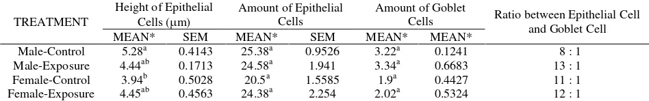
Result of trachea histopathology is presented in Table 2. The ciliated epithelial cell of the trachea in female rats was higher in exposed group compared to control, but it did not show significant difference in male rats. Numerical increase in goblet cells was also found in both male and female rats. Male control groups show higher epithelial to goblet cell ratio (8:1) compared to female control groups (11:1).
Table 3 shows result of histopathologic examination on bronchus. Similar to trachea, the ciliated epithelial cell in female rats was higher in exposed group compared to control, but it did not show significant difference in male rats. Numerical increase in goblet cells was also found in both male and female rats. Exposed male control group show slight decrease in epithelial cell to goblet cell ratio (4:1) compared to the control group (5:1).
Table 1. Average result of height measurements, epithelial and goblet cell counts, and its ratio to nasal sinus per 1000 µm mucous length
TREATMENT
Height of Epithelial
Cells (m) Amount of Epithelial Cells
Amount of Goblet
Cells Ratio between Epithelial Cell and Goblet Cell MEAN* SEM MEAN* SEM MEAN* SEM
Male-Control 3.62ab 0.4131 11.38b 0.3839 4.68a 0.7473 2 : 1 Male-Exposure 4.35a 0.1405 13.08a 0.3308 5.88a 0.3121 2 : 1 Female-Control 2.99b 0.1155 11.6ab 0.5874 3.74a 0.8727 3 : 1 Female-Exposure 3.96a 0.2619 12.92a 0.5928 4.52a 0.731 3: 1
* Different superscript shows significant difference for p<0.05
Table 2. Result of Average Epithelial height, Epithelial and Goblet cell amount, and its ratio in trachea per 1000 µm mucous length.
TREATMENT
Height of Epithelial
Cells (m) Amount of Epithelial Cells
Amount of Goblet
Cells Ratio between Epithelial Cell and Goblet Cell MEAN* SEM MEAN* SEM MEAN* MEAN*
Male-Control 5.28a 0.4143 25.38a 0.9526 3.22a 0.1241 8 : 1 Male-Exposure 4.44ab 0.1713 24.58a 1.941 3.34a 0.6683 13 : 1 Female-Control 3.94b 0.5028 20.5a 1.5585 1.9a 0.4427 11 : 1 Female-Exposure 4.45ab 0.4563 24.38a 2.254 2.02a 0.5324 12 : 1
* Different superscript shows significant difference for p<0.05
Table 3. Result of Average Epithelial height, Epithelial and Goblet cell amount, and its ratio in bronchus per 1000 µm mucous length.
TREATMENT
Height of Epithelial
Cells (m) Amount of Epithelial Cells
Amount of Goblet
Cells Ratio between Epithelial Cell and Goblet Cell MEAN* SEM MEAN* SEM MEAN* SEM
Male-Control 4.96a 0.6196 18.98bc 0.5739 3.6a 0.8385 5 : 1 Male-Exposure 4.52a 0.2723 21.2ab 1.187 5.2a 0.5119 4 : 1 Female-Control 4.29a 0.5335 18.08c 1.0495 3.58a 0.6127 5 : 1 Female-Exposure 5.03a 0.6691 22.18a 1.0428 4.42a 0.5987 5 : 1
Table 4 shows bronchiolus epithelial cell in male and female treated group undergo hyperplasia and hyper-trophy compared to the control group. Epithelial and Goblet cell ratio is also increased, even though it is statistically insignificant. There is no tissue cellular reaction difference between male and female treated groups.
From table 5 we see significant difference between male and female control and treated groups in increase
of macrophage, decreased pneumocyte type I, and increased pneumocyte type II amount (P<0.05).
Result for lung interstitial histopathology could be found in Table 6. The reaction found was thickening of interstitial tissue by lymphocytic inflammation cell. These conditions is known as interstitial pneumonia.
Table 4. Results of Average Epithelial height, Epithelial and Goblet cell amount, and its ratio in bronchiolus per 1000µm mucous length.
TREATMENT
Height of Epithelial
Cells (m) Amount of Epithelial Cells
Amount of Goblet
Cells Ratio between Epithelial Cell and Goblet Cell MEAN* SEM MEAN* SEM MEAN* SEM
Male-Control 3.02ab 0.5789 19.26ab 1.5449 3.22a 0.1241 6 : 1 Male-Exposure 4.55a 0.4704 22.7a 1.9026 3.34a 0.6683 8 : 1 Female-Control 2.54b 0.4311 16.36b 0.8635 1.9a 0.4427 7 : 1 Female-Exposure 3.3ab 0.5692 20.46ab 1.6148 2.02a 0.5324 10 : 1
* Different superscript shows significant difference for p<0.05
Table 5. Results of Average Alveolar Macrophage, and pneumocyte type I and II amount in 10 viewfinders far-ranging 1,6x1,3 µm2.
Treatment Amount of macrophage Type I Type 2
MEAN SEM MEAN SEM MEAN SEM
Male-Control 2.56b
0.10 6.69a 0.88 6.26b 0.66
Male-Exposure 4.38a 0.18 3.02c 0.23 11.39a 0.67
Female-Control 2.44b
0.05 5.01b 0.36 7.63b 1.31
Female-Exposure 3.99a
0.25 3.09c 0.17 12.43a 0.67
* Different superscript shows significant difference for p<0.05
Table 6. Results of Average lung interstitial tissue reaction scoring in 10 viewfinders far-ranging 1,6x1,3 µm2
Treatment Mean rank*
Male-Control 5.6ª
Male-Exposure 14.5b
Female-Control 6.9ª
Female-Exposure 15.0b
* Different superscript shows significant difference for p<0.05
DISCUSSION
From the study result we observed changes in respiratory tract due to cigarette smoke exposure. In the nasal sinus (Table 1) we can see the amount of epithelial cells in the group that was given smoke treatment was higher than control, especially in males. Treated groups tend to have taller epithelial cell compared to control groups, which was especially significant in females. The increase in cell amount is termed as
goblet cell increased numerically. Goblet cell reaction draws special attention because these cells can bring forth respiratory tract obstruction by provoking lumen occlusion through mucous plugs formation in periphery respiratory tract, and the mucous production can disrupt surface tension in respiratory tract, therefore causing instability in the periphery respiratory tract and the tendency to collapse.12
Ciliated trachea epithelial cell in treated female group underwent hyperplasia and hypertrophy compared to control, while in treated male group the inversion was found, although they were statistically insignificant (Table 2). Ciliated epithelial cell hyperplasia and hypertrophy in treated female group was provoked by active substances found in cigarette smoke. In treated male group, there was epithelial cell hypoplasia. Hypoplasia occurred in consequence of several process that affect cell genome, e.c degeneration process that makes a deceleration in epithelial cell fission, or decrease in epithelial cell because of cell death.9 Male control group showed higher epithelial to goblet cell ratio (8:1) than female control group (11:1). Exposure of cigarette smoke in male and female groups brought up different response. Although both groups showed a decrease in epithelial to goblet cell ratio. Female group underwent an increase compared to control with value of 12:1, while male group also underwent an increase with a value of 13:1.
Significant difference was found in amount of bronchus epithelial cell between treated male group and its control, and between treated female group with its control. Nevertheless, there is no significant differences between male and female treated group. Female mouse might have slower response than male. Generally, hipertrophy occurred at the first place as a result of irritants, then abridgement or atrophy occurred subsequently. Increased epithelial to goblet cell ratio found the in the male exposed group, were interpreted as metaplastic tendency of cilliated epithelial into Goblet cell. Metaplasia is a mutation of tissue into other kind of tissue with different function. Metaplasia was measured from the increased ratio of epithelial cell to goblet cell.13 This changes was in accordance with previous researches using white cigarette (Saetta et al, 2000), which discover chronic bronchitis and ventilation obstruction in consequence of significant increase in goblet cell in respiratory tract.12
Bronchiolus epithelial cell in male and female treated group underwent hyperplasia and hypertrophy, in comparison with the control group. Epithelial to goblet
cell ratio was also increased, although statistically insignificant. There were no tissue cellular reaction difference between male and female treated groups. Goblet cell hyperplasia in bronchiolus can be fatal, because it could easily clog the small bronchiolar lumen, just like in chronic obstructive pulmonary disease (COPD), as a consequence of goblet cell proliferation due to cigarette smoke.14
From the alveolar histopathology result we could see significant difference between male and female control and exposed groups in the increase of macrophage, decrease of type I pneumocyte, and increase of type II pneumocyte. The increase of alveolar macrophage in treated group was in accordance with previous researches where they found that chronic sputum production in smokers caused infiltration increase of neutrofil and macrophage.15-17
In treated group, decreased amount of pneumocyte type I is because the cell has a vast surface, absence of replication ability, and is highly sensitive to toxic or toksikan inhaled, including cigarette smoke. While increasesd amount of pneumocyte type II happened because the ability to replicate in alveol and will replace damaged or number of type I cell.10,18
The interstitial tissue was found to be thickened by the lymphocytic inflammatory cells. These condition is known as interstitial pneumonia. In control group, interstitial tissue reaction is generally found as a result of nonspesific contaminant exposure from unsterile environment. The Kruskal-Wallis test proved significant increase in male and female exposed groups. Thickening of lung interstitial tissue occurred due to proliferation of type II pneumocyte and macrophage, and infiltration of lymphocytic cell. This is also in accordance with previous researches.15,9 As a matter of fact, lung interstitial reaction is a common reaction found in research rat non-spf (specific pathogen free), therefore rarely found scoring value of 0 in control rat. However, the increase of interstitial pneumonia score in treated group indicates increased lung sensitivity. Similar changes was reported in an earlier research about cigarette smoke effects on guinea pig and white rat type SD.19
CONCLUSION
1. hyperplasia and hypertrophy of epithelial cells in the sinus, bronchus, and bronchiolus 2. increase of goblet cell number
3. significant increase on number of alveolar macrophages and type II pneumocyte, and 4. the type I pneumocyte is decreased significantly
REFERENCES
1. World Health Organization. 2000. Global Youth Tobacco Survey 2000. WHO Bulletin.78(7): 868–76.
2. Hanusz M. Kretek – The Culture and Heritage of Indonesia’s Clove Cigarettes. 1st Ed. Equinox Publishing (Asia) Pte. Ltd: Jakarta; 2000.
3. Hashim NH. Kesan Buruk Akibat Hisap Rokok Kretek. Pusat Racun Negara, USM Malaysia; 1996 [cited 2005 Jul 28]. Available from: http://www.prn2.usm.my/mainsite/ bulletin/racun/1996/ kretek.html
4. Church DF, Pryor W. The oxidative stress placed on the lung by cigarette smoke. In: Crystal RG, West JB, editors. The Lung. New York: Raven Press; 1991. p.1975–9. 5. Kritz H, Schmid P, Sinzinger H. Passive smoking and
cardiovascular risk. Arch Intern Med. 1995;155(18): 1942-8.
6. Susanna D. Penentuan Kadar Nikotin dalam Asap Rokok. Jurnal Ekologi Kesehatan. 2003;2(2).
7. American Thoracic Society. Cigarette Smoking and Health. American Journal Respir Crit Care Med. 1996;153:861-5. 8. Vanwye JE. Passive smoking. In: Hilman BC, editor.
Pediatric Respiratory Disease. 1st ed. Philadelphia: WB Saunders; 1993. p.794-806.
9. Czekaj P, Palasz A, Lebda-Wyborny T, Nowaczyk-Dura G, Karczewska W, Florek E, Kaminski M. Morphological changes in lungs, placenta, liver and kidneys of pregnant rats exposed to cigarette smoke. Int Arch Occup. Environ Health. 2002;75 Suppl: S27-35.
10. Herbert RA, Leininger JR. Nose, Larynx, and Trachea. In: Maronpot RR, Boorman GA, Gaul BW editors. Pathology of the Mouse Reference and Atlas. Illinois: Cache River Press; 1999.
11. Cheville N. Introduction to Veterinary Pathology. Second edition. Ames: Iowa State University Press; 1999. p.235-53. 12. Saetta M, Turato G, Baraldo S, Zanin A, Braccioni F, Mapp CE, et al. Goblet cell hyperplasia and epithelial inflammation in peripheral airways of smokers with both symptoms of chronic bronchitis and chronic airflow limitation. Am J Respir Crit Care Med. 2000;161:1016-21. 13. Verdugo P. Goblet cells secretion and mucogenesis. Ann
Rev Physiol 1998;52:157.
14. Maestrelli P, Saetta M, Mapp CE, Fabbri LM. Remodeling in response to infection and injury – airway inflammation and hypersecretion of mucus in smoking subjects with chronic obstructive pulmonary disease. Am J Respir Crit Care Med. 2001;164:S76-80.
15. Saetta M, Turato G, Facchini FM, Corbino L, Lucchini RE, Casoni G, et al. Inflammatory cells in the bronchial glands of smokers with chronic bronchitis. Am J Respir Crit Care Med. 1997;156:1633-9.
16. Amin K, Ekberg-Jansson A, Löfdahl C-G, Venge P. Relationship between inflammatory cells and structural changes in the lungs of asymptomatic and never smokers: a biopsy study. Thorax. 2003;58:135-42.
17. Woodruff PG, Koth LL, Yang YH, Rodriguez MW, Favoreto S, Dolganov GM, et al. A distinctive alveolar macrophage activation state induced by cigarette smoking. Am J Respir Crit Care Med. 2005;172:1383-92.
18. Bills RF, Christie BR. The experimental pathology of oxidant and air pollutant inhalation. Int Rev Exp Pathol. 1980;21:195-293