JOURNAL OFCLINICALMICROBIOLOGY, Sept. 1996, p. 2201–2204 Vol. 34, No. 9
0095-1137/96/$04.0010
Copyrightq1996, American Society for Microbiology
Identification and Molecular Characterization of Serological
Group C Streptococci Isolated from Diseased Pigs
and Monkeys in Indonesia
I. SOEDARMANTO,1F. H. PASARIBU,2
I. W. T. WIBAWAN,2
ANDC. LA¨ MMLER1*
Institut fu¨r Bakteriologie und Immunologie der Justus-Liebig-Universita¨t Gießen, Gießen, Germany,1
and Faculty of Veterinary Medicine, Bogor Agricultural University, Bogor, Indonesia2
Received 21 February 1996/Returned for modification 19 April 1996/Accepted 3 June 1996
The present study was designed to comparatively investigate 34 beta-hemolytic streptococci isolated from infected pigs and monkeys from various islands in Indonesia. According to the serological and biochemical data, all 34 isolates were Lancefield’s serological group C streptococci and could be identified asStreptococcus equisubsp.zooepidemicus. Of the 34 group C streptococci investigated, 28 grew on solid media in large, mucoid colonies, in fluid media at a uniform turbidity, and in soft agar in diffuse colonies. A decapsulation test with a hyaluronidase-producingStaphylococcus aureus strain revealed the hyaluronic acid nature of the capsular material. The remaining six streptococci grew on solid media in small, nonmucoid colonies, in fluid media as sediment with clear supernatant, and in soft agar in compact colonies. Determination of surface hydropho-bicity by salt aggregation revealed a hydrophilic surface for the encapsulated bacteria and a hydrophobic surface for the unencapsulated group C streptococci. To further analyze the epidemiological relationships, all 34 mucoid and nonmucoid isolates from pigs and monkeys were subjected to protein and DNA fingerprinting. The latter was performed by pulsed-field gel electrophoresis. The protein profiles of all 34 isolates and the DNA profiles of 32 isolates appeared to be identical, with the DNA profiles of 2 isolates being closely related, indicating that a single virulent clone is responsible for this disease outbreak in Indonesia.
At the beginning of 1994, a disease outbreak among pigs and monkeys was reported on the island of Bali, Indonesia. The diseased animals showed such clinical symptoms as painful swelling of the joints, respiratory disturbances, and diarrhea. Most of the animals died within a few days. The postmortem examinations of the pigs and monkeys revealed signs of pol-yarthritis, bronchopneumonia, pleuritis, epicarditis, endocardi-tis, and meningitis (6). Further bacteriological examinations resulted in the isolation of streptococci of serological group C. These bacteria seemed to be one of the major causative agents. These group C streptococci could be isolated from most of the pigs and monkeys in pure cultures.
The first cases were reported among animals of a pig owner in a small village on the island of Bali. In the following weeks and months, the outbreak spread rapidly to the surrounding districts in Bali and into a monkey population. Within 3 months, comparable cases were reported for diseased pigs on the islands of Sumatra and Sulawesi. No comparable disease was reported in Indonesia before 1994.
At present, little is known about the properties and epide-miological relationships of these group C streptococcal isolates from various places in Indonesia. The present study was de-signed to further characterize pig and monkey isolates from Bali and the islands of Sumatra and Sulawesi.
MATERIALS AND METHODS
Bacterial isolates.A total of 34 beta-hemolytic streptococci were investigated in this study. Thirty streptococci were isolated from diseased pigs from small farms in Bali, Sumatra, and Sulawesi, Indonesia, and four streptococci were isolated from diseased monkeys from two Bali monkey resorts over a time period of 4 months (April to July) in 1994. Before being sent to Germany, the bacteria were stored in liquid nitrogen. The bacteria were cultivated on Columbia sheep
blood agar plates (Oxoid, Wesel, Germany) and in Todd-Hewitt broth (Gibco Europe, Karlsruhe, Germany).
Serogrouping.Two commercial grouping kits (Slidex Strepto-Kit; bioMerieux, Nu¨rtingen, Germany; and Streptococcal Grouping Kit; Oxoid) were used.
Biochemical properties.Utilization of sugars was determined with phenol red broth (Merck, Darmstadt, Germany) containing 1% (each) arabinose, inulin, lactose, mannitol, melezitose, raffinose, rhamnose, ribose, saccharose, sorbitol, trehalose, and xylose. The Voges-Proskauer reaction was basically performed by a method described by Collins and Lyne (4). Hydrolysis of sodium hippurate was determined by the method of Hwang and Ederer (11).
Decapsulation test.For the determination of the presence of hyaluronic acid capsular material, the streptococci were cultivated on Columbia blood agar plates in close proximity to a hyaluronidase-producingStaphylococcus aureus strain (S. aureusSn 2440) (3).
Growth properties in soft agar.One loopful of the freshly cultivated bacteria was mixed with 10 ml of NaCl at 0.14 mol/liter. One loopful of this suspension was inoculated into 10 ml of soft agar medium (brain heart infusion [Gibco] containing 0.15% agar) and incubated for 18 h at 378C. The morphology of the bacterial colonies was evaluated as described previously (25).
SAT.A salt aggregation test (SAT) was performed by a method described by Jonsson and Wadstro¨m (12), with the bacteria being adjusted photometrically (10% transmission at 620 nm) in 0.002 mol of sodium phosphate buffer (pH 6.8) per liter. The bacteria were mixed with equal volumes of ammonium sulfate at various concentrations (0.2 to 3.2 mol/liter of distilled water). A visible aggre-gation was regarded as positive. To exclude the possibility of self-agglutination, a bacterial suspension in the buffer mentioned above was used as a control.
Whole-cell protein profiles.The bacteria were cultivated, centrifuged, and resuspended in distilled water to a protein concentration of approximately 2 mg/ml. The suspension was heated (958C for 5 min) in the presence of a mixture containing sodium dodecyl sulfate (SDS; 0.625 mol of Tris-HCl [pH 6.8] per liter, 4% SDS, 20% glycerol, 10% 2-mercaptoethanol, 0.002% bromphenol blue). Approximately 20ml of the solubilized proteins was subsequently examined by SDS–11% polyacrylamide gel electrophoresis (PAGE) with a Mini Protean II apparatus (Bio-Rad Laboratories, Munich, Germany) and stained with 0.25% Coomassie blue (R 250; Serva, Heidelberg, Germany).
Pulsed-field gel electrophoresis.The preparation and digestion of genomic DNA were performed according to the methods of Maslow et al. (16) and Thiele et al. (23). The cultivated bacteria (in 40 ml of Todd-Hewitt broth) were centri-fuged, washed in TE buffer (10 mM Tris-HCl, 1 mM EDTA [pH 8.0]), resus-pended in TE buffer (5% transmission at 620 nm), and mixed with the same volume of 1% low-melting-point and low-gelling-point InCert agarose (FMC Bio Products, Rockland, Maine), and then this mixture was solidified for 10 min on ice in special molds. The cells were lysed by subsequent incubation of the blocks in 200ml of ES buffer (0.5 M EDTA [pH 8.0], 1%N-lauroylsarcosine [Sigma,
* Corresponding author. Mailing address: Institut fu¨r Bakteriologie und Immunologie, Justus-Liebig-Universita¨t Gießen, Frankfurter Str. 107, D-35392 Gießen, Germany.
2201
by on June 6, 2010
jcm.highwire.org
Deisenhofen, Germany]) for 30 min at room temperature and then by the addition of 10ml of mutanolysin (10 U/ml; Sigma) for 1 h at 378C. After overnight treatment with proteinase K (final concentration, 0.5mg/ml; Boehringer, Mann-heim, Germany) in 200ml of TE buffer at 568C, the agarose blocks were washed twice for 30 min each time in TE buffer, incubated with phenylmethylsulfonyl fluoride (final concentration, 1.0 mM phenylmethylsulfonyl fluoride in 200ml of TE buffer; Sigma) twice for 1 h each time at 568C, and washed in TE buffer twice for 30 min each time. The genomic DNA of the isolates was digested by incu-bation of the agarose block with 6ml ofSmaI (48 U; Stratagene, Heidelberg, Germany) suspended in 40ml of 103universal buffer (Stratagene) and 354ml of twice-distilled water for 5 h at 258C.
Samples were electrophoresed at 148C through 1% chromosomal-grade aga-rose (Bio-Rad) (in a 13-by-14-by-0.55-cm gel) containing 0.5mg of ethidium bromide (Sigma) per ml in 0.53TBE buffer (45 mM Tris, 45 mM borate, 1.0 mM EDTA [pH 8.3]) in a CHEF-DR II PFGE electrophoresis cell (Bio-Rad).
Lambda Mix (MBI Fermentas, St. Leon-Rot, Germany) and a 50-kb ladder (l DNA concatemers at 50 to 1,000 kb; Sigma) served as standards.
The running conditions were as follows. Switch time ramping was for 0.1 to 11 s for 8 h at 5 V/cm and then for 9 to 45 s for 17 h at 6 V/cm. For control purposes,Streptococcus equisubsp.zooepidemicus500 and W60 were added. Both strains were kindly obtained from J. F. Timoney (University of Kentucky at Lexington).
RESULTS
All 34 isolates used in the present investigation were beta-hemolytic and could serologically be classified into Lancefield’s serological group C. Both serological grouping kits gave iden-tical results.
The biochemical properties of the 34 group C streptococci isolated from pigs and monkeys appeared to be identical. All group C streptococci utilized lactose, ribose, saccharose, and sorbitol but failed to utilize arabinose, inulin, mannitol, me-lezitose, raffinose, rhamnose, trehalose, and xylose. They were Voges-Proskauer and sodium hippurate negative and could be identified asS. equisubsp.zooepidemicus.
Upon cultivation of the bacteria on Columbia blood agar, 24 of the group C streptococcus isolates from pigs and all 4 iso-lates from monkeys grew in large mucoid colonies. The re-maining six group C streptococci grew in small, nonmucoid colonies on solid media. Cultivation of the bacteria in the presence of a hyaluronidase-producingS. aureusstrain caused a change in colony morphology. All mucoid colonies cultivated in proximity to the hyaluronidase-producing S. aureus strain appeared to be small and nonmucoid. The growth of the six unencapsulated group C streptococcal strains remained un-changed.
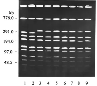
All 24 group C streptococci isolated from pigs and all 4 group C streptococci that were isolated from monkeys and that had mucoid growth on solid media grew to a uniform turbidity in liquid media and exhibited a broad, diffuse colony morphol-ogy in soft agar. In addition, these cultures did not react in the SAT, even at an ammonium sulfate concentration of 3.2 mol/ liter. In contrast, all six group C streptococci showing nonmu-coid growth on solid media grew in liquid media, producing sediment and clear supernatant, grew in soft agar in compact colonies, and aggregated in SATs at an ammonium sulfate concentration of 1 mol/liter. The protein fingerprints of all 34 of the S. equi subsp. zooepidemicus cultures tested revealed identical protein profiles. A typical protein fingerprint of the isolates is shown in Fig. 1.
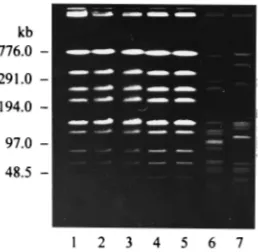
In addition, digestion of the chromosomal DNAs of 32 of the isolates with endonuclease SmaI revealed identical DNA patterns for these isolates. Digestion of the chromosomal DNAs of the remainingS. equisubsp.zooepidemicus isolates yielded DNA patterns with a difference in two fragments (Fig. 2). The DNA restriction patterns of theS. equisubsp.
zooepi-demicus control strains appeared as unrelated DNA profiles
(Fig. 3).
DISCUSSION
All streptococcal isolates used in the present investigation were isolated from diseased pigs and monkeys in Indonesia.
On the basis of their serological and biochemical properties, all 34 isolates from the islands of Bali, Sumatra, and Sulawesi belonged to Lancefield’s serological group C and could be identified asS. equisubsp.zooepidemicus. All 34 cultures, re-gardless of their place of isolation, had identical biochemical profiles.
S. equisubsp.zooepidemicusis well known from infections of
a wide variety of animals, including pigs, sheep, cows, goats, foxes, birds, rabbits, guinea pigs, and monkeys (21). All of these animals might be potential reservoirs for infections of humans. Cases of human infections withS. equisubsp.
zooepi-demicushave been reported, and such infections are frequently
associated with the consumption of homemade cheese or un-pasteurized milk (2, 5, 7). In addition, the isolation ofS. equi subsp.zooepidemicusfrom humans has been described in cases of endocarditis (15), pneumonia (17), meningitis (9, 14), septic arthritis (1, 10) and cervical lymphadenitis (13). However, pig infections withS. equisubsp.zooepidemicusare rare.
To our knowledge, this is the first report ofS. equi subsp.
zooepidemicus infections in pigs with bacteria being isolated
from a large number of infected animals.
In the past decade, the application of chemotaxonomic cri-teria and the use of techniques based on nucleic acid hybrid-FIG. 1. Typical SDS-PAGE patterns of protein extracts of eight S. equi subsp.zooepidemicusisolates from pigs and monkeys.
FIG. 2. Pulsed-field electrophoretic restriction patterns of chromosomal DNAs of nonmucoid (lanes 1, 2, and 8) and mucoid (lanes 3 to 7 and 9)S. equi subsp.zooepidemicusisolates from pigs (lanes 1 to 3 and 6 to 9) and monkeys (lanes 4 and 5) after the DNAs were digested with endonucleaseSmaI.
2202 SOEDARMANTO ET AL. J. CLIN. MICROBIOL.
by on June 6, 2010
jcm.highwire.org
ization and sequencing have led to important changes in strep-tococcal taxonomy. These techniques, which give information about the natural relationships of various streptococcal spe-cies, showed similarities between “S. equi” and “S. zooepidemi-cus.” According to a proposal of Farrow and Collins (8), “S.
zooepidemicus” should be reclassified asS. equisubsp.
zooepi-demicus.
The S. equi subsp. zooepidemicus isolates of the present
investigation displayed differences in growth properties on solid media and in liquid media and soft agar. Growth on solid media in large and mucoid colonies seemed to be related to a uniformly turbid growth of the bacteria in liquid media and to a diffuse colony morphology of the bacteria in soft-agar media. On the other hand, cultures with small, nonmucoid colonies grew as a sediment with a clear supernatant in liquid media and in compact colonies in soft agar. Comparable relationships in terms of microencapsulation, chain lengths, and the growth properties of the bacteria could be observed for various sero-types of the streptococci of serological group B (18, 25, 26). However, in contrast to those of group B streptococci which have neuraminic acid as a major part of the bacterial micro-capsule (26), the encapsulation and growth properties of S. equisubsp.zooepidemicusseem to be influenced by hyaluronic acid capsular material. This influence could be demonstrated by the decapsulation test with a hyaluronidase-producing S.
aureusstrain. In addition, this type of encapsulation seems to
influence the surface hydrophobicity of the isolates. This influ-ence could be determined by the SAT. Comparable to the neuraminic acid of group B streptococci (26), the hydrophilic hyaluronic acid ofS. equisubsp.zooepidemicusseems to mask hydrophobic surface structures.
The importance of group B streptococcal microencapsula-tion for pathogenicity could be demonstrated in adherence and phagocytosis experiments (18, 26). In addition, the hyaluronic acid capsule of group A streptococci is well known to be a major virulence factor (24). Further studies with encapsulated and unencapsulatedS. equisubsp.zooepidemicusmight eluci-date the role of this encapsulation for bacterial pathogenicity. To further characterize the epidemiological relationships of the bacteria, all 34S. equisubsp.zooepidemicusstrains isolated from pigs and monkeys from different islands of Indonesia were subjected to protein and DNA fingerprinting. The latter was performed by pulsed-field gel electrophoresis. Regardless of the origins of the fragments, whole-cell protein analysis and electrophoresis of large DNA fragments produced identical
profiles, indicating that a single virulent clone is the causative agent of the various pig and monkey infections on the island of Bali and the other islands of Indonesia. Further information about the trading connections among the islands and among the farmers and the monkey resorts might elucidate how these bacteria could spread in Indonesia.
However, the pulsed-field gel electrophoresis patterns of two pig isolates differed from the general outbreak pattern by two fragments. This difference was possibly caused by a dele-tion of DNA from the original DNA fragment. It was of inter-est that both strains were isolated from the same place but from two different pigs on the island of Bali. According to the criteria for interpreting the pulsed-field gel electrophoresis pattern described by Tenover et al. (22), this difference might be caused by a single genetic event, indicating that these twoS. equi subsp. zooepidemicus strains are closely related to the other isolates. The two isolates belonged to the group of six unencapsulated strains, indicating that the degree of encapsu-lation does not seem to be directly related to the differences in the DNA fingerprints.
These nonmucoid colonies, which also included the three pig isolates from Sumatra, might possibly represent genetically identical phase variants of the mucoid strains. Encapsulated and unencapsulated phase variants of group B streptococci of various serotypes have been described previously (18, 19).
The occurrence of phenotypic variations among S. equi subsp.zooepidemicusisolates might support the adaptability of these bacteria to different environmental conditions and might help in the understanding of the process of infection with this microorganism.
As was seen for other isolates in the studies reported by Skjold et al. (20), fingerprinting of the chromosomal DNAs of
theS. equisubsp.zooepidemicusisolates studied in the present
investigation proved to be a useful and informative technique for further analysis of the epidemiological clusters of these bacterial isolates.
ACKNOWLEDGMENTS
I.S., F.H.P., and I.W.T.W. received fellowships from the Deutscher Akademischer Austauschdienst.
REFERENCES
1.Barnham, M., A. Ljunggren, and M. McIntyre.1987. Human infection with Streptococcus zooepidemicus(Lancefield group C): three case reports. Epi-demiol. Infect.98:183–190.
2.Barnham, M., T. J. Thornton, and K. Lange.1983. Nephritis caused by Streptococcus zooepidemicus(Lancefield group C). Lanceti:945–948. 3.Carter, G. R., and S. W. Rundell.1975. Identification of type A strains of
Pasteurella multocidausing staphylococcal hyaluronidase. Vet. Rec.96:343. 4.Collins C. H., and P. M. Lyne.1985. Identification methods, p. 103–113.In C. H. Collins and P. M. Lyne (ed.), Microbiological methods. Butterworths, London.
5.Colman, G., and A. Efstratiou.1985. The investigation of outbreaks of infection caused by human strains of Lancefield group C or group G strep-tococci, p. 30–31.InY. Kimura, S. Kotami, and Y. Shiokawa (ed.), Recent advances in streptococci and streptococcal diseases. Reedbooks, Chertsey, Surrey, United Kingdom.
6.Dharma, D. M. N.1994. Wabah streptokokkosis pada babi dan kera di Bali. InlavetI(2):1–2.
7.Duca, E., G. Teodorovici, C. Radu, A. Vita, P. Talasman-Niculescu, E. Bernescu, C. Feldi, and V. Rosca.1969. A new nephritogenic streptococcus. J. Hyg.67:681–698.
8.Farrow, J. A. E., and M. D. Collins.1984. Taxonomic studies on streptococci of serological group C, G and L and possibly related taxa. Syst. Appl. Microbiol.5:483–493.
9.Ghoneim, A. T., and A. M. Cooke.1980. Serious infection caused by group C streptococci. J. Clin. Pathol.33:188–190.
10. Gorman, P. W., and D. N. Collins.1987. Group C streptococcal arthritis: a case report of equine transmission. Orthopedics10:615–616.
11. Hwang, M. N., and G. M. Ederer.1975. Rapid hippurate hydrolysis method for presumptive identification of group B streptococci. J. Clin. Microbiol.
1:114–115. FIG. 3. Pulsed-field electrophoretic restriction patterns of chromosomal
DNAs of mucoid (lanes 1, 4, and 5) and nonmucoid (lanes 2 and 3)S. equisubsp. zooepidemicusisolates from pigs after the DNAs were digested with endonucle-aseSmaI. The DNA restriction patterns ofS. equisubsp.zooepidemicus500 and W60, which were investigated for purposes of comparison, are shown in lanes 6 and 7.
VOL. 34, 1996 IDENTIFICATION OF SEROLOGICAL GROUP C STREPTOCOCCI 2203
by on June 6, 2010
jcm.highwire.org
12. Jonsson, P., and T. Wadstro¨m.1984. Cell surface hydrophobicity of Staph-ylococcus aureusmeasured by the salt aggregation test (SAT). Curr. Micro-biol.10:203–210.
13. Ko¨hler, W., and A. Cedeberg.1976. Case report:Streptococcus zooepidemicus (group C) as a cause of human infection. Scand. J. Infect. Dis.8:217–218. 14. Low, D. E., M. R. Young, and G. K. M. Harding.1980. Group C streptococcal
meningitis in an adult: probable acquisition from a horse. Arch. Intern. Med.
140:977–978.
15. Martinez-Luengas, F., G. M. Inclan, A. Pastor, M. Montejo, J. Barron, A. Baroja, and C. Aguirre.1982. Endocarditis due toStreptococcus zooepidemi-cus. Can. Med. Assoc. J.127:13. (Letter.)
16. Maslow, J. N., A. M. Slutsky, and R. D. Arbeit.1993. Application of pulsed-field gel electrophoresis to molecular epidemiology, p. 563–572.InD. H. Persing, T. F. Smith, F. C. Tenover, and T. J. White (ed.), Diagnostic molecular microbiology: principles and applications. American Society for Microbiology, Washington, D.C.
17. Rose, H. D., J. R. Allen, and G. Witte.1980.Streptococcus zooepidemicus (group C) pneumonia in a human. J. Clin. Microbiol.11:76–78.
18. Salasia, S. I. O., I. W. T. Wibawan, C. La¨mmler, and M. Sellin.1994. Phase variation in streptococci of serological group B. Characteristic properties of isolates from human and bovine infection. APMIS102:925–930.
19. Sellin, M., M. Linderholm, M. Norgren, and S. Hakansson.1992. Endocar-ditis caused by a group B streptococcus strain, type III, in a nonencapsulated
phase. J. Clin. Microbiol.30:2471–2473.
20. Skjold, S. A., P. G. Quie, L. A. Fries, M. Barnham, and P. P. Cleary.1987. DNA fingerprinting ofStreptococcus zooepidemicus(Lancefield group C) as an aid to epidemiological study. J. Infect. Dis.155:1145–1150.
21. Stableforth, A. W.1959. Streptococcal diseases, p. 589–650.InA. W. Stable-forth and I. A. Galloway (ed.), Infectious diseases of animals. Diseases due to bacteria. Academic Press, New York.
22. Tenover, F. C., R. D. Arbeit, F. V. Goering, P. A. Mickelsen, B. E. Murray, D. H. Persing, and B. Swaminathan.1995. Interpreting chromosomal DNA restriction patterns produced by pulsed-field gel electrophoresis: criteria for bacterial strain typing. J. Clin. Microbiol.33:2233–2239.
23. Thiele, D., H. Willems, G. Ko¨pf, and H. Krauss.1993. Polymorphism in DNA restriction patterns ofCoxiella burnetiiisolates investigated by pulsed field gel electrophoresis and image analysis. Eur. J. Epidemiol.9:419–425. 24. Wessels, M. R., A. E. Moses, J. B. Goldberg, and T. J. DiCesare.1991.
Hyaluronic acid capsule is a virulence factor for mucoid group A strepto-cocci. Proc. Natl. Acad. Sci. USA88:8317–8321.
25. Wibawan, I. W. T., and C. La¨mmler.1990. Properties of group B streptococci with protein surface antigens X and R. J. Clin. Microbiol.28:2834–2836. 26. Wibawan, I. W. T., and C. La¨mmler.1991. Influence of capsular neuraminic
acid on properties of streptococci of serological group B. J. Gen. Microbiol.
137:2721–2725.
2204 SOEDARMANTO ET AL. J. CLIN. MICROBIOL.
by on June 6, 2010
jcm.highwire.org