Vol 3, No 4, October-Deceuber 1994 Antitunor of Nonnal Intestinal
Microflora
213Antitumor
Activity
of
Normal Intestinal
Microflora
in Human and Animals
Mochammad Hatta
Abstrak
Pada penelitian ini dilakukatr penteriksaan aktifitas antitunrcr nùkroflora
nornal
usus nnnusia,nnnut
dan nencit, dan hasilnya nenuniukkan bahwa Eubacteriutn, Bifdobacteriun, dan Bacteroides nerupakan bakteri yang doninan pada usus nanusia. Sedangkan Clostridiwnilnupu,l
Enterobacreriaceae tidak ditenukan padanarnut.
Denûkianpula
Clostridiwn dan Bifidobacteriun tidak ditenukan pada nencit. Junlah bakteri pada ntencityang nengandung tuntor nenurun dibandingkan dengan nrencitnornal.
Khususnya pada ileuu dari nencit yang nengandunç tuntor terjadi penurunan junilah bakteri anaerob secara jelas. Dari bakteri yang ditenukanpada usus , sebany'ak 59 strain 1'ang hidup dan
nati diuji
aktifitas anlitutnornya terhadap " Ehrlich ascites tu,,tor't. Tanrpak bahwa II
srrain 1'ang diuii ntenpunyai aktivitas antitumor. Entpat diantaranya toksik terhadap "host", dan senua mencit yang diinjeksi dengan
Pseudontonas aeruginosa (TyH-8)
nati
dalam beberapa hari. Eubacteriun lentunt(TyH-ll),
Propionibacteriwn acnes (TyH- 2S), Proteusnirabilis
(TYM-7) dan Serratia,narcescens(Ty-
142)dalan
bentuk hidup naupun yang diberikanfornrclin nenunjukkanaktifitas antitunror. Kulrur supernatan Serratia,ilarcescens nenperlihatkan akriftas
antitutuor-Abstract
In order to investigate lhe antitunor aa1ivi1t of inteslinal nicroflora, the consrituûon of nonnalJlorav,as tested in hunan, guinea pig and uice.
It
u'c:s clari,fied that Eubacteriwn, Bif dobacteriunt and Bacteriodes u'ere the predominant bacterial generain huntans.
In additiott, neither Clostridium nor Enlerobacteriaceae v,as delected in guinea pigs and neither Clostridiun nor Bifdobacteriwn v,as Present in nice. Total bacterial count itr tunor-bearing uice v,ere reduced in conparison u,ith those in nornalnice.
Especially, in theileum oflwnor-bearing nice, the incidenae ofanaerobic bacterial generav,as sçikingb,decrea,sed. Frou the bacterialfound,fifq, nine (59) Iiving and killed strains isolated front intestinal nicroflora were e.raninedfor their
anlitunnr
activity against Ehrlich ascites tunrcr.II',vas observ'ed that I1 of the tested strains had antitunor activiS-. Four of these had
rcici4,to
the host, and especially, all nice injecred v'ilh PseudontoruLs aerugittosa (TYM-8), died u,ithin several days. Eubacleriuttt lentum (TYH-tl),
Propionibacreriun acnes (TYM-28),Proreus nirabilis (TYM-7) and Serratia,,mrcescens (Ty-142), in u,hich antitunnr actiuir;'N,as recogniTed
in
living andforrnaline-killed bacleria, cured the lutnor- bearing nice. The supernatant culture ofserratia narcescens contained altparenl a,1ûIu,troractiviry-Keywords : Antitunor actiti5., ttornal .flora, hunan, guinea pigs, ntce.
INTRODUCTION
It
iswell
known that in huntan nrany malignant tumors
occur
in
digestive
systent,except duodenum,
jejenum
and
ileunt which very few
tumors occur. Few animal
tunror
cell
lines have
been recognized
from
guineapigs,
while
manystrain are known
from
mice,
rats andrabbits. The
abovetwo fact
suggestthe
possibility
of
sonreform of antilumor activity
contmon to
both
the humansmall intestine
and guineapigs
from
theview-point of the intestinal microflora, although
it
is
alsonecessary
to
consider the structure and function
of
organs and tissues
of
the host.The intestinal microflora
has several effects
onthe host.
Also, the bacteria constituting the
normal
flora show various patterns
according to differences in
race, age,and
diet of
the host.l'2'3
Ëu.th"r.ore,
it
isthought
that the relation between typesof
food and the
microflora
may influence
the incidence of cancer.Hill
etal.4
investigated the
inteslinal microflora of British
and
American (high-risk bowel
cancer) subjects
and Japanese(low-risk)
subject, and
found that
the
high-risk
population
had amuch higher
proportion of
Bac-teriodes where as the low-risk group had
a
much
greater
proportion
of
Enterobacteriaceae
and
Strep-tococcus.
From
thesefindings,
it
was
suggested that2t4
Hattasuch differences
might
be related
to
the incidence
of
colon
cancer.Mastromarin
o
et
al.s
reported
that
bowel
cancerpatients have larger
number
of
Eubacterium
andClostridium than normal people,
but no
signif.icant
differences
fecal
anaerobic bacteria
andtotal
aerobic
counts are noted.
On the other hand,
Funchs
et
al.6
andTrock et
al1recognized an
increased
proportion
of
Clostridium,
Lactobacillus and
Streptococcus,
and
a
decrease
in
Eubacterium under
theinfluence
of
ahigh-fiber
diet.
Therefore,
it
can be thought that antitumor
ac-tivity
is produced by one strain and also bycooperation
of
somestrain of normal flora. In
the presentstudy, we
investigated
theconstitution
of bacteria of normalflora
of
humans, guinea
pigs
and
mice,
and
antitumor
ac-tivities
of
various
isolates
were
examined
in
animal
tumor experiments.
MATERIALS
AND METHODS
Fecal samples
:
Fresh
fecal
samples were collected
from
healthy
humans
(16
to
46
years
old)
and
ex-perimental animal
(guineapigs
andmice).
Animals
:Male
guineapigs wheighing
350 g and 5-weekold
malaICR
mice were obtainedfrom
SankyoLabo
Service Co.,
Ltd.,
Toyama,
Japan.The animals
were housed under standard
laboratory conditions
andwere given
a
commercial pellet
diet
and water
ad
libitunt.
Media
:
GAM
agar,BL
agar, Bacteroides
agar,modified
FM
agar (
Nissui
Co.,
Ltd., Tokyo,
Japan),
LBS
agar,TGC medium
(BBL Microbiology
Systems,Becton
Dickinson
and
Co., Cockeyville,
MD,
USA),
modified
VL-G,
Nagler's medium
andM10 were
usedfor isolation of
anaerobic bacteria,s andHI agar, Blood
agar,
Mannitol-salt agar (Nissui Co.,
Ltd.,
Tokyo,
Japan),NAC
agar, Sabouraud agar
(Eiken Chenrical
Co.,
Ltd.,
Tokyo,,Japan)
for
aerobic
bacteria.RPMI-1640 (Nissui
Co.,
Ltd., Tokyo,
Japan)supple-mented with l0% FCS was used
for tumor cell culture.
Bacteriological methods
:
One gram
of
freshfecal
samplewas
suspendedin
9
ml of TGC
medium
and
diluted
to I
:
10 concentration
with
diluent for
anaerobic bacteria under a CO2 atmosphere.Total
bac-terial
counts were determined
with
a
modified VL-G
using
the rolled tube method.eOrganisms
isolated
from
the plates were
iden-tified
on the
basis
of
colonial form, Gram
staining,
morphology, biochemical test and gas
chromatog-raphy. l0 The bacterial
numbers
were
represented
aslogro per
gramof feces.
Med
J
Uttiv IndottAntitumor activitv
: ICR
male
mice
were
sub-cutaneously inoculateâ
with
106
cells/animal
of
Ehrlich
ascites
tumor
in
RPMI-1640
supplementedwith l0% FCS. On day
5,
l0o cells/animal
of living
or
formalin-killed
bacterial samples
were
administrated
intratumorally
by
one-shotinjection
or
for
aperiod
of
5
days.
Another mice
were injected
0.1 ml/animal
of
supernatantculture,
which had been filtered
through
a0.45
pm millipore
filter
trsing
the same course.The
experimental
period was 80
days
from
theinoculation
of
tumor cells. Tumor weight
was
calcu-lated using
following
form-ula
:Tumor weight (mg)
={major
axis
x
(minoi
axis1211z.RESULTS
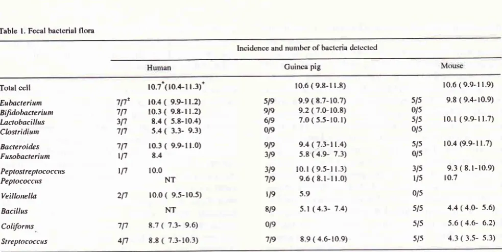
Fecal
microflora
No differences
in
total bacterial counts were
recog-nized
betweenhuman
andanimals
asshown
in
Table
1., although the
incidences
of
some
bacterial genera
showed
apparentdifferences.
Clostidium
was always
detected
in
humanfecal
samples, but notin
thosefrom
guinea
pigs
and
mice.
Conversely, Peptostreptococci
were predominant organism
in
guinea
pigs,
no
coliform
bacteria were
detected. Bacteroidaceae werepredominant bacteria
in
both
humans and animals.Influences
of
bacteria
on tumors
were
observedin the
ileum,
caecum andrectum
of
mice. As shown
in
Table2, total
bacterial counts
were reducedin
tumor-bearing
mice
comparison
with
normal
mice.
Par-ticlularly,
in
the
ileum
of
tumor-bearing mice, the
incidence
of
anaerobic bacteria was
decreasedstrik-ingly
andonly
Lactobacillaceae were
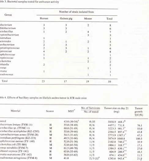
detectable.Antitumor
activity of
isolates
from feces
of
humans
and
animals.
The antitumor
activity
against solid Ehrlich
ascitestumor
bacteria
isolated from
humans and animals
isshown
in
Table
3.On day 5 as
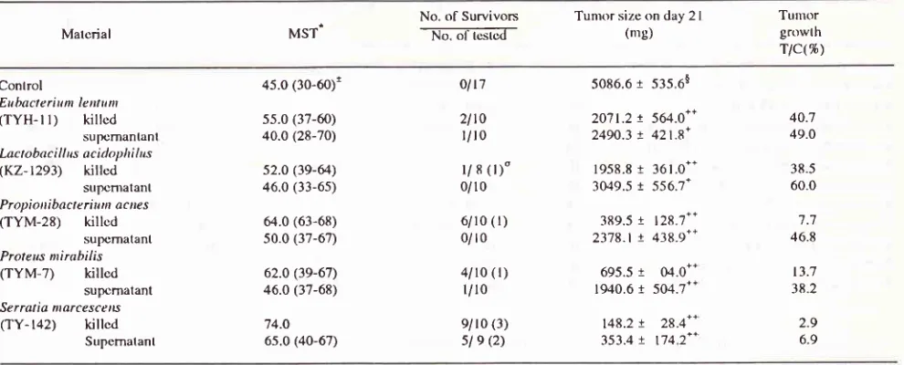
many as
106cells
of
various living
bacterial
strains weredelivered
intratumorally
by
one-shotinjection.
As the results in table 4,ho*"i"lËu.nl
Ibacterial
strains
of l0
species showed
antitumor
ac-tivity
againstEhrlich
ascitestumor.
Eubacteriunt
len-tum
(TYH-II)
andClostridiunt perfringens (KZ-233)
producedprolongation
of
thesurvival period in
tumor-bearing
n-rice. Lactobacillus (TYMC-
I
andKZ- 1293)
and
Propionibacterium
acnes(TYM-28)
showed
sig-nificant (p < 0.01)
suppression
of
tumor growth.
Onthe other hand,
disappearanceof tumor was observed
in-Vol 3, No 4, October-Decenber 1994
tratumorally for
5 daysstarting
from
day
5.As shown
in table
5,
Eubacteriunt lentum, Propionibacterium
acnes
and Serratia
ntercescens
(TY-L42)
showed
remarkable antitumor activity,
with
a cured rate
of
more than 50%. On the other hand,
living
Pseudo-monas
aeruginosa
(TYM-8)
showed
toxic effect
onnice
which
diedwithin l0 days
afterinoculation
of thebacilli.
Clostridiunt perfringetts,
Staphylococcus
aureus
(TY-148)
and Klebsiella oxytoca (TY-141)
showed signs of
toxicity
which debilitated
mice butdid
not kill them.
Finally, the antitumor activity
of
for-malin-killed
cells
and supernatantsof
5strains
which
not show toxicity
were tested against
Ehrlich
ascitestumor. As
shown
in
table
6, killed bacilli of
4 strains
except
Lactobacillus acidophilus
showed aremarkable
effect.
In
particular, killed Propionibacteriunt
acnes,but
not its
supernatantculture
,showed
a cure rateof
more than
50%. With
regard
to
Serratia
marcescens,killed
bacteria and
supernatant
culture
showed
cure ratesof 90% and
33%, respectively.
DISCUSSION
Roe
andGrantll
reportedthat germ-free
statussignif-icantly inhibited
the early development
of
tumors
fol-lowing
exposure to 7, I 2-d i methyl benzanthracenegiven
shortly
afterbirth,
and that theinduction
of
tumors may beinfluenced
by
gut
microflora.
Hill
et a/.4 postulatedthat nuclear
dehydrogeneration
of
steroids
by
Clostridiunt paraputrificnu might play
on
important
role
in the
induction of colon
cancer, sincethis
metabo-lite
is abundantin
the fecesof high-risk
subject. t2Ho*-ever,
Finegold.
et
a/.lr
reported
that there was
nodifference
in
intestinal bacterial
betweeneither
cancer patients andcontrol
patients,or
between subjectswithn
Japanese diets and those onAmerican
diets.Our
present results showed that
Eubacterium,
Bifidobacterium and
Bacteriodes
were
predominant
bacterial
genera
in
human
feces,
but were
of a
low
incidence ofPeptostreptococci
and ahigh incidence
of
Clostridium. Although, Mitsuoka et
alt4
reported a
high incidence
of the
fornrer
and alow
incidenceofthe
latter.
In
thisexperiment,
no Clostridium was observed in either guineapigs
orntice.
With
regard to thisresult,
it
was
pointed out that Clostridium perfringens
and,Enterobacteriaceae were
not
detected
in
guinea
pigs,and that Clostridiunt perfringens given orally
could
exist
for only
ashort
tin.re inintestinal tract
of
guineapigr.
lsMice
had neither Clostridium
nor
Bifidobac-terium in their intestinal microflora, but
had ahigher
incidence and
high
counts
of Lactobacillus
than
humans and
guinea pigs
(Table
l).
These resultsindi-Attlitunror of Nontnl Intestinal
MicroJlora
215cated
an
apparent
difference
of microflora
between humans andexperimental animals. However,
therela-tion
between these
differences and the
incidence
of
colon
cancerin
humansis unknown.
Total
countsof
intestinal
flora
were
decreasedin
tumor-bearing mice, but
no changein
the componentsof
the microflora was
observed except
in
the ileum
(Table
2). This
result
suggest
that the
intestinal
microflora
is
slightly
affected by the
presence
of
tumor.
We
then
screened
the antitumor
activity of
bacterial strains isolated
from
intestinal
flora. At
present,
the antitumor activities of various
bacterial
strains are
being studied by
many researchers,l6-19 butmost
of
them
do
not
exist
in
normal
intestinal
microflora. In the
present
paper, we recognized
II
bacterial strains
which
showed antitumor activity
(Table
4),
and
four
of
these
strains of
these
had a
striking
effect
on Ehrlich
ascites tumor (Table
6).However,
we
were unable
to
conclude whether all
strains
of
these specieshave
anantitumor effect,
be-causeno activity
was observed
as to differentstrains
or
sourcesin
the same species.As
mentioned above,
four
species,Eubacteriunt
lentunt, Propionibacteriunt
ecnes,
Proteus mirabilis
(TYM-7)
and
Serratia ntarcescenq showed
indis-putable
antitumor
activity
againts
Ehrlich
ascitestumor
following
administration
of
both
living
andfor-malin-killed
cells, and also showedlittle
to-xicity on thehost
. As for Serratia ntercescens, supernatantculture
showed
apparent
antitumor
activity.
The
antitumor
activity
of Serratia
nlarcescens
had already
beenreported
asColey's toxin by
Natus
et a/..20The
anti-tumor
activity
of
Propiotribacterium acnes, also
known
asCorynebacterium
pervum,
wasindicated by
Rossol et
el.''
and the
antitumor activity
of
proteus
ntirabilis
againstEhrlich
ascitestumor
was reportedby
Murata
et a1.22On
theother
hand, as to theantitumor
activity
of
Eubacterium lentum,
we havenot found
anyreport
uptill now. This
paper
is
therfore the
first descriftion
of
theantitumor
activity of
Eubacterium
lentum.Mizutani
and Mitsouka23 reported
that
liver
tumorigenesis
is
markedly promoted
with
a
bacteriacon.rbination
of
Escherichia
coli,
Streptococcus!ae-calis
and
Clostridiunt
paraputrificunr,
and
that
this
promoting
effects
is suppressed
by
addition
of
Bffidobacteriunt longunt, Lactobacillus acidophilus
andEubacterium rectale
in gnotobiotic
CrH/FIe
mice.In the
presentexpriment,
Bifdobacteriunr
longunt had
no antitumor
activity
and
Lactobacillus acidophilus
had a mild
effect
againstEhrlich
ascitestumor.
These .results suggested
that the
apperent
an-titumor activity
of
abacterium could
alsovary
2r6
HattaTable l. Fecal bactcrial flora
Med
J
Univ IndonIncidcnce and nunrbcr of bactcria dclcctod
Guinca pig
Total cell
Eubacteriunt Biftdobacteriunt Lactobacillus Clostrirtiunr Bacteroides Fusobacteriunt Peptostreptococcus Peplococcns Veillonella Bccillus Coliforms Streplococcns 5/s 0/5 s/5 0/s s/s 0/s 3ls l/s 0/s sls sls 5/-5 717' 717 3l'l 717 111 tl7 t17 211
7 l'l 411
ro.7ilo.4-l 1.3).
r0.4
(
9.9-r 1.2) 10.3(
9.8-r 1.2)8.4 ( 5.8-10.4) 5.4
(
3.3- 9.3)10.3
( 9.9-11.0)
8.4
10.0 NT
10.0
( 9.5-10.5)
NT8.7 ( 7.3- 9.6) 8.8 ( 7.3-10.3)
sle 9le 6le ole ele 3le 3le 719 tle 8/e 0/e
lle
10.6 ( 9.8-l1.8)
9.9 ( 8.7- ro.7) 9.2 ( 7.0-10.8) 7.0 ( 5.s- r0. r)
9.4 (7,3-tt.4) 5.8-( 4.9- 7.3) r0.l ( 9,5-l r.3)
9.6( 8.1-1r.0)
5.9
5.1 (4.3- 7.4\
8.9 ( 4.6-10.9)
r0.6(9.9-l1.9)
9.8 ( 9.4-r0.9)
l0.l
(9.9-11.7)r 0.4 (9.9- l 1.7)
9.3 ( 8.1-10.9)
10.7
4.4 ( 4.0- s.6)
5.6 ( 4.6- 6.2) 4.3 ( 3.-s- s.3)
*
Figures indicate the average nunrbcr olbactcria cclls in onc grattr of l'ecal sanrplc and arc shown by logro.+
Figures in parenthcscs indicate the range of ccll nuntbcrs dctcctcd.t
Number of positlf sample(s) / nunrhcr of samplcs tcs(cd.NT : Not tested
Minimal levels for delcction of organism is 3.0O.
Tahle 2. Bacterial flora in lntestine of ICR micc
Incidance and nuntbcr of bactcria dotcctcd
Nornral mouse Tumor-bcaring nrouso
Cccunr llcunr Rcclutn
Total
ccll
9.5'(9.3-9.7)lEubacterium
4/5$ 8.8 (8.6-8.9)BiJidobacterium
0/5ktctobncillu,s
-5/5 9.5 (9.3-8.9)Propionibacterium 215 8.2(8.0-8.4)
Closrridium
0/5Bacteroides
315 7.7 (6.7-8.3)Fusobacterium
0/5Pcptostrepîococckr 315 8.0(6.3-9.0)
Peplococcus ll5
8.9 (7.9-9.2)Veillonella
0/5Stttlthylococcus
215 3.9 (3.8-4.0)Streptococcils
415 4.9 (3.7-6.4)Corynebateriunt
0/5Bacillu-s
5/5
4.4 (3.5-5.4)Escherchia
5/5
4.5 (3.5-4.7)Proreus
0/5Pseutlontonts l/5
3.3Yeast
ll5
7.33ls 0/s ,5/-s 0/s 0/-5 5/S 0/5 t/-5 l/s 0/s s/-s 4ls 0/-5 5/5 -s/-5 0/-5 2ls 0/-5 s/s 0/5 s/s t/s 0/-s 5/-5 0/s l/s t/s 0/5 415 5/5 0/s s/-5 -5/-s 0/-s 2ls 0/-s 0/s 0/5 5/s 0/s 0/s tls 0/5 0/s 0/s 0/s 5/s 3ls 0/s s/s sls 0/s t/s 0/5 s/s 0/s 5is l/s 0/s s/5 0/s 3ls l/5 0/s Jls s/5 2ls s/s 5/s 0/5 us l/5
r0.6(9.9- l l .9)
9.8(9.4- 10.9)
10.0(9.5- r0.e) 9.6
l0 4(9.9- l r.7)
9.3(8. r- l0.e)
t0.7
3.7(3.s- 4.0)
4.3(3.5- 5.3)
4.6(3.7- 5.s)
4 4(4.0- s.6)
5.6(4.9- 6.2)
3.1 1.7
r0.6(r0.2-r 1.5)
3/s
9.8( e.4- l0.6) 0/5s/s lo.2( 9. l - 10.6)
tls
9.30/s
5/s r0.4(r0.r-l r.3) 0/5
2ls
9.s( 9.3- 9.-5)2ls
9.3( e.0- 9.6) 0/5s/5
4r(
3.0- 5.s)s/5
4.5( 3.3- s.6) 0/ssls
4.4( 4.2- 4.8)s/s
6.0( s.0- 7.0)l/5
4.0l/5 5.0
0/s8.2( 7.0- 8.8)
8.2( 7.0- 8.9)
7.2
4.2(1.O- s.4)
4.0( 3 8- 4.9)
4.6( 3 0- 4.3)
4.6( 3.0- 5.7)
4.1
9.9(9.0- r0.-5)
9 r (8.3- 10. r )
8.7(8 0- 10.0) 9.'l
95(9 !-r0. l)
8.3
83
4.8(3.6- 6 .5\
5 0(1.9 -5.9)
42(36- 49)
s.0(4 3- s.6)
3.9(3.0- 4 7)
9.8(9.3- 10.2) 9.4(8.9- 9.7)
8.8(8.
r-
9.2)9.ô(9. l- r0. r )
9.9
9.2
4.s(3 0- 5.3) 4.6(1 2- 4 9)
4.3(3.3- -s. r)
5.7(.1.1- 7.t)
3.e(3.0- 4.8)
*
Micc wcrc givcn on intrapcritonoal transplant of 106 cclls of Ehrlich a-scitcs ttrltror and wcrc sacrilicod on day 7+
Figurcs indicate the avcragc nunrhcr of bactcrial coll in one granr o[ fccal sanrplc antl arc sltown by log ro.t
Figurcs in parcnthcscs indicatc thc range o[ccll nunrbcs dctcctcd. $ Nunrbcr of Jxxitive sanrplcs(s)/ nunthcr ol sanrplcs tcstùd. [image:4.595.51.543.74.321.2]Vol 3, No 4, October-Decenber 1994
Table 3. Bacterial samples tested for antitumor activity
A,llitutnor of Norrnal It[eslinal
Micro.flora
217Number of strain isolated from
Genus
Human Guinea pig Mouse Total
Eubacteriunt Bifidobacreriunr krctobacillus Propioni bacteriunr Clostridiuttt Baderoides Fusobacreriutn
Peptosl re ptococcus Peptococcus Staphl'lococcus Streptococcus Escherichia Klebsielln
Proteu.s
Se rratia Psc. utltt ttt otttts
3 3
I
I
2 2
2
4
I
I
I
3 32
4
3
I
2
I
2
3 3
2
8
6
3
I
5 56
8
1
2 2 2
I
I
I59
l9
l7
23
[image:5.595.58.545.92.618.2]Total
Table 4. Elfects of bacillary sarnples on Ehrlich ascites tumor in ICR male mice
Material Source MST
No. of
Survivors
Turnor size on day2l
TumorNo. of tested (me) growth
rlc(%)
Control
En bact c r i u t rt le ntu
n
('lYH- 1l)
ktcrobac i I I us.sp.
(TYMC-l)
ktc t o bac i I I us ac id ophi lus (KZ-1293) P ro pi otti bac te ri wn ac nes
(IYM-28)
C I o s t r id i un pe rfri ng ens Q<Z-223) St a p hy I oco cc us aureus (TY- 148)
Esc he ri chia coli (TY-M4) P rc te us n t i rabi Ii s (TYM-7) K I e bs i ee I o.t)'to ca (TY-l 4
l)
Se r rot ia, ilar cesc e ns (TY- 142)P se ud o ntonas ae ru gi tt osa (TYM-8)
H M H M H H M M H H
M
43.0 (30-54)1 55.0 (38-69) 60.0 (51-69) s5.0 (39-66) 54.5 (33-65) 48.s (33-66) 52.0 (5 l-56) s2.o (43-s4)
6l.s
(4e-70)65.0 (s5-69) 56.0 (43-62) 41.0
0 /35
ol6
ol6
ol6
ol6
ol4
Ll6
rls
rls
rls
rls
213(1)"
5858.9
468.1$4457.r
73r.83223.3
s}t.1*
2564.5
891.4"
2'713.9
t387.t'
5874.9 1068.8
2lo2.g
740.2** 1004.1 31g.7'+ 1584.5 630.1+'604.9
280.5**674.r
454.0*' 1285.6 945.4'*76.1
55.0
43.8
47.3
100.3 35.9
11
.t
27.O 10.3 11.5 21.?
Mice were inoculated with 106 cells of Ehrlich ascites tumor subcutaneously and injected intratumorally with 106 cells of bacteria by
one-shot on day 5.
*
Median survival time indicates mice except those alive on day 80.t
Figures in parentheses indicate the range ofsurvival time.$
Figures indicate the averange tumor size on day2l
with the mean + standard enor.o
Figures in parentheses indicate the numbers of tumor-bearing mice.+, ++ : Statistical significance from the control at p < 0.05 and p < 0.01, respectively.
-2t8
HartaTable 5. Effects of bacillary samples on Ehrlich ascitqs tumor in ICR male mice
Med
J Univ Indon
Material Mst
No. of Survivots
No. of tested
Tumor size on day 2 I
(ns) Tunrorgrow(h
Ttc(16)
Control
Eubaoerium /zrrrrnr (TYH-l l)
Lactobaci IIus sp. (TYMC-
l)
ktctobac i lltrs ac i do phi I us (KZ- 1293) Propio ni bact e ri ilm ac,re s (TYM-28)
C losr r i d i n m pe rfr i n g e rc (KZ-223)
St ap hy lococ c u s arrraus (TY- I 48)
Esc heric hia coli (TY-M4)
Prote us mi rabilis (TYM-7)
Klebsieel otyroco (TY-14 l)o
Se r rat i a m arc e s ce,Ls (TY- 142)
Pse u do monas ae r u g i tnsa (TYM-8)
45.0 (30-55)!
46.0 (3 l-60) 43.0 (32-68) 49.0 (40-66)
s8.5 (3e-63) 46.5 (38-6s) 5 1.5 (44-65)
4e.s (40-69)
4't.s (32-10)
52.0 (45-70)
5s.0 (43-66) 9.5
0 /31
9/r8 2lt8 Q)o
2lt6 rolt6 (2,
2/10
(l)
4/r8 (3)
4lt8 (2)
4lt8 (2) 3/18 (r) r0/17 (r)
0/r0
5027.4
!
436.9çt3\7.O
!
440.7" 2874.r! U9.4"
1428.3
!
282.7"691.2
!
240.4*'1468.4
t
340.5'* 2t5t.g!
40t.7"t317.4
!
376.6*' 825:l!
2cil..7"llll.6 1
l8t.2t*475.5
r
185.6*-NT
26.2 57.2 28.4 13.7 29.2 42.8 26.6
t6.4
22.1 9.5
Micewereinoculaledwith 106cellsof Ehrlichascitestunror subcutaneously and injectcd intratumorally with l06cclls of bactcria for5 days
from day 5.
*
Mediunr survival timc indicates nrice exccpt those alive on day 80.I
Figures in parcn(hescs indicate the range ofsurvival time.$
Figures indicate the averange tumor size on day 2 I with the mcan+
standard error.o
Figures in parentheses indicate lhe numbers of lurnor-bearing mice.O These bactcria showed toxicity : Dcath, Weakncss.
++ : Slatistical significance from lhe control at p < 0.01.
NT : Not tested
Table 6. E[lccts of bacillary sanrplcs on Ehrlich ascilos lunror in ICR nralemco
Matcrial MST
No. of Survivors
-To.
oI rù".t6if Tunror size on day (mg) 2 ITutnor growth
rlc(%)
Control
Eubacletirm Ientilnt
(TYH-l
l)
killcdsuprnantanl
Lactobac i I I us ac idophi I us
(Kz-t293)
killedsurx)nratant Propioni bacte riu m ac nes
(TYM-28)
killcdsur)cmatant Proteus mirabilis
(TYM-7)
killcdsupcmatant
Serralia narcesccns
(TY-
142)
killcdSupematant
45.0 (30-60)r
ss.0 (37-60) 40.0 (28-70)
s2.o (tg-64.)
46.0 (33-6s)
64.0 (63-68) 50.0 (37-67)
62.0 (3e-67)
46.0 (37-68)
74.O 6s.0 (40-67)
oltT
2lt0
r/r 0
l/
s (r)"0/10
6/10 (r) 0/ r0
4/10 ( r) l/10
e/10 (3)
sl e (2)
5086.6
t
535.6$20'71.2
!
564.0*'2490.3
!
42t.8*1958.8
r
36t.0t.3049.5
t
556.7*389.5
t
128.7*. 2378.1r
438.9..695.5
r
ù1.0*.1940.6
!
504.7**t48.2
!
28.4*. 353.4!
t742"
40.7 49.0
38.5 60.0
't.7 46.8
t3.7 38.2
2.9 6.9
Mice wcrc inoculatctl wi th 106 cclls of Ehrlich ascitcs tumor sunuu tanoously an<l injcctctl intratumorally with lO6 ccl ls of killcd bactcria anil 0. I ml or culturcd supematânt.
*
Mcdian survival linrc indicatcs nrice cxccpl thosc alive on day 80.1
Figurcs in paronthcscs indicalc thc rangc ol'sûrvival tinrc.$
Figurcs indicatc tho avcrângc tunror sizc on day 2l with the nrcan slandanl orrttr.o
Figurcs in parcnthcscs indicatc thc numbors oflutnor-hcaring nricc. [image:6.595.65.565.94.280.2] [image:6.595.71.566.454.654.2]Vol 3, No 4, October-December 1994
Acknowledgments
I
am most grateful
toDr.
K.
Kawai
for
expert
help
in
the preparation
of
the manuscript and
to
Dr.
T.Hayashi
for
financial support through a grant from
JIMA.
REFERENCES
l.
Kono S, Imanishi J, Shinchi K, Yanai F. Relationshipof Dietto
Small and Large Adenomasof
the
Sigmoid Colon.Japanese J Cancer Res 1993; 83: 13-9.
2. Moore WEC, Holdeman LV. Discussion of current bacterio-logical investigations of the relationships
between
intes-tinal
flora, diet and colon cancer. Cancer Res 1975; 35:3418-20.
3. Nomura
A,
Stemmermann GN, Chyou PH, KatoI,
Perez-Perez
GI,
Blaser MJ.Helicobacterpylory
infection andgastric carcinoma among Japanese Americans in Hawaii. N Eng J Med I99l1'325:. 1132-6.
4.
Hill
MJ, Drasar BS, Aries V, Crowther JS, Hawksworth G,Williams REô. Bacteria and aetiology
of
cancerof large
bowel. Lancet; 1971: 16:95-100.
5. Mastromarino A, Reddy BS, Wynder EL. Fecal profiles
of
anaerobic microflora
of
large bowel cançer patients withnonhereditary large bowel polyps. Cancer Res 1978; 38:
4458-62.
6. Funchs HM, Dorfman S, Floch HM. The effect of dietary fiber supplementation in man. II. Alteration in fecal physiol-ogy and bacterial flora. Am
I
Clin Nutr 1976;29:1443-7. 7. Trock B,Lanza E, Greenwald P. Dietary fiber, vegetables,and colon cancer: critical review and metanalyses
of
theepidemiologis evidence.
I
Natl Cancer Inst 1990; 82:650-61.
8. Mitsuoka T, Morishita Y, Terada A, Yamarnoto S. A simple methode ("plate-in-bottle method") for cultivation
of
fas-tidious anaerobes. Japan J Microbiol 1969; 13: 383-5. 9. Cadwell
D,
Bryant MP. Medium without rumenfluid
fornonselective enumeration and isolation of rumen bacteria.
Appl Microbiol 1966; 14: 794-801.
10. Holdeman
LV,
Moore EEC editors. Anaerobic laboratory manual. The Virginia Polytechnic Institute and StateUniver-sity Anaerobe Laboratory: Balckburg. 1978.
I
l.
Roe FI, Grant GA. Inhibition by germ-free status of develop-ment of liver and lung tumours in rnice exposed neonatallyto 7, I 2-dimethylbenz(a)anthracene : Inrplications in relation to test for carcinogenicity. Int J Cancer 1970; 6: 133-44.
Antitutnor of Nortnal Intestinal
Microflora
2L912. Aries VC, Goddard P,
Hill
MJ. Degradation of steroids by intestinal bacteria III..3-oxo-5 beta-steroid theta-l dehydro-genase and 3-oxo-5 beta-steroid theta-4 dehydrogenase.,
Biochim Biophys Acta 1971; 248: 482-8.13. Finegold SM, Attebery HR, Sutter
VL.
Effectof
diet on human fecal flora. Comparison of fapanese and American diets. AmI
Clin Nutr 1974;27 : 1456-69.14. Mitsuoka T, Kaneuchi C. Ecology of the Bifidobacteria. Am
J Clin Nutr
l97l;
30: 1799-1810.15. Yamagishi T, Konishi
K,
SakamotoK,
Sakurai S, IshisakaS. Fate
of
Clostridium perfringensin
intestinalof guinea
pigs. Iapan J Bacteriol 1981; 36: 757-65.
16. Ebina T, Murata K,'Tamura K. Antitumor Effect of Intratu-moral Administration of Biological Respon Modifiers:
In-duction
of
Immunosuppressive Acidic Protein, a Typeof
alpha-l Acid Glycoprotein, in rnice. fapanese J Cancer Res
1994; 85: 93-100.
17. Matsuzaki
T,
ShimizuY,
YokokuraT.
Augmentation ofantimetastatic effect
on
Lewis lung carcinoma(3LL)
in C57BL|6 mice by primiting with Lactobacillus cassei.llledMicrobiol Immunol 1990; 179: 161-8.
18. Fujimura H, Nanjo M, Tamura K, Saito M, Ebina T, Ishida N. Effect of OK-432, a Streptococcal preparation, on serum
immunosuppresive acidic protein in tumor-bearing mice. J
Clin Exp Med 1990; 152:675-9.
19. Kataoka
T,
Yamamoto S, YamamotoE,
Kuramoto E,Kimura Y, Yano O, et al. Antitumor Activity
of
Synthetic Oligonucleotideswith
Sequencesfrom cDNA
Encoding Proteinof
Mycobacteriun bovis BCG. JapaneseI
CancerRes 1992;3:244-1.
20. Natus HC, Swift rù/E, Coley BL. The treatment of malignant
turnors by bacterial toxins as developed by the late William B. Coley, M.D., reveiwed in the light
of
modem research. Cancer Res 1946; 6: 205-216.21. Rossol S, Voth R, Brunner S, MLrller WE, Buttner M, Gallati
H, et al.
Corynebacteriutn parvutn (Propionibacteriwnacnes) i An induce r of tumor necrosis factor-alpha in human peripheral blood mononuclear cells
in
vitro. Eur J Immun l99O;2O:l76l-5.
22. Murata T, Arakawa M, Sugiya Y, Inazu Y, Hattori Z, Suzuki
Y, et al. Oncolytic effect of Proteus nirabili.ç upon tumor-bearing animal. Life Sciences 1965; 4: 1055-67.