Intrauterine Growth Restriction in Twin-Twin Transfusion
Syndrome Caused by Placental Insufficiency:
A Case Report Diagnosed by Histopathology
So e kim in, Dja fa r Sid d ik*, A nto nius Ha rking to Wib iso no , Sufid a
De p a rte me nt o f Ana to mic a l Pa tho lo g y – De p a rte me nt o f O b ste tric a nd G yna e c o lo g y*
Fa c ulty o f Me d ic ine Unive rsity o f Suma te ra Uta ra , Me d a n
Abstract: Objective: to get the correct information concerning the caused of dead of one fetus
(intrauterine fetal death/IUFD ) and intrauterine fetal growth restriction (IUGR) of other fetus of twin pregnancy of 34-36 weeks gestation. Materials and methods: the whole placenta together with the amniotic membrane and umbilical cords of twin pregnancy were examination macroscopically and histopathologic analyzed. Pathologic features: monozygotic twins placentation of monochorionic monoamniotic membrane with twin-twin transfusion syndrome were found. There was a large thrombus and larged old infarct formation at intermediate area of the placenta suspected the cause for the IUFD of one fetus. There were also many fibrine, capillaries occlusion and multiple small infarct of villous vessel suspected the cause for IUGR for the second fetus of twin pregnancy. Results: a big thrombus suspected as the caused for IUFD for one fetus and placental insufficiency as the cause for IUGR on the second fetus. Conclusions: a twin pregnancy of 34-36 weeks gestation with twin-twin transfusion syndrome, a big thrombus caused IUFD for one fetus and placental insufficiency caused IUGR on other fetus.
Keywords: intrauterine growth restriction, twin-twin transfusion syndrome, placental insufficiency
Abstrak: Tujuan: untuk mendapatkan informasi yang berhubungan dengan peyebab kematian dari
satu janin (kematian janin dalam kandungan/KJDK) dan intrauterine fetal growth restriction (IUGR) dari janin yang lain pada kehamilan 34 – 36 minggu. Bahan dan cara: keseluruhan plasenta beserta selaput janin dan kedua tali pusat dari kehamilan kembar diperiksa secara makroskopis dan histopatologi. Gambaran patologi: dijumpai kehamilan kembar monozigot plasenti monoamniotik dengan twin-twin transfusion syndrome (TTTS). Di daerah tengah plasenta dijumpai trombus yang besar dan formasi infark lama yang diduga sebagai penyebab KJDK dari satu janin. Juga tanpak banyak fibrin, oklusi kapiler dan infark kecil yang multipel pada pembuluh darah jonjot sebagai penyebab IUGR pada janin kedua dari kehamilan kembar ini. Hasil: satu trombus yang besar diduga sebagai penyebab KJDK dari satu janin dan insufisiensi plasenta sebagai penyebab IUGR janin kedua. Kesimpulan: kehamilan kembar usia 34 – 35 minggu dengan TTTS dengan sebuah trombus besar sebagai penyebab KJDK satu janin dan insufisiensi plasenta sebagai penyebab IUGR pada janin yang lainnya.
Kata kunci: intrauterine growth restriction, twin-twin transfusion syndrome, placental insufficiency
INTRODUCTION
Twin pregnancy is one of the most frequent and well-known causes of IUGR. In all series of IUGR, twin pregnancy account for 20-30 per cent of the cases. Twins may be monozygotic, from one fertilized egg, or dizygotic, from two different eggs.1 Chorionicity can be determined by ultrasound, on the assessment of fetal gender, on the number of placenta and characteristics of the membrane between the two amniotic sacs.
membrane is composed of a central layer of chorionic tissue as a sandwiched between two layers of amnion, whereas in monochorionic twins there is no chorionic layer. Consequently, the inter-twin membrane is thicker and more echogenic in dichorionic than monochorionic pregnancies.2,3
Approximately 20 percent of all twin pregnancies are monochorionic, and the incidence of Twin-Twin Transfusion Syndrome in monochorionic diamniotic gestation is approximately 10 to 20 percent. Twin-Twin Transfusion Syndrome is a phenomenon almost exclusively of monochorionic twin pregnancies. Clinically, twin-twin transfusion syndrome is typically first recognized by acute hydramnios that develops at the midsemester of gestation. When sonograms or radiographs are obtained after the hydramnios appears, the twins are recognized and their size may already be significantly different.5
The subject of intrauterine growth restriction ( IUGR ) is a very important topic of concern, for if not early recognized, it can lead to a severely compromised fetus as well as fetal and / or neonatal demise.4 IUGR is the most common generic term that is used to describe the fetus with a birthweight at or below the 10th percentile for gestational age and sex.4,6 IUGR fetus doesn’t reach his potential of growth. A fetus who has a potential of growth at the 50th percentile but because of maternal, fetal or placental disorders occurring alone or in combination, becomes growth restricted ( birthweight < 10th percentile ) and he is at risk for adverse perinatal outcome.4
IUGR is a major cause of morbidity and mortality in the perinatal period.6 IUGR may be considered the consequence of a disease process within one or more of three compartments that sustain and regulate fetal growth : the maternal compartment, the placenta or the fetus.4,7
Since most cases result from placental insufficiency, it would be useful to find a better prenatal correlates between placental pathology and predictors of poor pregnancy outcome in this condition so that therapeutic trials may be designed to optimize placental function in those at greatest risk.8
CASE REPORT
A 42 year old woman was presented with hydramnion and history of twin pregnancy. At the followed up the clinician found that there were the problem of fetal growing of IUGR of both fetuses. Then that woman deliveried at
34-36 weeks gestation by section caesarian with one fetus death intrauterine of 500 grams weight, length 27 cm and Apgar score 0. The other fetus alived and weighed 1900 grams, body length 41 cm and the Apgar score was 7/8/9. Then the placenta was sent for histopathologic examination to find the cause of the IUGR and fetal death of this twin pregnancy.
GROSS EXAMINATION
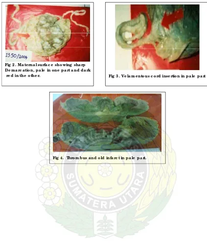
Macroscopically, the placenta with two cords weighs of 750 grams, measuring 15 cm in diameter, approximately its thickness 3,5 cm. Maternal surface showing sharp demarcation with two unequal contrastry portions ( Fig 2 ). The two halves have different color, pale in one part and dark red in the other. We also found white plague which resemble thrombus and old infarct measuring 1 cm at the pale portion. There was no abnormality in the fetal surface. The two cords inserted at the opposite at each halves. At pale portion of the placenta the cord insertion is velamentous with 2,5 cm spaces ( Fig 3 ). That cord length 35 cm, thickness 1,5 cm with four clockwise spiraling and also have four false knots. The other cord insertion is lateralis at the red portion, length 35 cm, thickness 1 cm with one false knot and nine clockwise spiraling. Placental membrane is yellowish in color with chorealis insertion and consists only one sac. But there is an area which looks like the site of the T-Zone, then the membrane is took and rolled for histology examination.
HISTOLOGY EXAMINATION
DISCUSSION
Twins tend to occur in families, the familial tendency is apparent in dizygotic twinning, but this appears to be on the maternal side only. The rate increase with parity but seems only to apply to dizygotic twinning and also has age linked, with an increasing in twinning in the older mothers. Multiple pregnancy is common following the use of drugs to induce ovulation. The use of fertility drugs account for 10-15 per cent of all multiple pregnancies, and has therefore significantly altered the incidence of multiple pregnancy.2
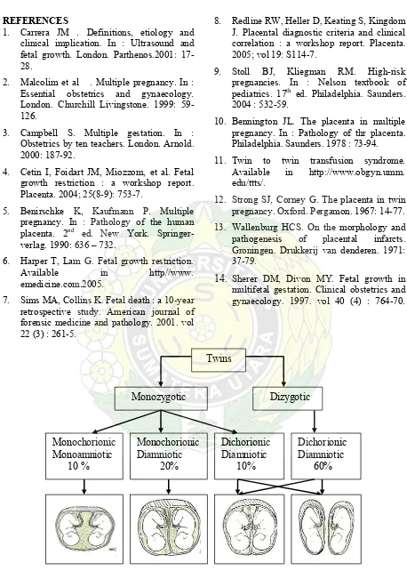
The form of placentation of monozygotic twins depends upon the stage at which duplication occurs. It is believed (Corner 1955) that if the single fertilized ovum separates into two during the first three days after fertilization, that is before the differentiation of the trophoblast. Two separate embryos will develop, each having its own placenta and amniotic sac. Thus if the two embryos implant at some distance from each other there will be two separate placentae and amniotic sacs and placentation will obviously be of the dichorionic-diamniotic variety. If the two developing embryos implant in close proximity to each other there may be fusion of the two placentae but two amniotic sacs; as with dizygotic twins, the fusion is not absolute and placentation is still of the dichorionic-diamniotic type. If splitting occurs during the blastocyst stage, i.e. between the third and eighth day after fertilization, when the trophoblast, but not the amniotic cavity, has differentiated, the twins will develop a single placenta with two amniotic sacs; the placentation is then categorized as monochorionic-diamniotic. The amniotic cavity differentiates between the eight and thirteenth day after fertilization, if splitting occurs during this period there will be only one placenta and one amniotic sac, the placentation being of the monochorionic-monoamniotic form. If splitting occurs at or after the thirteenth day of development this will result in conjoined twins.5,9,10
Twin-Twin Transfusion Syndrome (TTTS) is a serious complication in 6-18 percent of monochorionic multiple pregnancies ( including twins and triplets ). In TTTS, the blood flow is unbalanced resulting in a ‘donor’ twin donating blood through the placenta to a ‘recipient’ twin. Since the donor twin is pumping blood, not only for itself but also across to the other twin, it has less energy to use in growing. This results in a smaller than average baby (sometimes noted as
suffering from intrauterine growth retardation (IUGR) with a smaller than average amniotic fluid level (oligohydramnios). When examined by ultrasound the baby's bladder will be small or unable to be seen due to the lack of growing being done by the baby. Sometimes the baby will be almost wrapped in its amniotic membranes due to the lack of amniotic fluid. Hence the condition is occasionally referred to as stuck twin syndrome. The recipient, however, is trying to do too much growing due to the excess blood and fluid being sent from the donor twin. This extra work results in the baby urinating more causing an abnormally large amount of amniotic fluid around them (polyhydramnios). This extra work can also cause heart failure. The recipient twin's body cavities may accumulate fluid and can result in a condition called 'hydrops'. If the recipient twin develops hydrops, its life is seriously threatened.11 Schatz (1882) had already suggested that the fetal vessels were anastomostic through a ‘third circulation’ which proceeding from one of the donors’ terminal arteries into a shared cotyledon, then it drained into the venous circulation of the recipient.5
TTTS can happen at anytime during a monochorionic pregnancy. It can also occur in triplet or higher order pregnancies that include monochorionic twins. The risks of TTTS to the twins depend on when the condition occurs. If the condition occurs late in the pregnancy, the risks are usually minimal. If one twin threatens to develop hydrops, the babies may be delivered. Of course the later in the pregnancy the delivery occurs, the safer the babies are. Today, comprehensive ultrasound assessment of monochorionic twin gestations provides the opportunity for early prenatal detection and accurate prognostic assessment of this potentially treatable condition.11
Placental or umbilical cord causes of IUGR include twin – twin transfusion syndrome (TTTS), decreased placental weight and / or cellularity, decreased in surface area, villous placentitis, infarction, tumor, placental separation and abnormal cord insertion.6,9
A recent study from S. Kaeting and J. Kingdom of 60 cases of severe early onset IUGR found lesions in the parenchyma or villi were common : 70% accelerated villous maturation, 40% excess perivillous fibrin deposition, 53% villous infarction, 7% intervillous thrombi, 5% fetal thrombotic vasculopathy. So in 93% cases either with one or more of these histopathologic abnormalities.8
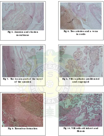
Gross examination at the maternal surface of placenta will showing sharp demarcation into unequal contrastry portions. The two halves differed in color, that of the anaemic baby being pale and bulky, and of the plethoric baby dark red. The histological appearance of the placenta territory of the plethoric baby is essentially normal, though the capillaries are engorged and dilated, but the chorionic villi in the territory of the anaemic (donor) twin is bulky, pleomorphic and evidently oedematous. The trophoblast here is thicker than in the plethoric territory. The foetal capillaries are narrow and often filled with nucleated red – cell precursors.5,12 The gross examination describes above are similar to the placenta of this case which help in diagnosing twin-twin transfusion syndrome.
Normally, blood vessels run from the placenta via the umbilical cord to the baby. Velamentous insertion means that these veins travel across the amniotic membranes before they come together into the umbilical cord. Velamentous insertion happens in 1-2% of all pregnancies. Benirschke (1972-1975) and Robinson et al (1983) reported the velamentous cord insertion frequency is 1,5% of singleton term deliveries. Because of its relative frequency and importance to the course of pregnancy and delivery, membranous insertion of the umbilical cord has been studied by many investigation. Shanklin (1970) suggested that abnormal cord insertion correlates with low birth weigh.5
Infarct is an area of ischemic necrosis resulting from an arrest of circulation in an artery supplying an area without adequate collateral circulation. Placental infarct results from interruption of maternal blood supply through a spiral artery to a fetal cotyledon. Such an interruption may be attributed to vascular occlusion originating from primary thrombosis.
The incidence infarcted placentas is about 28 %. More than half the number of infarct has been found located in the marginal area of the placenta. Infarcts have been classified as recent, rather recent and old infarct (Wallenburg 1971).13 Fetal development and function will be dependent at least partially on a sufficient maternal blood supply to the placenta. Little (1960) reports a progressive decrease in fetal weight as the extent of placental infarction is increased. He also find a high rate of stillbirth and perinatal death in cases with severely infracted placentas.5,14
Intrauterine death of a fetus in a twin pregnancy may be associated with a poor outcome for the co-twin. Death of one fetus with the onset of acute hypotensive episodes in monochorionic twins leading to death of the co-twin in about 25 per cent of cases. The mechanism is acute haemodynamics shifts from the live to the dead fetus.3 Prompeler et al analyzed fetal outcome of the surviving twin in 43 sets of twins in gestations complicated by fetal demise of one fetus. Pregnancies in which the loss occurred before 16 weeks were not affected, whereas pregnancies greater than 16 weeks of gestation were associated with significant morbidity, including prematurity in 50 per cent of cases, growth restriction of the surviving twin in 22 per cent of cases, and a perinatal mortality rate of 13 per cent.14
Conclusions
REFERENCES
1. Carrera JM . Definitions, etiology and clinical implication. In : Ultrasound and fetal growth. London. Parthenos.2001: 17-28.
2. Malcolim et al . Multiple pregnancy. In : Essential obstetrics and gynaecology. London. Churchill Livingstone. 1999: 59-126.
3. Campbell S. Multiple gestation. In : Obstetrics by ten teachers. London. Arnold. 2000: 187-92.
4. Cetin I, Foidart JM, Miozzom, et al. Fetal growth restriction : a workshop report. Placenta. 2004; 25(8-9): 753-7.
5. Benirschke K, Kaufmann P. Multiple pregnancy. In : Pathology of the human placenta. 2nd ed. New York. Springer-verlag. 1990: 636 – 732.
6. Harper T, Lam G. Fetal growth restriction.
Available in http//www. emedicine.com.2005.
7. Sims MA, Collins K. Fetal death : a 10-year retrospective study. American journal of forensic medicine and pathology. 2001. vol 22 (3) : 261-5.
8. Redline RW, Heller D, Keating S, Kingdom J. Placental diagnostic criteria and clinical correlation : a workshop report. Placenta. 2005; vol 19: S114-7.
9. Stoll BJ, Kliegman RM. High-risk pregnancies. In : Nelson textbook of pediatrics. 17th ed. Philadelphia. Saunders. 2004 : 532-59.
10. Bennington JL. The placenta in multiple pregnancy. In : Pathology of thr placenta. Philadelphia. Saunders. 1978 : 73-94.
11. Twin to twin transfusion syndrome. Available i edu/ttts/.
12. Strong SJ, Corney G. The placenta in twin pregnancy. Oxford. Pergamon. 1967: 14-77.
13. Wallenburg HCS. On the morphology and pathogenesis of placental infarcts. Groningen. Drukkerij van denderen. 1971: 37-79.
14.
Sherer DM, Divon MY. Fetal growth in multifetal gestation. Clinical obstetrics and gynaecology. 1997. vol 40 (4) : 764-70.Twins
Monozygotic Dizygotic
Monochorionic Diamniotic 20%
Dichorionic Diamniotic 10% Monochorionic
Monoamniotic 10 %
Dichorionic Diamniotic 60%
Fig 2. Ma te rna l surfa c e sho wing sha rp De m a rc a tio n, p a le in o ne p a rt a nd d a rk
re d in the o the r. Fig 3. Ve la m e nto us c o rd inse rtio n in p a le p a rt
Fig 5. A m nio n a nd c ho rio n m e m b ra ne
Fig 6. Two a rte rie s a nd a ve na in c o rd s
Fig 7. The lo o se n p a rt o f the la ye r o f the a m nio n
Fig 8. Villi c a p illa rie s p ro life ra te d a nd e ng o rg e d