PAC Studies of BSA Conformational Changes
J. GRYBOS´1,*, M. MARSZAL= EK1, M. LEKKA1, F. HEINRICH2 and W. TRO¨ GER2
1
The Henryk Niewodniczan´ski Institute of Nuclear Physics Polish Academy of Sciences, Radzikowskiego 152, 31-342 Krako´w, Poland; e-mail: [email protected]
2
Institute of Experimental Physics II, University of Leipzig, Linne´straße 5, 04103 Leipzig Germany
Abstract. The structure of biological molecules plays an important role in many biological
processes. The variation of ambient parameters such as pH, temperature or pressure can influence important properties of protein molecules like conformation or stability. By means of the perturbed angular correlation (PAC) technique and atomic force microscopy (AFM) a conformation change of bovine serum albumin (BSA) molecules has been studied as a function of the pH value of the BSA solution. The observed decrease of the rotational correlation time and the increase of molecule diameter as a function of pH were attributed to conformational changes of bovine serum albumin induced by different pH values of the BSA solution.
Key Words:albumin conformation, atomic force microscopy, AFM, dynamic hyperfine
interaction, nuclear quadrupole interaction, NQI, rotational correlation time, perturbed angular correlation ofg-rays, PAC.
1. Introduction
Bovine serum albumin is one of the most abundant proteins in blood circulatory systems. They provide many functions like, e.g., maintaining osmotic pressure or pH. Albumins participate in the transport of a variety of ligands whose affinity depend on the state of the protein which is affected by the pH and the calcium concentration in the blood. Since these conditions vary in different tissues and organs, the characterization of albumin isoforms may help for a better understanding of its functionality [1].
The primary structure of BSA is constituted from a single polypeptide chain of 582 amino acid residues and its amino acid sequence is well known [2]. Its secondary structure, in the native form, is formed by 67% of a-helixes with six turns, 33% ofb-sheets, and 17 disulfide bridges. The protein was modeled as an ellipsoid with axes of 14 nm4 nm, with three domains in line.
It is known that albumin undergoes pH dependent conformational changes in alkaline and acidic solutions [3]. These conditions influence the forms of the BSA molecules and they are classified as expanded (E, below pH 2.7), fast (F, below pH 4.3), native (N, pH 7.0), basic (B, above pH 8.0) and aged (A, above pH 10.0). These five forms are characterized by a different content of a-helixes, b-sheets and random coils [4]. At a pH value between 2.7 and 4.3, albumin is in the fast form which is characterized by an increase of the intrinsic viscosity, and a significant loss of the helical content is observed [5]. The native form with a globular shape is predominant in the pH range 4.5Y7.0 [3].
In this paper, two different techniques, perturbed angular correlation ofg-rays and atomic force microscopy, were applied to study the size and dynamics of BSA molecules in aqueous solutions at different pH. By AFM the diameters, by PAC the rotational correlation times of the BSA molecules were determined. Both parameters depend on pH.
2. Samples preparation and experiment
The BSA molecules for the AFM imaging were immobilized on glass coverslips activated with a 2.5% glutaraldehyde (supplied by Sigma) aqueous solution for 30 min. Before activation, they were silanized in 4% solution of 3-amino-propyltriethoxysilane (APTES, Sigma) in toluene. The protein immobilization was done by substrate immersion in 0.5 mg/ml aqueous protein solution for one hour.
For the PAC experiments a radioisotope, the PAC probe, has to be attached to the BSA molecule. Here, we used the PAC probe111In(EC)111Cd, which decays from 111In to 111Cd via an electron capture (EC) before the g-g-cascade occurs which is used for the PAC experiments. The samples for the PAC experiments were prepared in the following way: first, a 200 mM aqueous solution of the bovine serum albumin (Sigma,Mw= 69 kDa) was prepared which was incubated with a solution of 111InCl3(no carrier added) at 30-C for 1 h in order to assure better binding of the In3+ ions. The sample activity was in the range of 100 to 400 kBq. The total sample volume was about 1Y2 ml. The pH value of the BSA solution varied from 2 to 10 and it was checked before and after each measurement. PAC experiments were performed at room temperature and at liquid nitrogen temperature.
The AFM images were recorded using silicon nitride cantilevers with a spring constant of 0.03 N/m and the radius of curvature was about 50 nm. The measurements were carried out at room temperature in deionized water using a
(assumed to be circular) and their real diameterD, taking into account the radius
R of curvature of the AFM tip:
S¼2
The angular correlations of the 172Y245 keVg-ray cascade in the PAC probe 111In(EC)111Cd were measured using a 6 detector PAC spectrometer equipped with 6 BaF2 detectors [7]. Thirty coincidence spectra Ni(Q,t) (i=1,. . .,30) were
collected simultaneously at anglesQ= 90-and 180-between the detectors. For a detailed description of the data analysis see [8].
In the case of a static nuclear quadrupole interaction the data were fitted by the perturbation functionG22static(t):
Gstatic22 ð Þ ¼t X 3
n¼0
s2nð Þcos ð!nð Þ tÞexpð!nð Þ tÞ ð2Þ
where s2n are the amplitude coefficients and wn are the frequencies, which are
functions of the asymmetry parameter h=(VxxjVyy)/Vzz of the electric field
gradient (tensor componentsVii(i=x,y,z)).dwnis the half width at half-maximum
of the Lorentzian frequency distribution around the mean value wn. Such
distributions arise due to variations of the charge environment of the probe nuclei. This is typical for frozen solutions, where the EFG is determined not only by the regular charge distribution in the molecule, but also by random arrangements of the solvent molecules.
In the case of molecules dissolved in a liquid a relaxation effect is produced by the rotational diffusion of the molecules. This can be described by the following perturbation function:
G22ð Þ ¼t elst
Gstatic22 ð Þt ð3Þ
with the slow relaxation constantls ¼t1
s and with the assumption thatwnts> 1. In the case of a very rapid molecular motion which corresponds to the condition
wnts < 1, the perturbation function G22(t) becomes in accordance with the Abragam and Pound theory [9] a simple exponential form:
G22ð Þ ¼t elft ð4Þ
where lf is the fast relaxation constant. For the nuclear spinI ¼52 of the used
PAC probe 111In(EC)111Cd, lf becomes 1f 100:8!2
Qtc, where tc is the
3. Results and discussion
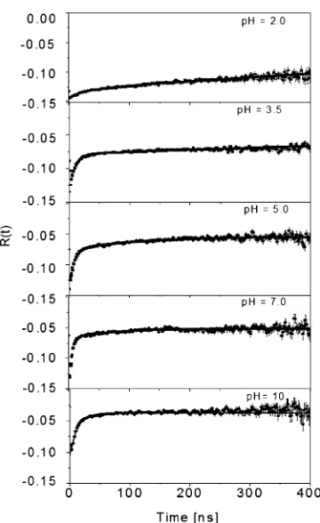
The PAC spectra recorded at room temperature and at different pH values are shown in Figure1. The spectra were fitted with a linear combination of formulae (3) and (4) and an unperturbed, time independent PAC signal (a0):
G22;totalð Þ ¼t a0þa1e 1st
G22staticð Þ þt a2e1ft ð5Þ
Figure 1. The PAC spectra of the 111In-BSA complex at different pH measured at room temperature.
Table I.The hyperfine parameters for111In-BSA samples at different pH value
pH f0[%] f1[%] ls[nsj 1
] ts[ns] f2[%] lf[nsj 1
] tf[ns]
The amplitudes a0,a1 anda2 were treated as free parameters. These amplitudes contain the anisotropy of the cascade, the solid angle correction factors, and the population of the different states by the PAC probe. In order to compare the populations directly the fractions fi¼
ai P2 i¼0
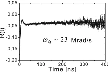
ai were calculated and shown together with the hyperfine parameters obtained from the fitting procedure in TableI. The fractionf1of the slowly rotating species was assigned to In3+ions bound to BSA molecules whereas the fractionf2with the fast rotating species was attributed to In3+ ions bound to hydroxyl groups or other small molecules. The unperturbed fraction f0, which changed only little with pH, was assigned to unbound probe ions in the solution. In order to determine the fast rotational correlation timetc, it is necessary to know the parameters of the static nuclear quadrupole interaction. These parameters were determined from PAC measurements at the liquid nitrogen and were very similar for all pH values: quadrupole frequency wQ $
22Y23 Mrad/s, asymmetry parameterh $0.5 and a large Lorentzian frequency distribution. The static PAC spectrum of the 111In-BSA complex at pH 7 measured at 77 K is shown in Figure 2. The rotational correlation time of slow molecular motion is simply given as a reciprocal of the ls value. The values of
both rotational correlation times are also shown in Table I.
The decrease of the fraction of slow relaxation (f1, ls) accompanied by the increase of the number of indium ions bound to hydroxyl groups (f2, lf) with
Figure 2. The PAC spectrum of the111In-BSA complex at pH 7 measured at 77 K.
increasing pH can be explained by conformational changes of the BSA molecules. The only amino acid of BSA which can bind In, cysteine 34, is located in the center of the elongated BSA structure, and for the F-conformation of albumin this binding site is easily accessible for the indium probe. However, an increase of the pH leads to a conformational change of BSA into the more oval structure (N), which results in a reduced accessibility of the cysteine 34 for In3+ ions. Such a structural change is also reflected by the rotational time tf which decreases with the increase of pH. For pH 10, the parameters f1, lsand ts,
indi-cate that the indium ions did not bind to the BSA molecule at all. Previously reported rotational correlation times for 111In-BSA system were different from those obtained here [11]. The discrepancy can be explained by the different con-ditions of the PAC experiments (shorter time scale and less statistical quality of the PAC spectra of [11]) and by different procedures of the sample preparation. The second fraction f2 represents 111In3+ ions bound to hydroxyl groups or other smaller molecules. These smaller molecules may result from the electron capture after-effects accompanying the radioactive decay of 111In to 111Cd leading to a partial or entire disintegration of the BSA molecules. This fractionf2 increases with pH at the expense of the fractionf1and becomes dominant for pH values larger than 5 since the amount of hydroxyl groups in the solution increases with increasing pH.
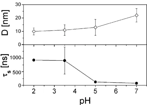
The AFM images of albumin molecules immobilized on a glass surface for three chosen pH values are presented in Figure 3. The average value of the diameter of a single albumin molecule was determined by fitting Gaussian distributions to the histograms of the molecule diameters. Taking into account the effect of a topography convolution with AFM tip (Equation 1), the real diameter of the BSA molecule is 10 T2.5 nm, 11T4 nm, 13T6 nm and 22T5
nm for pH 2.0, 3.5, 5.0 and 7.0, respectively. It was observed that the molecule diameter increases as a function of pH. Figure 4 shows the comparison of the BSA molecule diameter and rotational correlation times.
These results obtained by two different methods confirmed that albumin molecules undergo conformational changes at different pH value. These struc-tural properties were reflected by the decrease of the rotational correlation time (PAC) and the increase of the molecule diameter seen in AFM images.
Acknowledgments
The authors acknowledge the support of a PolishYGerman Scientific and Technical Collaboration (WTZ No 4440/2002) from the Bundesministerium fu¨r Bildung und Forschung and State Committee for Scientific Research.
References
1. Carter D. C. and Ho J. X.,Adv. Protein Chem.45(1994), 153.
2. Burova T. V., Grinberg N. V., Golubeva I. A., Mashkevich A. Y., Grinberg V. Y. and Tolstoguzov V. B.,Food Hydrocoll.13(1999), 7.
3. Harmsen B. J. M. and Braam W. J. M.,Int. J. Protein Res.1(1969), 225.
4. Reed R. G., Feldhoff R. C., Clute O. L. and Peters T. Jr.,Biochemistry14(1975), 4578. 5. Harrington W. F., Johnson P. and Ottewill R. H.,Biochem. J.62(1956), 569.
6. Engel A., Schoenenberger C. A. and Mu¨ller D. J.,Curr. Opin. Struct. Biol.7(1997), 279. 7. Butz T., Saibene S., Fraenzke T. and Weber M.,Nucl. Instrum. Methods A284(1989), 417. 8. Butz T.Z., Naturforsch.51a(1996), 396.