The extract of "shoe lower" (
Hibiscus rosea sinensis L) leaves inhibit
the spermatogenesis of ddy strain mice
Margaretha Mawuntu
Abstrak
Penelitian ini dilakukan dalam rangka mencari bahan kontrasepsi pria yang bersumber dari tanaman, khususnya daun kembang sepatu (Hibiscus rosea sinensis L). Penelitian ini bertujuan untuk melihat apakah ekstrak daun kembang sepatu (Hibiscus rosea sinensis L) dapat menghambat proses spermatogenesis mencit strain ddy. Penelitian ini dilakukan pada tiga kelompok yang masing-masing terdiri dari delapan ekor mencit. Kelompok kontrol diberikan karboksimetil selulose (CMC) 1% dalam 0,5 ml aquabides, kelompok perlakuan I diberikan ekstrak daun kembang sepatu (Hibiscus rosea sinensis L), dosis 700 mg/kg BB ditambahkan CMC 1% dalam 0,5 ml aquabides dan kelompok perlakuan kedua diberikan ekstrak daun kembang sepatu (Hibiscus rosea sinensis L), dosis 800 mg/kg BB ditambahkan CMC 1% dalam 0,5 ml aquabides. Perlakuan ini diberikan selama 40 hari sesuai dengan siklus spermatogenesis. Setelah itu dilakukan pem buatan preparat histologis testis mencit, diikuti pengamatan preparat testis dengan mikroskop cahaya pembesaran 100x dan 400x untuk menghitung sel-sel spermatogenik. Terakhir, dilakukan pemotretan tubulus seminiferus ketiga kelompok yang terdiri dari sel-sel
spermatogenik melalui mikroskop cahaya dengan pembesaran 100x dan 400x dengan memakai kamera Fuji dan ilm Fuji, ASA 200. Hasil
menunjukkan perbedaan yang sangat bermakna antara kelompok kontrol dan kelompok perlakuan I dan II, yaitu terjadi penurunan jumlah sel-sel spermatogonia, spermatosit primer pakhiten dan spermatid pada kelompok perlakuan (P<0,01). Dari hasil penelitian ini, dapat disimpulkan bahwa ekstrak daun kembang sepatu (Hibiscus rosea sinensis L) menghambat proses spermatogenesis. Hasil penelitian ini diharapkan dapat dikembangkan sebagai bahan kontrasepsi pria. (Med J Indones 2008; 17: 157-62)
Abstract
This study was conducted in order to develop male contraception from plants, namely the "shoe lower" (Hibiscus rosea sinensis L) leaves. The objective of this study was to ind out whether the extract of "shoe lower" leaves could inhibit the process of spermatogenesis
on ddy strain mice. This research was performed in 3 groups and each group consisted of 8 mice. The control group was given 1%
carboxy methyl cellulose (CMC) in 0.5 ml aquabides. The treatment group I was given the extract of "shoe lower" leaves 700 mg/kg BW and 1% CMC in 0.5 ml aquabides, and the second treatment group was given the extract of "shoe lower" leaves, 800 mg/kgBW and 1%
CMC in 0.5 ml aquabides. The treatment were given for 40 days in accordance with the spermatogenesis cycle. Then, the production of
histological slides of the mice testis and the observation of the slides using light microscope with magniication of 100x and 400x were done. Further, counting of the spermatogenic cells was done. At last the pictures of seminiferous tubulus cross-section of the three groups which consisted of spermatogenic cells were taken through light microscope with magniication of 100x and 400x using Fuji camera and Fuji ilm, 200 ASA. The results showed signiicant differences between the control, treatment I, and treament II group. There were
decreased numbers of spermatogonia, pachyten primary spermatocytes and spermatids in treatment groups (P<0.01). The result of
this study showed that the extract of "shoe lower" (Hibiscus rosea sinensis L) leaves, inhibited the process of spermatogenesis of ddy
strain mice. It is hoped that the result of this study can be developed into a male contraception. (Med J Indones 2008; 17: 157-62)
Keywords: spermatogonia, Pachiten primary spermatocytes, spermatids
Indonesia is the fourth largest populated country in the world after China, India, and USA, with 215 millions inhabitants, and the growth rate in 1990-2000 was 1.48%.1 The national birth control program has
Department of Biology and Andrology, Faculty of Medicine
Sam Ratulangi University, Manado, Indonesia
participation rate is due to social, cultural, and religious factors. Among the reasons is the limited selection of male contraception available.2 With wide selections of contra-ception methods there would be more alternative for men. Therefore it is necessary to promote research on male contraception.3 Further development of male contra-ception has large expectation in the future.4
The WHO has form a task force to develop a save, effective, reversible, and acceptable method of male contraception. One of the strategies is to develop herbal male contraception from plants containing active infertility substrates.3,4 Indonesia is rich in tropical botanic resources. Botanic contraception has advantages
including low toxicity, accessibility, cost eficient and
less side effects.4,5
Hibiscus (Hibiscus rosea sinensis L) has promising
features. The lower, leaves, bark, and roots contain hibiscetin glycoside, an anti-spermatogenesis agent, favourable for male contraception. In addition, the bloom contains hibiscetin glycoside and calcium oxalate, and the leaves contain some kind of alkaloid. However, the information of its application is still scarce.6,7 Previous reports described that Hibiscus rosea sinensis L had anti-spermatogenesis features on male mice.5,6 Purwaningsih (1998)7 performed administration of 300 mg/kg Hibiscus rosea sinensis bloom extract on AJ strain mice, and the decrease in spermatogenia was
signiicant compared to the control group. However,
the treatment group receiving group receiving 200 and
250 mg/kg did not show signiicant results.
Although the mechanism of the active substrate in
Hibiscus rosea sinensis L in reducing spermatogenesis is still unknown, it is generally presumed that the bloom and leaf of Hibiscus rosea sinensis L inhibit germinal cells through cytotoxic or anti-androgen effect.5,6
The aim of this study was to ind out the effect of
Hibiscus (Hibiscus rosea sinensis L) leaves extract in inhibiting spermatogenesis in ddy strain mice.
METHODS
This study had a randomized post test control group design, and conducted in Sam Ratulangi University, Faculty of Medicine, Joint Research Laboratory. The research period was July to November 2004.
Healthy male mice (mus musculus) ddy strain, 10-16 weeks of age,8 30-45 gr was used in this setting. The sample number was based on Kirk’s equation, 1982,9 that concluded that each treatment required minimal 8 mice.
Extraction of hibiscus leaves
The hibiscus leaves were obtained from home yards of Manado and surroundings. The leaves selected were mature and dark green ones.
Extraction: the leaves were cleaned and dried not under direct sunlight or oven dried at 37-400C for 5 days until the leaves disintegrated. The dry leaves were blended, shaken, and then weighted. The powders were extracted with 90% ethanol in Sochlet and Weaton instrument for 4-5 hours at 50-600C. The liquid extract was distillated until free from alcohol.10,11
The extract weighted and then kept in bottles in a refrigerator. The extract was weighted according to the necessity based on the body weight and then carboxy
methyl cellulose (CMC) 1% was added and liqueied
with aquabides 0.5 ml.
ddy strain mice selection and treatment
Selection
Male mice, 10-16 weeks, 30-45 grams (weighted with special scale, Tanita, Japan). The selected mice are caged in a plastic basin, one for each. The basins were labeled according to the treatment start, weight, and age.
Feeding
The mice were fed with pellets containing corn starch,
soy starch, grain powder, ish powder, rice beads,
vitamin C and B, non fat milk, coconut oil, and water. Sometimes the menu included carrots and long bean. Tap water was used for hydration.
Treatment
Hibiscus leaves (Hibiscus rosea sinensis L) extract was administered orally to male ddy strain mice in treatment groups, i.e.:
Second treatment group: 800 mg/kgBW with CMC 1% in 0.5 ml aquabides.
Control group: received CMC 1% in 0.5 ml aquabides, orally.
The treatment of treatment and control groups was continued for 40 days.
Spermatogenic cell-counting and sizing of the tubulus seminiferus diameter
Mice testicle slides, was made and the preparation was observed under light microscope with 100 and 400
times magniication.
Every slices containing cross sectioned seminiferus epithelium stage VII were examined. This selection is due to the long duration of the stage VII and the large number of the spermatogenic cells. The spermatogonia, pakhiten primary spermatocytes, and spermatids were counted, and their diameters were measured.
Determination of spermatogonia, primary spermatocytes and spermatids
Spermatogonia are attached on the basement membrane.
In spermatogonia A, the nucleus exhibits ine smooth
chromatin. Spermatogonia B exhibits condensed irregular chromatin.12
Primary spermatocytes perform meiosis to become secondary spermatocytes, and the processes include: a long profase leptoten, zigoten, pakhiten, diploten, and diakinesis. In pakhiten phase, the chromosomes wave shortened presenting rough and solid appearance. The pakhiten stadium is the longest (175.3 hours).13 The primary spermatocytes demonstrate a large and obvious nucleus. Generally the nucleus is located in the center. The oldest generation of the spermatocyte is the pakhiten.13 The spermatid determination was based on the groups and cap that is generally eccentric. The head cap gains the maximum size on stage VII.13
Taking picture of the tubulus seminiferus
The pictures were taken from 100 and 400 magniication
of the microscope using Fuji camera and Fuji ASA 200
ilms, printed on 3R paper.
Data Analysis
Normality assessment was done using Kolmogrov-Smirnov test and variance homogeneity using F test.
If the data is normal and the variance homogenous, analysis of variance (ANOVA) is used to test the research hypothesis followed by Dunnett test when the
result is signiicant.
If the result is the contrary from above, Kruskal-Wallis test are used followed by multiple comparison test
when the result is signiicant.
RESULTS
Spermatogonia number in the three groups
The data of spermatogonia did not meet the criteria for ANOVA.
The median and the Kruskal-Wallis test result are presented in Table 1.
Spermatogonia median of the control group was higher than the 700 mg/kgBW and the 800 mg/kgBW group (P < 0.000).
Pakhiten primary spermatocyte number in the three groups
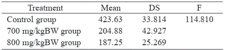
The mean and standard deviation (SD) of the pakhiten primary spermatocyte and the result of analysis of variance are presented in Table 2.
Table 1. Kruskal-Wallis test: spermatogonia number difference in the 3 groups
Table 2. ANOVA: pakhiten primary spermatocyte number difference in the 3 groups
Treatment Mean DS F
Table 3. Dunnet test : treatment to control group difference
The F in table 2 indicated a statically signiicant difference,
therefore Dunnet test was performed.
The test results in Table 3 show signiicant difference
of the control group compared to the 700 mg/kgBW group and the 800 mg/kgBW group (P < 0.01).
Spermatid number of the three groups
The median and the Kruskal-Wallis test result are presented in Table 4.
Spermatid median of the control group was higher than the 700 mg/kgBW and the 800 mg/kgBW group (P < 0.000).
Table 4. Kruskal-Wallis test : spermatid number difference in the 3 groups
Treatment Median Mean Rank
X2 P
Control group 700 mg/kgBW group 800 mg/kgBW group
546.0 187.0 206.5
20.50 8.19 8.81
15.411 0.000
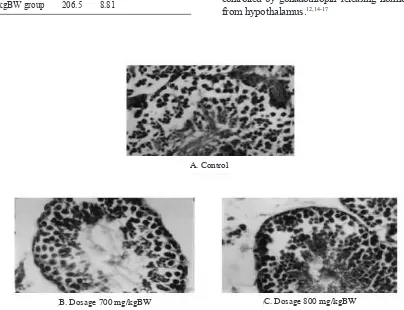
Microphotographs of seminiferous tubules in the 3 groups are presented in Figure 1.
DISCUSSION
From this study we observed that administration of the
Hibiscus rosea sinensis L leaves extract reduced the number of the spermatogenic cells (spermatogonia, pakiten primary spermatocyte, and spermatid) very
signiicantly.
The hibiscetin glycoside in the leaves of Hibiscus rosea sinensis L might cause these anti-fertility/anti-spermatogenesis effect as was supposed by Purwaningsih (1998, 2003) and Kholkute (1977).5-7
The spermatogenic cell reduction due to hibiscetin glycoside in the Hibiscus rosea sinensis L extract might be effected by hormonal mechanism through hypothalamus-pituitary-testis axis. Hormones that directly have effects on the spermatogenesis are gonadotropins, i.e. follicle stimulating hormone (FSH) and lutenizing hormone (LH) secreted by pituitary gland that is controlled by gonadothropin releasing hormone (GnRH) from hypothalamus.12,14-17
Figure 1. Spermatogenic cells in seminiferous tubulus transverse section of mice testis with H. E. stain, 400x
A. Control
Hibiscetin was supposed to inhibit FSH and LH, and to cause disruption in testosterone/androgen production in the testis. Lutenizing hormone stimulates Leydig cells to synthesize testosterone and FSH stimulates Sertoli cells to secrete andogen binding protein (ABP).18 The ABP binds testosterone/androgen to form ABP-androgen complex. Bound testosterone/ABP-androgen was carried into the cytoplasm of the target cells. Dissociation process caused the testosterone/ androgen to be released from the complex. Free testosterone/androgen binds cytoplasm receptor forming a new complex, the receptor testosterone/androgen-complex. Dissociation process in the cell releases testosterone/androgen that enters the cell nucleus.15,19
One of the functions of Sertoli cells is to support mitosis and meiosis.17,20
Reduced Sertoli cells in the tubulus seminiferus due to environmental pressure inhibits spermatogonia development. This is because the Sertoli cells can not support the nutrition for all spermatogonia.
During spermatogenesis process, spermatogenic cells are very active, and changes include morphology, bio-chemistry, and genetic transformation of the cells. The spermatogenic cells depend on glucose for energy. However, pakhiten primary spermatocytes and spermatids depend on lactic and piruvic acid as energy provided by Sertoli cells. Lactic and piruvic acid
production is inluenced by FSH.15,18,21
Hibiscetin glycoside contained in Hibiscus rosea sinensis L extract might inluence FSH production
and the Sertoli cells causing spermatogenic cell development inhibition.
The mechanism of hibiscetin glycoside in Hibiscus rosea sinensis L extract in inhibiting spermatogenesis is still unknown. However previous studies presumed that Hibiscus rosea sinensis L extract inhibited germinal cell metabolism and proliferation through cytotoxic or anti-androgen effects.6,7
Hibiscetin glycoside in Hibiscus rosea sinensis L
extract also has steroid-like features due to cyclo-pentanoperhydrophenanthrene steroid ring. With the feature, androgen receptor of the spermatogenic cell will identify and match with the hibiscetin in the lock and key manner. Androgen and hibiscetin competes to bind with the receptor. If hibiscetin binds the receptor, meiosis, spermiogenesis, and mitosis will be inhibited.15,20
Therefore, hibiscetin glycoside content in Hibiscus rosea sinensis L extract might inluence spermatogenesis
by: cytotoxic effect or hypothalamus-pituitary-testis axis.7
However, it is necessary to study whether puriied
hibiscetin glycoside will cause spermatogenic cell reduction or not.
In conclusion, Hibiscus rosea sinensis L leaves extract 700 and 800 mg/kg BW decreased spermatogenic cell number, i.e. spermatogonia, pakhiten primary spermatocytes, and spermatids compared to control (P = 0.000).
Suggestion
It is necessary to evaluate the side effects on other organs such as kidney and liver.
Acknowledgements
Special thanks to Prof. Dr. dr. Wimpie Pangkahila, SpAnd, FAACS and Prof. Dr. dr. O.S. Tendean, SpAnd. I also would like to thank Dr. Julius H. Lolombulan for his assistance in statistical analysis, dr. Jan Nainggolan, DAF (), Basic Laboratory, University of Sam Ratulangi, and Head of Pathology Anatomy Laboratory, Faculty of Medicine, University of Sam Ratulangi for their help. Finally, thanks to Frans Mintardjo, SKed, Christoffel Mintardjo, SE, Dimas, and Frits Lasut for the supports.
REFERENCES
BKKBN Pusat.
1. Sumbangan pemikiran untuk pemimpin
bangsa pengelolaan kependudukan dan pembangunan keluarga. Jakarta: BKKBN; 2004.
Moeloek N.
2. Perkembangan kontrasepsi pria tahun 2002. In: Buku Acuan dan Kumpulan Abstrak. Pertemuan Ilmiah tahunan XIV Perkumpulan Andrologi Indonesia (PANDI); 2002 July 18-19; Denpasar, Indonesia. Denpasar: PANDI; 2002.
Moeloek N.
3. Perkembangan andrologi tahun 2002 dan masa
datang. In: Buku Acuan dan Kumpulan Abstrak. Pertemuan Ilmiah tahunan XIV PANDI; 2002 July 18-19; Denpasar, Indonesia. Denpasar: PANDI; 2002.
Wang C, Waites GMH. Research strategy of the world 4.
health organization task force on methods for regulation of male fertility and need for sperm function assays. In: Oshima, Henry ED. Current topics in andrology. Tokyo: Japan Society of Andrology; 1993. p.249-99.
Purwaningsih E.
5. Pengaruh tanaman obat tradisional
Kholkute SD. Effects of Hibiscus rosea sinensis on 6.
spermatogenesis and ascesory reproduction organs in rats. Planta Medica. 1977;31:129-35.
Purwaningsih E.
7. Pengaruh pemberian ekstrak bunga Hibiscus
rosea sinensis Linn terhadap proses sper matogenesis mencit
strain AJ dan jumlah anak hasil perkawinannya. [Thesis] Fakultas Kedokteran Universitas Yarsi Jakarta, 1998. van Denberg J. Vaccine technology transfer center. RIVM 8.
(research for man and environment). In: Rijksinstituut voor volksgezondheid en Milieu National Institute of Public Health and Environment. International course on laboratory animal science and husbandry in vaccine production; 1998 June 15-26; Bilthoven, Netherland. Bilthoven: The Institute; 1998.p.8.
Kirk RE. Experimental design: procedurs for the behavioral 9.
sciences. 2nd ed. California: Brooks/Cole; 1982.
Clermont Y, Leblond CP. Spermatogenesis of rat, mouse, 10.
hamster and guinea pig as reveated by the periodic acid funchsin sulfurous acid technique. Anatomic Journal. 1990: 90 (Suppl.2): 167-215.
Dixit VP, Khanna P, Bhargava SK. Effects of Momordica 11.
charantia L fruits extracts on the testicular functions of dog. J Med Plant Res. 1978; 34:280-4.
Holstein AF, Schulse W, Davidoff M. Understanding 12.
spermatogenesis is a prerequisite for treatment. Repro-ductive biology and endocrinology. 2003. Available from: http//www.Rbej.Com/content 1/1/107 [cited 2008 Mar 14]. Fitzgerald PA. Endocrinology. In: Tierney LM, Mc Phee SJ, 13.
Papadakis MA. Current medical diagnosis and treatment. 42nd ed. New York: Lange Medical Books; 2003.p.1137-40.
Purwaningsih E.
14. Pengaruh ekstrak bunga hibiscus rosea
sinensis L terhadap kesuburan (fekunditas) mencit jantan
strain AJ. Jurnal Kedok YARSI. 2002;10 (3):1-7.
Weinbauer GF, Gromoll J, Simoni M, Nieschlag. Physiology 15.
of testicular function. In: Nieschlag E, Behre HM, eds. Andrology male reproductive health and dysfunction. 2nd
ed. Berlin: Springer; 2000.p.34-56. Tendean OS.
16. Terapi sulih testosteron pada gangguan
spermatogenesis. In: Buku kumpulan makalah/ abstrak. Kongres PANDI IX PERSANDI I. Andrologi: sesuatu yang hilang dalam kesehatan reproduksi untuk meningkatkan kualitas hidup manusia; 2005 April 19-23; Jakarta, Indonesia. Jakarta: PANDI/PERSANDI; 2005. p.225-8. Arjatmo Tjokronegoro H.
17. Mungkinkah fertilitas se seorang
bisa menurun. In : Buku kumpulan makalah/ abstrak. Kongres PANDI IX/PERSANDI I. Andrologi: Sesuatu yang hilang dalam kesehatan reproduksi untuk meningkatkan kualitas hidup manusia; 2005 April 19-23; Jakarta, Indonesia. Jakarta: PANDI/PERSANDI; 2005.p.235-8. Moeloek N, Ilyas S.
18. Pengaturan fertilitas pria secara
hormonal terkini. In: Pertemuan Ilmiah PERSANDI ke-IV and PANDI ke-IX. Dengan fertilitas dan fungsi seksual yang baik mencapai kebahagiaan keluarga yang lebih baik; 2006 April 22-23; Palembang, Indonesia. Palembang: PERSANDI/PANDI; 2006.p.153-7.
Liao S, Liang T, Fang S, Castaneda E, Shao TC. Steroid 19.
structure and androgenic activity. Speciicities involved in the receptor binding and nuclear retention of various androgens. J Biol Chem. 1973; 248:615.
Martini FH. The reproductive system of the male. In: 20.
Martini FH. Fundamental of anatomy and physiologi. 5th ed. New Jersey: Prentice Hall; 2001.p.1021-34.
Sherwood L. The reproductive system. In: Sherwood L. 21.
Human physiology from cells to system. 5th ed. California: