Vol 9, No 2, APril
-
June 2000 Histopathology of BCC andSCC
87Histopathological study
on basal
cell carcinoma and
squamous
cell
carcinoma of the skin
Mpu Kanokol, Achmad
Tjartal,
Masato Ueda2,Mochtar
Hamzah3, Herman Cip-to3, Evert Poetiraya,Rrman Mukhtara,
SantosoCornainr,
Joedo Prihartonos,SetyawatiBudiningsihs,
Yoshiyuki
Ohno6,Nobuo
idunakata?, Masamitsu Ichihashi2Abstrak
penelitian knnker kulit Indonesia - Jepang dilaksanakan untuk menelaah
faktor
isiko
dan karakteristik gambaran klinikopatologi pada orang Indonesia dan Jepang. Pada penelitian ini, dilakukan analisa gambaran histopatologik tumor ganas kulit non-melanoma 'yaitukarsinoma sel basal (KSB) dan knrsinoma sel skuamosa (KSS) pada pendertn
di Indonesia.
Menyadaribalwa
radiasi sinar ultraviolet(uV)
memegang peranan pada terjadinya lcanker kulit non-melonoma, dicart hubungan antara gambaran histopatologikdengan loknsi tumor, yang berhubungan dengan daerah yang terpajan sinar matahari. Pada umumnya, ukuran tumor penderita di Indonesia lebih besar dari pada
di
Jepang. Karenaitu dalam penelitian
ini ditinjau pula
hubungan antara besar tumor dengangambaran histopatologik. SeLama tahun /996 sampai 1998 dikumpulkan 40 kasus l<nrsinoma sel basal dan 16 kasus karsinonta sel skuamosa. Berdasarkan difurensiasi sel, dari 40 kasus knrsinoma sel basal, 28 kasus dari jenis solid, 5 kasus jenis adenoid, 2 kasus
jenis keratotik dan 5 kasus jenis campuran (solid-adenoid atau keratotik-adenoid).
Dari 16
kasus karsinoma sel skuamosa,I2
lcasusmerupakan derajat I (berdiferensiasi baik) dan sisanya 4 kasus dengan derajat
II
(berdiferensiasi sedang). Karena dari 40 kasus, 39diantaranya terdapat pada daerah yang terpajan matahari, maka perbandingan antara jenis histopatologik dengan perbedaan lolcasi
tumor berdasarkan pajanan sinar matahari tidak dapat dinilai. Dari 16 kasus !"arsinoma sel skuamosa, 9 kasus terdaPat Padt daerah yang terpajan sinar matahari, 7 kasus lainnya pada daerah yang terlindung dari sinar matahari. Tidak terdapat perbedaan yang nyata yang dapat dibuktikan dengan jumlah lcasus yang lerbatas ini. Berdasarkan ukuran tumor dari 40 kasus knrsinoma sel basal, 9
kasus berdiameter
I
cm atau kurang (KSB kecil) dan3I
kasus berdiameter lebih dari I cm (KSB besar).Ij
kasus diantara KSB besarini
nrcnunjukkanlipe pertumbuhan
infiltratif
dengan seklerosis. Agalorya karsinomasel
basal dengan ukuran besar sering ,berhubungan dengan sifat pertumbuhan infilftatif dengan seklerosis. Diantara 16 kasus l<arsinoma sel skuamosa, 7 kasus berdiameterl
I
cm atau kurang (KSS kecil) dan 9 lainnya berdiameter iebih dariI
cm (KSS besar). Ke dua jenis ini tidak m,enunjukkan perbedaan tingkat difurensiasi sel yang jelas. Pengaruh sinar ukraviolet pada perubahan patologik kahker kulit iron--rnelanoma aknn diteliti lebih lat{ut dengan menggunakan jumlah kasus yang lebih banyak.Abstract
A collaborative study on skin cancer h.as been conducted to analyze the risk factors and clinico-pathological characteristics of skin cancers among both thc Indonesian and the Japanese. In this sndy, we have analyzed the histopathological features of non-melanoma
skin cancer (NMSC), basal cell carcinoma (BCC) and squamous cell carcinoma (SCC) of Indonesian patients. Considering the fact
that uhravioLet light (UV) radiation is the major cause of NMSC, we focus on studying the relationship between the histopathoLogical
fnding
and the location of the tumors, whether they were developed on swt-e4tosed sites or sun-protected sites. In addition, becauseit
has been revealed thot the size of NMSC in Indonesian was larger than those in Japanese, we also attempted tofind
the relationshipbetyveen the tumor size
of
NMSC and the pathological characteristics. From periodof
1996to
1998, we could analyze thehistopathological feotures
in
40 cases of BCC and 16
casest
Department of Anatomic Pathology, Facuby of Medicine,University of Indonesia, Jaktrta 10430, Indonesia 2 Department of Dermatology, Kobe University School
of
Medicine, Kobe 650-0017, Japan
t
Department of Dermatology, Faculty of Medicine, University of I ndonesia, Jakarta I 0430, Indonesiaa Department of Surgery, Faculty of Medicine, university
of
Indone sia, Jakarta I 043 0, Indonesias Department of Community Medicine, Facuity of Medicine, University of Indonesia, Jakarta 10320, Indonesia 6 Department of Preventive Medicine, Nagoya University
School of Medicine, Nagoya 466-8550, Japan
7 Radiobiology Division, National Cancer Cenler Research
Instirute, Tolcyo 104-0045, Japan
of
SCC. Among BCC, the pathological typing accordingto
thedifferentiation revealed that 28 cases were solid,
5
caseswere adenoid, 2 cases were keratotic and the remaining 5 cases
were mixed type
(solid-adenoidor
keratotic-adenoid). Among SCC, 16 cases were classifed as well-differentiated type inI2
cases and moderate-dffirentiatedtype in 4 cases. Since 39 of 40 cases of BCC developed on
sun-exposed siles, the compaison of pathological fndings in relation to the effect of UV-exposure was not possible. Of 16 SCC, 9 cases were
from
sun-exposed sites (face and arm) and 7 cases were f'rom sun-protected siles. No cleardffirence of pathological
fndings
between SCC on88
Kanoko et al Med J Indonessclerosing infiltrative type pathologically. The effect of IJV on pathological changes in NMSC will be further examined using a higher number of cases. Of 16 cases of SCC, 7 cases were
I
cm or less in diameter (small SCC) and 9 cases were more thanI
cm (large SCC). There was no difference in differentiation of the cells betvveen small SCC and large SCC.Keywords: skin cancer, basal cell carcinoma, squamous ceII carcinoma, histopathological type
In
Indonesia, according
to
a nearly nation-wide
datacollected
from
13
Pathology Laboratories
by
theNational
Cancer Registry,
the
non-melanoma
skin
cancer
(NMSC)
ranked thefirst
among males and thefourth
among females
in
1988
until
t992.1
The
incidence of
NMSC
in
1988-1989 was obtained basedon
a
population-based cancer
registry
in
Semarang,Middle Java. It
ranked second among males (6.62per
100,000)
and third
among females
(16.54
per 100,000)t
Hirtologi.ally. BCC
and SCC were
themost
cornmon
types.
Many
studies,
including
ours,showed that ultraviolet
light (UV)
is
an
important
factoi in the
carcinogenesisof NMSC
due to mutageniceffect
on cells and imunosuppresiveeffect on host.'
The
collaborative study
on
skin
cancer has been
conducted
to
analyze
the risk
factors and
clinico-pathological
characteiistics
of
skin
cancerof both
theIndonesian and the Japanese.
This
study has revealed
that the amount
of UV
exposure
was
fairly
high in
Indonesia compared
to that
in
Japan.a Taken together,it
is postulated that
UV
exposuremight
also becrucial
for
skin
cancer development
among the
Indonesian.As a
matter
of
fact,
the sizeof NMSC of
Indonesiair's
are often larger comparedwith
those of Japanese.In
this
study,
we
analyzed
the
histopathological
featuresof
NMSC in
Indonesian, i.e. 40
casesof
BCC
and
16 cases
of
SCC,
by
focusing on unveiling
therelationships
of
pathological
findings,
the
sites
of
tumor
(sun-exposed vs. sun-protected) and tumor size.MATERIALS
AND METHODS
During
the
period
of
1996
to
1998,
we
observed 40
casesof
BCC and
16 casesof
SCC. Formalin-fixed,
paraffin-embedded
tissueswere sectioned and stained
with
hematoxylin-eosin.
Microscopic examination
fcr
squamouscell
carcinomaincluded
the
description
of
the type
of
cells
andgrading of malignanôy.5
Grade
|
:
well dffirentiated (mild
anàplasia,
pearl
formation
andintercellular
bridges )Gratle
II
:
moderatelydffirentiated
(peerlformaticn
a
nd
i nd ividual
c elI
ke ranit iTat ion) GradeIII
'.poo,Iy dffirentiated
(severe
anaplasic
and
afew individual
ceIIkeratinization)
Grade
[V
: undffirentiated
(severe anaplasia
and
no
individual
cell
keratinization)
The diagnosis
of
basal
cell
carcinoma was
confirmed
by
describing
the
differentiation
of
the cell
andgrowth
pattern
of
the
tumor. According
to
thedifferentiation
of
the cells, the tumor was
classified
into solid type
if
it
vrasundifferentiated, adenoid type
if it
was differentiated toward ecrine gland,
cystic
type
if it
was
differentiated
to
sebaceousgland,
and kei'atotic typeif
it
was differentiated to hairfolicle.
The nature
of
growth
in
relation
to
the possibility of
recurrence
after the
excision
was also
described,namely nodular,
nodular with invasive
f'eaturein
theperipheral
area,infiltrative
sclerosing,
infiltrative
non-sclerosing, andmultifocal
type.o We have
to
describe whether the marginof
theoperation was free
from
thetumor cells or not.
RESULTS
Forty
casesof
BCC were
analyzed
in
this study,
18were males
and22 were
females.
The
results
of
thehistopathological
analyzes aregiven
in Table
l.
Classification according
to
differentiation
pattern showedthat
28 cases
(707o)were solid type, 2 cases
were
keratotic type
(5Vo),5
caseswere
adenoid
type(12.5Vo) and 5 cases were
mixed type
(l2.5%o).There was
no
srgnificant difference
of tumor
types
of
BCC
between
males and
females,
and between
theyoung
and the old cases.The growth type
of
BCC
(Table
2)
was
classified
asfollows:
8
cases
of
nodular,
4
cases
of
nodular
infiltrative,
14 cases of sclerosinginfiltrative,
1 1 casesof non-sclerosing
infiltrative
and 3 casesof multifocal
types.
No difference
was
found
between males
andVol 9, No 2, APriI
-
June 2000The location
of BCC
and thetype
of
tumor according
to
differentiation
areshown
in
Table
3'
Since
almostall
of
the
cases developed
on
sun-exposed
sites,characteristics
of
pathological
features related
to
theUV-exposure
wasnot
determined.Histopathology of BCC and SCC 89
When BCC was divided
into
small
BCC
(diameter lessthan
I
cm) and large BCC,
9
caseswere
small
BCC
and 31 caseswere
largeBCC
(Table 4).
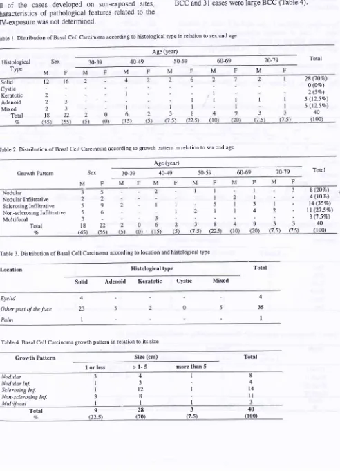
Table 1. Distribution of Basal Cell Carcinoma according to histological type in relation to sex and age
Age (year)
60-69 M M M M M
Sex 30-39 40-49 50-59 't0-79 Total
Histological Type Solid Cystic Keratctic Adenoid Mixed Total 7o l6
l2
(s) 3 3 22 55 2 2 2l8
45 I 3(7 5) 5)
I
3
2 (5Vo) 5 (l2.5Vo) 5 (l2.5Vo)
40 I
Ttble2.Distribution of Basal Cell CarcinoÛra according to growth pattern in relation to sex end age
28 (1OEo)
O (lVo)
Age (year)
Growth Pattcrn Sex 30-39 40-49 50-59 60-69 't0-'79 Total
Nodular
Nodular Infiltrative Sclerosing Infiltrative Non-sclerosrng Infiltrative Multifocal
Total
4o
Table 3. Distribution of Basal Cell Carcirroma according to location and histological type
I l 3 4 9 I I 5 I 8 5 2 9 6 22 55 3 2 5 5 J l8 (4s
8 (2OEo) 4
(ll7o)
t4 (3570)ll
(27.SVo) 3 (7.57o)40 I
Histological type Total
Location
Solid Adenoid Keratotic Cystic Mixed
EyeIid
Other part of the face
Palm 4 35 1 5 0 5 4 23 I
Table 4. Basal Cell Carcinoma growth pattern in relation to its size
Growth Pattern Size (cm) Total
I
or less>1-5
more than 5 i,lodularNodular Inf. Sclerusing Inf.
Non-sclero.sing Inf. Multifttcal 8 4 3 t2 8 I t4
ll
3 Total Vo928340
[image:3.550.31.523.93.774.2]90
Kanoko et alLarge BCC
consisted
oî 5
casesof
nodular type,
3cases
of
nodular
infiltrative
type, l3
cases of
sclerosing
infiltrative type, 8
casesof
non-sclerosinginfiltrative
type,
and 2 casesof muitifocal
type.In
contrast, small
BCC
consistedof
3 casesof
nodular
type,
I
case
of
nodular sclerosing type,
i
case
of
sclerosing
infiltrative
rype,
3
casesof
non-sclerosinginfiltrative
type, and
I
caseof multifocal
type.The
relationship between age, sex and
the
patho-logical
types
of
SCC are shown
in Table
5.
Among
the
16
cases,
12
caseswere well-differentiated
type
Med J lndones
(grade
I)
and
4
cases
were
moderate-differentiated
type (grade
II).
In
respectto
the sun exposure, 9 caseswere
fiom
sun-exposed
sites and
7
cases
were
from
sun-protectedsites (Table
6). No
pathological differences
werefounci between
SCC
from
sun-exposed sitesand
thosefrom
sun-protected sites.When
the casesof
SCC were
divided into small
SCC(less
than
I
cm
in
diameter)
andlarge SCC (more
thanI
cm),
there was nodifference
in
difTèrentiation
of
thecelis between
small SCC
andlarge SCC (Table
7).Table 5. Distribution of Squamous Cell Carcinoma according to histological grading in relation to sex and age
Age (year)
40-49 -50-59 Totrl
Cnde I
Gmde lI
t2
(75Va\
4
(25Eo)
Gnde III 0 0
Gmde lV 0 0 Toial
7)0r0t00r14
(E
)
43.15
56.25 (0) (6.2s) (0) (6.2s) (0)
(0)
(62s)
(625)
(25 0) (r 222
-5)
(12 5)2
(12.5) 0
(0)
(r2.5\
216 (100)Table 6. Distribution of Squemous Cell Carcinoma according 1o location and histologicâl grading
Location Histological graCing
Grade
I
GradeII
GradeIII
GradeIV
Face
Arm Foot
Truink Peni.s
kg
4
)
I 3 I I
3 0 I 0 0 0
0 0
c
0 0 0
0 0 0 0 0 0
7 2
)
3
I
I
Table 7. Distribution of Squamous Cell Carcinoma according to histological grading and size
Grade Sizc (cm) Total
I
or less>l-5
more than 5Grade
I
GradeII
GradeIII
Grade IV6 I
3 2
3
I
12 (75Vo)
4 (25Eo)
Total
7Vol 9, No 2, April
-
June 2000DISCUSSION
Basal
cell
carcinoma
is
locally
invasive, slowly
spreading
tumor
which
rarely
metastasize'
Histo-patologically, the
characteristic
cell
of
the
basal
cell
carcinoma,
have a'large, oval
or
elongated
nucleusand
relatively little
cytoplasm'
For a long time, basal
cell
carcinoma has been considered
to be developed
from
basal
cell
of
epidermis,
but
nowadays,
it
hasbeen suggested
that basal
cell
carcinoma
might be
developedfrorn
a morepluripotent
stemcell'
According
to
the
differentiation
of
cells,
BCC
is
divided into
solid type
if
it
is undifferentiated, adenoid
type
if
it
stic
type,
wi
andkelatotic
of
keratiniz
this
classification
was
believed
to
be a good criteria for
predicting the
prognosis
of
the tumor,
since usually
ihere is
correlation
betv,'eenthe grade
of
malignancy
and thedifferentiation of
tumor cells,
as seenin
other
malignancies.Recently,
classification
of
basal
cell
carcinoma
according
to the growth
pattern
has been consideredto
be
môre correlated
to
the aggressiveness
of
thetumor
and ispotential
to recurrence.Squamous
cell
carcinoma
may
show considerable
variation. The cells usually show
grade
variation in
size.
Variants
of
squamous
cell
carcinoma
areadenoid
squarnous
cell
carcinorna
and spindle cell
type
squamouscell
carcinoma.Biodëis
has
divided
squamouscell
carcinoma using
the percentage
of the
undifferentiated cells
into
gradeL
U;
III
and[V.
However,
it
is not
easy tocount
thecells,
soin
later publication
thecriteria
wasmodified'
Sun-exposure
is
a
crucial
carcinogen
of
NMSC'
CC
to
TT
mutations
andC to
T
mutations
at dipyrimidinesites are
frequently
observedin
p53 tumor
suppressor geneof
NMSC developed on sun-exposed
sites'7In
the
current study,
we
attempted
to
analyze the
pathological characteristics
of
BCC
and SCC,
in
ielation
to
the effect
of
sun-exposure.
In
previous
report, adenoid
type)
of SCC
hasun-exposure.t SCC
in this
study.Histopaîhology of BCC and
SCC
91Our
data showed
that
sclerosing
infiltrative
type of
BCC was frequently found
in
large
BCC.
This
isconsistent
with
the previous study that
showed that
the
sclerosing
infiltrative type
of
BCC
was
moreaggressive
as
measured
by.
the high
number
of
AgNOR
andPCNA
positivitY.'''
Larger number
of the
casesof NMSC
will
be necessaryfor
evaluating
the
histopathological
characteristicsassociated
with
UV
exposure.
In
addition
tomorphoiogical analysis
on
IIE
stained section,
studyon
pSf or Ki67
by immunohistochemistry may help
todistlnguish the difference
in
relation
to
uv-induced
molecular changes.Acknowledgement
Central
GeneralHospital, we would like to
thank
histechnical
assistance.This work
hâs been supportedby
Fund and the
Terry Fox Foundation,
Canada.REFERENCES
l.
Cornain S, MangunkusumoR,
NasarIM,
Prihartono J' Ten most frequent cancerin
Indonesia' Pathology Based Cancer Registry Dataof
1988-1992.In:
Cancer Registryin
Indonesia, National Cancer Registry Center, Jakarta 1997.2.
Sarjadi. Cancer Incidence i985-1989
in
SemarangIndonesia. Indonesian Cancer Society 1990'
3.
UedaM,
KanokoM,
Comain S, HamzahM,
Poetiray E'Ichihashi
M,
Prihartono
J,
Ohno
Y'
Molecularpathological changes induced
by
ultravioletlight
during ihe developmentcf
skin cancer. Med.I.
Indonesia, 2000;9:ll8-22.
4.
MunakataN,
ComainS,
Mulyadi
K,
IchihashiM'
Prihartono J, Ohno
Y,
HamzahM,
KanokoM,
PoetirayE, Budiningsih
S,
TJartaA,
UedaM'
TJiptoH'
Td
Mukhtar A.-Biologically effective doses of ambient
92 Kanoko et al
Lever WF, Lever GS. Histopathology of the skin. 7th ed.
JB Lippincott Comp. Pbilladelphia. 1990.
Sloane JP.
The
valuetyping
basalcell carcinoma in
predicting
recurrenceafter
surgical
excision.BR
JDermatol. 197 7, 96:127 -32.
Nakazawa H, Englis[r D, Randell PL, Nakazawa K, Martel N, Armstrong BK, et al.
UV
and skin cancer specific p53gene mutatiorr
in normal
skin as a biologically relevantexposure measurement. Proc Natl Acad Sci 1994;91:3('C4.
Ieksono
P,
Nilai
AgNOR
sebagaipptunjuk
untuk menentukanresidif
pada basalioma. Thesisin
partial fulfillment of the specialty examination in pathology. 1995.Med J Indones
Kanoko M. Index
of
Proliierating Cell Nuclear Antigen(PCNA)
in
Basalioma;correlation
with the
histo-pathological type. Presented
in
the 4th Congress Asia-Pacihc Association of Societiesof
Pathologists. Beijing, China. 1995.Ro YS,
Kim
JH. Immunohistochemical analysis of p53protein expression
in
benign and malignant skin tumors using a panelof anti-p53 antibodies.
J
Korean Med Sci1993; 8: 361-6,
Ro YS. Oncogene interaction
in
basal cell carcinomasof
human skin. J Korean Med Sci 1995;
l0:
85-925. 9.
10.
I
l.
6
7.