Sunlight–derived vitamin D affects interleukin-4 level, T helper 2
serum cytokines, in patients with Graves’ disease: a prospective
cohort study
Keywords: Graves’ disease, IFN-γ, IL-4, sunlight exposure, Th1 and Th2 pathway, vitamin D
pISSN: 0853-1773 • eISSN: 2252-8083 • http://dx.doi.org/10.13181/mji.v24i4.1270 • Med J Indones. 2015;24:228–33 • Received 20 Agu 2015 • Accepted 02 Dec 2015
Correspondence author: Dyah Purnamasari, [email protected]
Copyright @ 2015 Authors. This is an open access article distributed under the terms of the Creative Commons Attribution-NonCommercial 4.0 International License (http://creativecommons.org/licenses/by-nc/4.0/), which permits unrestricted non-commercial use, distribution, and reproduction in any medium, provided the original author and source are properly cited.
Dyah Purnamasari, Pradana Soewondo, Samsuridjal Djauzi
Department of Internal Medicine, Faculty of Medicine, Universitas Indonesia, Jakarta, Indonesia C l i n i c a l Re s e a rc h
ABSTRAK
Latar belakang: Penyakit Graves (GD) merupakan penyakit autoimun paling sering dijumpai yang menyebabkan hipertiroidisme. Peran keseimbangan jalur Thelper 1 (Th1)/T helper 2 (Th2) pada GD masih diperdebatkan. Vitamin D dilaporkan memiliki efek terhadap kedua jalur tersebut. Studi ini bertujuan mengetahui efek pajanan sinar matahari terhadap kadar vitamin D 25(OH) dan kadar sitokin jalur Th1/Th2 pada pasien GD.
Metode: Studi ini menggunakan desain kohort prospektif untuk membandingkan efek pajanan matahari terhadap kadar vitamin D 25(OH) dan sitokin jalur Th1 serta Th2 pada 32 pasien GD yang belum pernah remisi, yang dibagi menjadi 2 kelompok: kelompok GD pajanan dan non-pajanan. Kelompok pajanan mendapatkan intervensi berupa pajanan sinar matahari 3 kali seminggu dengan durasi 30 menit/kali, pukul 09.00–11.00 WIB, selama satu bulan. Data laboratorium berupa kadar thyroid stimulating hormone (TSH), free thyroxin (fT4), vitamin D 25(OH), interferon-γ (IFN-γ), dan interleukin-4 (IL-4) dinilai sebelum dan sesudah pajanan. Analisis data menggunakan uji t berpasangan atau uji Mann Whitney.
Hasil: Pajanan sinar matahari selama satu bulan meningkatkan kadar vitamin D 25(OH) sebanyak 27,90% pada kelompok GD pajanan (15,34 ng/mL ke 19,62 ng/mL, p<0,001), sementara kadar vitamin D kelompok GD non-pajanan turun dari 20,48 ng/mL menjadi 18,86 ng/mL (p<0,001). Dari dua kelompok, kenaikan kadar vitamin D 25(OH) kelompok GD pajanan tidak diiringi dengan peningkatan kadar IL-4 setelah pajanan matahari. Kesimpulan: Pajanan sinar matahari meningkatkan kadar vitamin D 25(OH) serum dan mempengaruhi kadar IL-4, sitokin jalur Th2, pada pasien GD. Peran vitamin D yang berasal dari pajanan sinar matahari pada kadar IFN-γ pasien GD tidak dapat disimpulkan dalam studi ini.
ABSTRACT
Background: Graves’ disease (GD) is the most common autoimmune disease leading to hyperthyroidism. The role of Th1/Th2 pathways balance in GD is still controversial. Vitamin D is reported to have an effect on those pathways. This study aims to examine the effect of sunlight exposure on vitamin D 25(OH) level and Th1 and Th2 pathway-derived cytokines in GD patients.
Methods: A prospective cohort study was conducted on 32 GD patients to compare the effect of sunlight exposure on vitamin D level and cytokines of Th1 and Th2 pathways between exposed and unexposed groups. Exposed group received sunlight exposure three times a week for 30 minutes each between 9–11 a.m for 1 month. Thyroid stimulating hormone (TSH), free thyroxin (fT4), vitamin D 25(OH), interferon-γ (IFN-γ) and interleukin-4 (IL-4) serum levels, were investigated before and after one month of sunlight exposure. Paired t-test or Mann Whitney test were used to analyze the difference between exposed and unexposed GD groups before and after sun exposure.
Results: One month of sunlight exposure increased vitamin D 25(OH) level by 27.90% among exposed GD group (15.34 ng/mL to 19.62 ng/mL, p<0.001). Meanwhile, unexposed GD group’s vitamin D 25(OH) level decreased from 20.48 ng/mL to 18.86 ng/mL (p<0.001). Increased vitamin D 25(OH) level in exposed group was not accompanied by the increase of IL-4 level after sunlight exposure.
Graves’ disease (GD) is the most common cause of hyperthyroidism and is the second most prevalent endocrine diagnosis after diabetes mellitus. Its prevalence is higher among female than that of male (0.5–2% vs 0.2%).1 GD may affect upon all age groups, but the peak age is 20–49 years old. The incidence of GD is estimated to be 0.8/1,000 per year.2
Hyperthyroidism in GD was caused by autoantibody binding to thyroid stimulating hormone receptor (TSHR) on basolateral membrane of thyroid epithelium. When TSHR is stimulated, thyroid hormone will be secreted in excessive amount,
causing hyperthyroidism.3 Until now, the
pathogenesis of GD has not been fully defined. The combined effect of environmental factors and genetic predisposition is believed to disrupt the tolerance toward self-antigen, resulting in autoimmune reaction. The regulation of immune system involves balanced work of T helper 1 (Th1) and T helper 2 (Th2) pathways. Both Th1 and Th2 pathways secrete different kinds of cytokines which have significant roles in the pathogenesis of diseases, including autoimmune diseases. These two cell types directly affect immune response via different pathways. Th1 pathway cytokines, interferon-γ (IFN-γ), tumor necrosis factor-α (TNF-α), interleukin-2 (IL-2), contribute to cellular immunity responsible for fighting viruses and other intracellular pathogens, eliminating cancer cells and stimulating slow-type hypersensitivity reaction on the skin. Th2 pathway cytokines (4, 5, IL-6, IL-9, IL-10, IL-13) are more dominant in humoral immunity, regulating the production of antibodies to fight organisms outside the cell. Excessive activity of either pathway may lead to disruption of the other.4
Cytokines are considered as environmental factors which may play important role in the natural history of GD. Cytokines of both Th1 and Th2 pathways in hyperthyroidism have been studied, both in animal model and human. Several studies in human showed that Th2 pathway is predominant in GD, although both of those pathways were said to have similar role in animal model. A study by Nagayama et al5
showed roles of Th1 pathway cytokine (IFN-γ) and Th2 pathway cytokine (IL-4) in hyperthyroidism in two different animal models (TSHR-adeno model vs. TSHR-M12 model). As opposed to Nagayama et al5 a study done by Phenekos, et al6 showed that GD patients had higher Th2 pathway cytokine (IL-4) level than IL-4 level in Hashimoto thyroiditis
patients. Several factors contribute to the activities of Th1/Th2, one of them is vitamin D.
Active form of vitamin D, calcitriol (1.25-dihydroxyvitamin D3), affects the action of T lymphocytes by inhibiting Th1 proliferation which leads to decrease on macrophage activation and IFN-γ and IL-2 production. Meanwhile, calcitriol increases the amount of Th2 cells by facilitating dendritic cells/antigen presenting cells (APCs) to produce cytokines (IL-4, IL-5, IL-10) that support T cell differentiation towards Th2.7,8
As immune regulator, low vitamin D level is related to a number of autoimmune diseases, one of them being GD.7 The study of Yamashita et al9 revealed that vitamin D 25(OH) level was lower in patients with GD compared to that of healthy subjects. Furthermore, vitamin D 25(OH) level in female patients with GD was lower than in male patients (31.8±13.3 vs 41.3±15.0 nmol/L, p<0.001). This finding was strengthened by the study of Yasuda et al10 which showed significantly lower vitamin D 25(OH) serum level in GD patients who did not have remission compared to those in patients with remission, or normal subjects (14.5±2.9 ng/mL in non-remission vs 18.2±5.1 ng/mL in remission p<0.005).10 Both studies suggest the role of vitamin D level in the pathogenesis of GD.
Eventhough, patients with GD are known to have lower vitamin D 25(OH) level than that of healthy subjects and vitamin D theoretically plays a role in the activity of Th1 and Th2 pathways, the benefit of vitamin D intervention in altering the action of Th1 and Th2 in GD has not been identified. This study was aimed to determine vitamin D 25(OH) serum level in patients with GD and examine the effect of sunlight exposure as natural vitamin D source on vitamin D 25(OH) serum level and Th1/Th2 pathway-derived cytokines (IFN-γ and IL-4) level.
METHODS
interview, physical examination, laboratory tests [free thyroxin/fT4, thyroid stimulating hormones (TSHs), and TSH receptor antibody (TRAb)] and thyroid scintigraphy when necessary. Inclusion criteria were female patients with non-remission GD who consented to take part in the study. Patients who were pregnant, had severe liver or kidney problems, cancer, other autoimmune diseases and vitamin D supplementation were excluded.
Grave’s disease patients were recruited by consecutive sampling and divided into two groups, exposed group and unexposed group. Exposed group received intervention in the form of sunlight exposure three times a week for one month. Subjects were asked to expose themselves (face, both arms, and legs) under the sun without using sun-block agent between 9 to 11 a.m (GMT+7) for approximately 30 minutes. This intervention took place between August to October 2013, during which ultraviolet (UV) power equaled 1.0-1.5 MED (measured by Solarmeter®). Unexposed GD patients were not requested to expose themselves to sunlight but were asked to go about their day as per usual. The protocol of this study has been approved by the Ethical Committee of the Faculty of Medicine, Universitas Indonesia (No. 441/ H2.F1/ETIK/2013).
Study procedures
All subjects underwent clinical interview, physical examination, and blood sample drawing. Clinical interview aimed to gather data about identity, disease, and medication history, as well as to assess sun protection score and sun exposure score. Sun protection scoring is an evaluation of how subjects protect themselves from sun exposure, measured with a questionnaire featuring questions about outdoor clothings and attributes (sun-block agents, veil, hat, long-sleeved shirt, gloves, trousers, long skirt, umbrella). Positive use of each item is valued one point. Sun exposure scoring is a means to predict the duration of sunlight exposure on subjects in one week. Total score is calculated by multiplying the amount of daily exposure (minute/day) with the amount of exposure per week (day/week).
Blood parameters included TSH, fT4, vitamin D 25(OH), IFN-γ, and IL-4 serum level. Vitamin D 25(OH)2 (Euroimmune®), IFN-γ, and IL-4 (Quantikine, R&D system) were measured using
enzyme-linked immunosorbent assay (ELISA) human kit, before and after one month of sunlight exposure.
Data analysis
Numerical data were presented in mean (standard deviation) or median (minimum–maximum) based on the data distribution. Bivariate analysis using paired t-test was used to analyze the difference between exposed and unexposed GD groups before and after sun exposure. Data were processed using SPSS for Windows v.12.
RESULTS
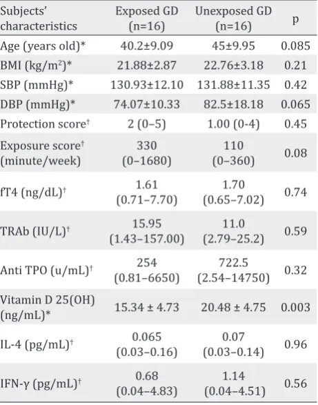
During study period there were 32 subjects with GD. Among 32 patients with GD, there were 14 hyperthyroid subjects and eight euthyroid subjects. Table 1 shows that there were some significant differences of several parameters
Subjects’ Age (years old)* 40.2±9.09 45±9.95 0.085 BMI (kg/m2)* 21.88±2.87 22.76±3.18 0.21
SBP (mmHg)* 130.93±12.10 131.88±11.35 0.42 DBP (mmHg)* 74.07±10.33 82.5±18.18 0.065 Protection score† 2 (0–5) 1.00 (0-4) 0.45
Exposure score†
†Mann-Whitney test, data is presented in median (min-max);
*Paired t-test, data is presented in mean±SD; BMI=body mass index; SBP=systolic blood pressure; DBP=diastolic blood pressure; fT4=free thyroxin; TRAb=thyroid stimulat-ing hormone receptor antibody; Anti TPO=anti thyroperoxi-dase; IL-4=interleukin-4; IFN-γ=interferon-γ
between exposed GD group and unexposed GD groups before this study commenced. At the beginning of study, exposed GD group had higher sun exposure score, but lower vitamin D 25(OH) level compared to unexposed GD group.
Vitamin D, IL-4 and IFN-γ level before and after sun exposure in GD group
Though at the beginning of this study, the vitamin D 25(OH) level of exposed GD group was lower than unexposed GD group, it increased in GD group after one month of sun exposure, while it decreased in unexposed group (Figure 1).
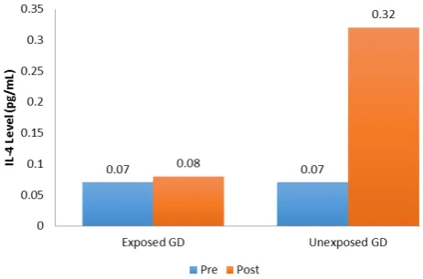
At the end of the study, cytokine IL-4 significantly increased in unexposed GD group, but did not increase in exposed GD group. Both exposed GD and unexposed GD had significantly higher IFN-γ levels at the end of this study (Figures 2
Figure 1. Vitamin D 25(OH) level pre- and post- sun expo-sure in exposed and unexposed GD Groups
Figure 2. IL-4 level pre- and post- sun exposure in exposed and unexposed GD groups
Figure 3. IFN-γ level pre- and post- study period in exposed
and unexposed GD groups. The IFN-γ level increased signifi -cantly in both groups after one month
and 3).
DISCUSSION
Graves’ disease patients with normal hormone level were included as subjects to examine the effect of sunlight exposure to varying GD population in outpatients of CMH. Mean age of subjects with GD is around 42 years old. This finding is linear to Vanderpump2 data which showed peak age of GD incidence to be from 20 to 49 years old.2 In Japan, Yamashita et al9 and Yasuda et al10 study showed mean age of 37 to 39 years old.
In this study, vitamin D level of exposed and unexposed groups are 15.34±4.73 ng/mL and 20.48±4.75 ng/mL respectively. This finding is similar to the results of studies done in Japan. Yamashita et al9 showed that vitamin D level of male and female GD subjects are 16.54±6.00 and 12.74±5.33 ng/mL. Yasuda et al10 also showed that vitamin D level of new onset female GD patients of 14.4±4.9 ng/mL.
Vitamin D 25(OH) and Th1 and Th2 pathway-derived cytokines among GD group
Exposed GD group had lower vitamin D 25(OH) level compared to unexposed GD group at the initiation of this study, but there were no differences found regarding IFN-γ and IL-4 levels between exposed and unexposed GD groups.
to 18.86 ng/mL). Although both groups did not have similar range of vitamin D 25(OH), sunlight exposure to exposed GD group may elevate vitamin D 25(OH) level significantly while in unexposed GD group this vitamin D 25(OH) level diminished instead. This finding is in line with the study of Setiati et al11 on elderly population in Jakarta, which showed that three times weekly sunlight exposure for 30 minutes each at one MED might significantly increase vitamin D 25(OH) level by 42.37% among elderly people who live in institutionalized care. Our study showed that the same method of sunlight exposure was associated with an increase of vitamin D 25(OH) level in GD patients by 27.90%.
While Th1 and Th2-derived cytokines profiles have been reported several times, the effect of sunlight exposure on GD patients through vitamin D 25(OH) level has not been yet reported. After one month of sunlight exposure, IL-4 level did not increase in exposed GD group (0.07 pg/mL to 0.08 pg/mL, p=0.293), while it significantly increased in unexposed GD group (0.07 pg/mL to 0.32 pg/mL, p=0.001). Non-increase of IL-4 level in exposed GD patients may be related to elevated vitamin D 25(OH) level in response to sunlight exposure. Vitamin D promotes Th2 cytokines pathway, however several studies reported inconsistency. Based on our study, vitamin D tends to be an immune balancer, which will suppress the overactive pathways. Since there is predominance of Th2 cytokines pathway in GD, vitamin D will prevent the increment of Th2 cytokines pathway. Up until now, there is no study which reported the inverse relationship between vitamin D and IL-4 level. Nonetheless, a longer observasional cohort study using more sensitive tool is needed to evaluate the effect of vitamin D 25(OH) on IL-4 cytokine.
After one month of sunlight exposure, IFN-γ level elevated significantly, in both exposed GD and unexposed GD groups (Figure 3). The elevation of IFN-γ level can not be clearly explained in this study. Among exposed GD patients, the elevation of IFN-γ level could be due to the suppression effect of sunlight-derived vitamin D on Th2 pathway.
To the best of our knowledge, the study about the effect of sunlight exposure, on vitamin D 25(OH) and Th1/Th2 pathways-derived cytokines levels has not been yet reported. Although one month
intervention using sunlight exposure increased vitamin D 25(OH) level in GD group, this study could not show direct effect of sunlight to Th1 and Th2 pathway cytokines. Out of two groups, there was only in exposed GD group that increasing level of vitamin D 25(OH) not accompanied by IL-4 level increment after one month of sunlight exposure. This finding showed the likelihood of sunlight effect on Th2-derived cytokines through vitamin D 25(OH) level.
This study has some limitation in terms of study subjects and sampling technique. Inclusion of GD subjects with normal fT4 at the beginning of the study hinders optimal results to represent patients with active hyperthyroidism. Consecutive recruitment of study subjects has lead to non-balance distribution in the two groups. Patients who wanted to participate but could not follow the exposure program were allocated for unexposed group. Sampling strategy for cytokine testing (serum, monocyte isolates, or peripheral blood mononuclear cell/PBMC) also affects study results. Cytokine analysis on blood serum is inferior because Th1/Th2 cytokines are also produced by other immune cells (CD8+, B cells, macrophages), therefore rendered unspecific.
In conclusion, sunlight exposure increases vitamin D serum level and may affect the level of IL-4, Th2 pathway-derived cytokine, in patients with GD. The role of sunlight-derived vitamin D on IFN-γ, Th1 pathway-derived cytokine, in GD patients can not be concluded in this study.
Acknowledgments
This study is supported by research grant of Universitas Indonesia, 2013. The content is solely the responsibility of authors and does not necessarily represent general view of Universitas Indonesia.
Conflict of interests
The authors affirm no conflict of interest in this study.
REFERENCES
1. Orgiazzi J. Thyroid autoimmunity. Presse Med. 2012;41(12P2):e611–25.
3. Prabhakar BS, Bahn RS, Smith TJ. Current perspective on the pathogenesis of Graves’ disease and ophthalmopathy. Endocr Rev. 2003;24(6):802–35.
4. Kidd P. Th1/Th2 balance: The hypothesis, its limitations, and implications for health and disease. Altern Med Rev. 2003;8(3):223–46.
5. Nagayama Y, Saitoh O, Mclachlan SM, Rapoport B, Kano H, Kumazawa Y. TSH receptor-adenovirus-induced Graves’ hyperthyroidism is attenuated in both interferon-gamma and interleukin-4 knockout mice; implication for the Th1/ Th2 paradigm. Clin Exp Immunol. 2004;138(3):417–22. 6. Phenekos C, Vryonidou A, Gritzapis AD, Baxevanis CN,
Goula M, Papamichail M. Th1 and Th2 serum cytokine profiles characterize patients with Hashimoto’s thyroiditis (Th1) and Graves’ disease (Th2). Neuroimmunomodulation. 2004;11(4):209–13.
7. Wahono CS, Rusmini H, Soelistyoningsih D, Hakim R, Handono K, Endharti AT, et al. Effects of 1,25(OH)2D3 in immune response regulation of systemic lupus
erithematosus (SLE) patient with hypovitamin D. Int J Clin Exp Med. 2014;7(1):22–31.
8. Antico A, Tampoia M, Tozzoli R, Bizzaro N. Can supplementation with vitamin D reduce the risk or modify the course of autoimmune disease? A systematic review of the literature. Autoimmun Rev. 2012;12(2):127–36.
9. Yamashita H, Noguchi S, Takatsu K, Koike E, Murakami T, Watanabe S, et al. High prevalence of vitamin D deficiency in Japanese female patients with Graves’ disease. Endocr J. 2001;48(1):63–9.
10. Yasuda T, Okamoto Y, Hamada N, Miyashita K, Takahara M, Sakamoto F, et al. Serum vitamin D levels are decreased in patients without remission of Graves’ disease. Endocrine. 2013;43(1):230–2.