The application of human umbilical cord blood mononuclear cells in
the management of deep partial thickness burn
Yefta Moenadjat,1 Maurin Merlina,2 Camy F. Surjadi,2 Caroline T. Sardjono,2 Yuyus Kusnadi,2 Ferry Sandra2
1 Burn Unit, Cipto Mangunkusumo Hospital, Jakarta, Indonesia
2 Stem Cell Clinical Study Division, Stem Cell & Cancer Institute, PT Kalbe Farma Tbk., Jakarta, Indonesia
Abstrak
Latar belakang: Penyembuhan luka bakar merupakan proses rumit dan penutupan luka sering dihadapkan pada berbagai masalah. Berkembangnya bukti-bukti keberhasilan terapi sel punca pada regenerasi kulit menarik perhatian banyak klinisi yang mengelola kasus luka bakar. Diantara sumber sel punca alogenik yang tersedia, darah tali pusat mudah diperoleh dan tidak dihadapkan pada masalah etik, dan mengandung sel punca multipoten dengan imunogenisitas rendah. Penelitian ini ditujukan untuk evaluasi potensi terapi sel mononuclear darah tali pusat manusia (hUCBMNCs) pada proses re-epitelialisasi luka bakar derajat dua dalam.
Metode: Dua puluh pasien dengan luka bakar derajat dua dalam diberikan 2 x 10⁷ hUCBMNCs secara topikal. Sebagai kontrol, setiap pasien juga menerima olesan silver sulfadiazine (SSD) untuk luka sebanding yang terdapat pada lokasi tubuh lain. Kedua perlakuan diberikan sebanyak enam kali berturut-turut dengan selang waktu satu hari. Luas luka diukur menggunakan Visitrak® pada hari ke-0, 7, dan 11. Intensitas nyeri pada pergantian balutan dinilai berdasarkan skala Wong Baker. Dilakukan pemeriksaan histologi pada beberapa sampel biopsi kulit pada luka yang sudah mengalami re-epitelialisasi pasca perlakuan. Dalam evaluasi keamanan terapi, dilakukan pemeriksaan HLA pada beberapa sampel. Kecepatan penyembuhan luka dianalisis dengan uji Wilcoxon Signed Rank.
Hasil: Enam belas pasien menunjukkan penutupan luka lebih cepat secara bermakna pada luka yang mendapatkan hUCBMNCs dibandingkan SSD; pada hari ke-7 (p = 0,041) dan 11 (p = 0,021). Penurunan intensitas nyeri dijumpai lebih sering pada kelompok perlakuan hUCBMNCs dibandingkan SSD; dari rata-rata skala 3 ke 1/0 pada hari ke-7 dan 11. Tidak ada reaksi alergi, penolakan, dan infeksi yang ditemukan pada studi ini, menunjukkan bahwa terapi hUCBMNCs tergolong aman, meski dijumpai ketidakcocokan HLA. Studi histologi menunjukkan terbentuknya penjuluran–pertautan epidermis ke lapis dermis menyerupai konigurasi kulit normal.
Kesimpulan: hUCBMNCs dinilai aman dan berpotensi dalam re-epitelialisasi luka lebih cepat dan baik pada luka bakar derajat dua dibandingkan dengan terapi konvensional. (Med J Indones. 2013;22:92-9)
Abstract
Background: Wound healing in burn is a complex process and early complete wound closure still enfaces many problems. Application of stem cells is found to be the future method of wound healing. Among the available sources of allogenic stem cells, umbilical cord blood is quite easy to be obtained, has less ethical issue, and contain multipotent stem cells, which are characterized by low immunogenicity. The study aims to evaluate the potential of human umbilical cord blood mononuclear cells (hUCBMNCs) treatment in the management of deep partial thickness burns.
Methods: Twenty patients with deep partial thickness burns were treated with topical application of 2 x 10⁷ hUCBMNCs
and silver sulfadiazine (SSD) cream on the comparable wound size in the other sites. The treatments were applied for six times in every two consecutive days. Wound surface area was measured with Visitrak® on day 0, 7, and 11. Pain intensity
was evaluated using Wong Baker’s faces scale on each wound dressing change. Histology examination was performed in some samples of collected skin biopsy of the newly re-epithelialized area of hUCBMNCs and SSD-treated wound at the end of treatment. HLA typing is used to evaluate the issue of safety. Wilcoxon signed rank test was used to compare the rate of wound healing.
Results: Sixteen patients of hUCBMNCs-treated showed a signiicant wound closure in faster than SSD-treated; measured
on day 7 (p = 0.041) and day 11 (p = 0.021). Number of patients with reduced pain intensity, from approximately scale 3 to 1/0 on day 7 and 11, were higher in hUCBMNCs-treated compared to SSD-treated wound. In spite of the HLA-mismatch, no allergic reaction, rejection, and infection found on hUCBMNCs-treated wound suggested the safety of this therapy. Histology examination found the formation of dermal-epidermal junction and rete ridges equal to the normal skin on hUCBMNCs-treated wounds.
Conclusion: hUCBMNCs are effective and safe to promote re-epithelialization in deep partial thickness burns.
(Med J Indones. 2013;22:92-9)
Keywords: Deep partial thickness burn, mononuclear cells, re-epithelialization, umbilical cord blood
Correspondence email to: [email protected]
Following exposure to thermal source, disintegration of skin and the tissue beneath leads to circulatory derangement and impaired perfusion to the local traumatized site, which in turn will decrease the capability to provide its normal functions, i.e. control
of evaporation, barrier protection, etc.1 With this defect,
scar formation in a later date is the major problem in burns. For such a reason, aggressive effort to obtain on time schedule of wound surface re-epithelialization is one policy in the management of deep partial and full thickness burns, as the structure of skin integuments is damaged and spontaneous re-epithelialization is almost impossible.2
The aggressive wound management in burns includes the concept to remove non-vital tissues such as eschar following resuscitation and after stable hemodynamic condition has been achieved. In standard conventional burn management, topical cream of silver sulfadiazine applied on the wound surface and waited for days to obtain information of wound progress. Afterwards, the clinician decide the method of wound closure, either spontaneous or surgically intervention.3 Mostly,
the management of deep partial thickness burns in the hospital unit referred to conservative treatment. In this kind of treatment, a various modern dressings of biological and biosynthetic materials have been applied.4
The development of cell-based technology in burn management started as cultured epithelial auto graft (CEA) proposed as the better option to solve the problem in major burns as there is limitation of skin graft donor.5,6 Nevertheless, there were disadvantages found with its application in clinical setting;7 such conditions has provoked the researchers to ind
comprehensive and better skin regeneration as well as skin replacement. This was found paralleled to the discovery of stem cells. Following the huge numbers of researches in stem cells following its discovery, it was then found that umbilical cord blood have the potency of renewal or to regenerate certain types of
cells, including keratinocytes; both in vitro8 and in
vivo.9
The goal of this study is to ind information of
re-epithelialization of applied human umbilical cord blood mononuclear cells (hUCBMNCs) on the deep partial thickness burns, compared to silver
sulfadiazine (SSD). The objective is to ind the safety and eficacy of hUCBMNCs in treatment of
deep partial thickness burns.
METHODS
Study design
This comparative study evaluates the treatment of deep partial thickness burns using hUCBMNCs (Stem Cells Institute, Jakarta, Indonesia) and SSD (Burnazin, Darya–Varya, Jakarta) in enrolled patients who were
admitted to the Burn Unit, Cipto Mangunkusumo Hospital in Jakarta from 2008 to 2009. The study has been approved by the Ethical Committee of Research in Faculty of Medicine, Universitas Indonesia (Ethical Approval no 307/PT2.FK/ETIK/2008).
Patient selection
The subjects were patients with deep partial thickness
burn (lame and scald). Patient selection was based on the inclusion criteria, i.e. young adults (age of ≥ 17 years old) with burns less than ≤ 30% of total body
surface area. Patients with inhalation injury, chemical, and electric burns and with unstable hemodynamic condition were excluded as well as patients, who had any disease prior to trauma, secondary infection, or history of hypersensitivity to citrate or dimethyl sulfoxide (DMSO) and pregnant women. Two equal wound sizes in different comparable sites in each recruited patient were selected for treatment using hUCBMNCs and SSD.
Preparation of hUCBMNCs
Human umbilical cord blood (hUCB) was obtained by aspiration following delivery through mother’s consent, and was processed within 48 hour after collection. From each collected hUCB, aliquots were set apart for bacterial and fungal screening. Human UCB was processed by differential centrifugation (Sepax®, Biosafe, Eysins, Switzerland) in combination
with hydroxyl ethyl starch (HemoHes, B.Braun, Melsungen, Germany) to obtain mononuclear cells. The hUCBMNCs were marked by CD34+ and counted
using low cytometry. Viability was tested by means
of trypan blue exclusion method. The hUCBMNCs
were kept in cryotubes in the presence of 10% DMSO,
placed in controlled rate freezer to reach -80°C and then transferred to -196°C in vaporized liquid nitrogen cryotank. Prior to each clinical application, cryotubes were taken out from the cryotank and thawed in 37°C water bath for 2 minutes. Immediately after thawing the cell suspension was washed with phosphate buffered saline (PBS). Total hUCBMNCs of 2 x 10⁷ with viability of more than 70% were prepared in 1 mL
PBS and transported to the burn unit at low temperature (2-8°C) for each single application.
HLA typing
DNA from donor and recipient was ampliied using
HLA typing kit (SSP UniTray kit, InvitrogenTM, Carlsbad, CA) and run for gel electrophoresis. Briely,
µL DNA templates. Polymerase chain reaction (PCR) was carried out in a 96-well polycarbonate PCR tray
containing 5 µL/ well of optimized HLA allele-speciic primer overlaid with parafin oil. Soon after cycling was completed, the amplicons were loaded onto a 2%
agarose gel for electrophoresis. The ethidium bromide-stained gel was photographed and the visible bands were interpreted using software (UniMatch® Plus,
InvitrogenTM, Carlsbad, CA) to determine the HLA-A,
-B, -DR, -DQ types.
Treatment
Two selected wounds from each patient were prepared. After surgically derided, wound surface area was drawn with red colored permanent ink marker pen in a grid lined of 0.5 cm square sterile plastic transparent sheet. The wounds were covered with nonpetroleum-based tulle (Atrauman®, Hartmann,
Heidenheim, Germany). One mL solution containing hUCBMNCs was topically applied on the wound surface. Accordingly, SSD cream was applied to the other wound surface. The wound was then covered with sterile moist gauze to obtain a suitable wound environment for re-epithelialization. The dressing change as well as the procedure was re-applied for 5 times, every two consecutive days.
Clinical evaluation
Clinical parameters i.e. wound surface area; pain
intensity during dressing change, sign of infection, and rejection were subjects to evaluation and recorded. Wound surface area was measured using electronic wound measurement device (Visitrak®, Smith &
Nephew, St. Laurent, Canada) on day 0, 7, and, 11 of application. Decreasing size of wound surface area represented re-epithelialization progress. Pain intensity was measured using Wong Baker’s pain intensity scale, which is ranged from 0-5. Bacteriological swab (BBLTM CultureSwabTM Plus, Becton Dickinson
and Co., Franklin Lakes, NJ) from each treated surface was collected and further microbial test was performed for the presence of aerobic and anaerobic bacteria. Increasing exudates and clinical signs of acute
inlammation of surrounding wound was monitored as
a parameter of rejection.
Histological examination
Skin biopsy from the hUCBMNCs- and SSD-treated wounds was carried out on the end of the treatment
(day 11). The samples were ixed in 4% formaldehyde
freshly prepared from paraformaldehyde and
prepared for parafin block. Slices of 4 mm thickness
were mounted on object glass and stained with hematoxylin-eosin.
Statistical analysis
Wound surface re-epithelialization of hUCBMNCs- and SSD-treated on day 7 and 11 were subjected to analysis. For statistical analysis, Wilcoxon Signed Rank test was used, since the data involved 2 groups, related, numerical type, and not normally distributed according to Saphiro-Wilk test. Computation was done
using SPSS Statistics 17.0 software; p value of < 0.05 is considered statistically signiicant.
RESULTS
Twenty patients were enrolled in this study. Two patients were excluded from the trial for self-withdrawal. One patient died before the treatment was completed and another one was excluded from the analysis due to incomplete data. In the end of the study, sixteen cases were analyzed.
Re-epithelialization
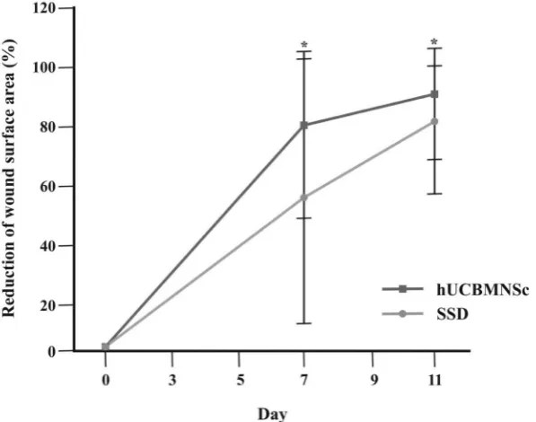
As shown in Figure 1, hUCBMNCs treatment results in better progress of wound re-epithelialization (i.e. decreased wound surface area) on day 7 after application compared to SSD treatment. The difference
is statistically signiicant (hUCBMNCs = 77.2% ± 28.8%, SSD = 56.8% ± 47.3%; p = 0.041). This was
consistently observed also on day 11, i.e. at the end
of treatment (hUCBMNCs = 89.7% ± 23.6%, SSD = 78.8% ± 28.0%; p = 0.021).
Issue of pain during dressing change
Pain was the issue in the irst day of application (mode
= 3, hurts even more). The number of patients feeling ‘no hurt’ (scale 0) and ‘hurts little bit’ (scale 1) were noted on day 7 and 11 to compare pain issues between hUCBMNCs- and SSD-treatment. There were more patients with pain relieve on hUCBMNCs-treated compared to SSD-treated wounds (Figure 2).
Rejection and signs of infection
The differences of healing process in wounds treated by hUCBMNCs and SSD were observed. Crust formation was noted in all SSD-treated, but not in hUCBMNCs-treated wounds (Figure 3, arrowed). There was
no excessive inlammation clinically observed in
Figure 1. Wound surface area of hUCBMNCs- and SSD-treatment on days 7 and 11. Data represent mean values ± SD (n = 16)
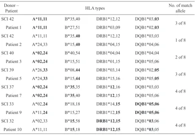
The HLA types in this study represent MHC class I (HLA-A and -B) and MHC class II (HLA-DR and -DQ). Highest matching degree found was 4 of 8 and the lowest matching degree was 1 of 8 (Table 1). However, no GvHD symptom was observed in the patient treated with hUCBMNCs from the donor with the lowest matching degree.
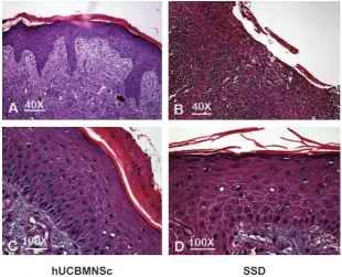
Figure 4 shows histological representation of hematoxylin-eosin-stained samples of hUCBMNCs- and SSD-treatment shows epidermal layers of recovered wounds with objective magnification of 40x and 100x. Dermal-epidermal junction is clearly reconstituted on day 11 in hUCBMNCs treatment and cellular rearrangement of recovered wounds in both of hUCBMNCs and SSD treatment. White bars
(upper panel): 100 mm, black bars (lower panel): 25 mm.
Histological examination
Histology examination on day 11 showed enhanced re-epitheliazation, both in SSD- and hUCBMNCs-treated wounds. Human UCBMNCs-treated wounds showed
clearly identiiable dermal-epidermal junction with the
rate ridges protruding to dermis close to the appearance in normal skin (Figure 4A). SSD-treatment indicated unclear dermal-epidermal junction (Figure 4B). Focused on cellular rearrangement, both treatments indicated similar numbers: 8-13 epidermal layers in hUCBMNCs-treated (Figure 4C) and 8-10 layers in SSD-treated wounds (Figure 4D).
Figure 2. Wong Baker’s pain intensity scale (a) and the number of patients (%) with reduced pain intensity (scale 0-10) on days 7 and 11of treatment (b)
DISCUSSION
In this study, hUCBMNCs application is compared to SSD, which is a widely used topical antibiotic for wound care in burn management. Silver sulfadiazine contains silver ions that bind to the microorganisms’ nucleic acid releasing sulfadiazine, which then interferes with the microbial metabolic activity.10 SSD
has an excellent broad–spectrum antibacterial coverage against Pseudomonas aeruginosa and other Gram-negative enteric bacteria, although some resistance
had been reported.11,12 However, systemic toxicity
was found in daily topical application, once or twice, indicated by the development of leukopenia.13 Since
SSD is only absorbed within the surface of epidermal layer, its effectiveness in severe injuries might be limited.14
Human UCBMNCs used in this study were isolated from crude umbilical cord blood and processed with minimum manipulation (without culture). The term ‘mononuclear cells’ was used instead of ‘stem cells’ to
Table 1. Result of HLA typing
Donor –
Patient HLA types
No. of match allele SCI 42 A*11,11 B*35,40 DRB1*12,12 DQB1*03,03
3 of 8 Patient 1 A*11,11 B*27,51 DRB1*03,09 DQB1*02,03
SCI 42 A*11,11 B*35,40 DRB1*12,12 DQB1*03,03
1 of 8 Patient 2 A*24,33 B*15,40 DRB1*04,15 DQB1*04,06
SCI 40 A*02,24 B*40,54 DRB1*04,04 DQB1*04,04
2 of 8 Patient 3 A*02,24 B*15,51 DRB1*01,15 DQB1*05,06
SCI 39 A*26,33 B*08,44 DRB1*03,14 DQB1*02,05
3 of 8 Patient 5 A*24,33 B*15,44 DRB1*15,16 DQB1*05,05
SCI 37 A*02,24 B*35,35 DRB1*12,16 DQB1*03,03
4 of 8 Patient 7 A*02,24 B*35,40 DRB1*12,15 DQB1*05,06
SCI 33 A*02,24 B*18,18 DRB1*14,15 DQB1*05,06
4 of 8 Patient 9 A*11,24 B*15,27 DRB1*12,15 DQB1*05,06
SCI 32 A*02,33 B*15,58 DRB1*12,15 DQB1*03,06
4 of 8 Patient 10 A*11,11 B*15,18 DRB1*12,15 DQB1*03,05
avoid misperception, since stem cell characterization and sorting were not performed in this clinical study. However, many studies proved that mononuclear fraction of human umbilical cord blood contain at least two types of stem cells: i.e. hematopoietic and mesenchymal stem cells. Hematopoietic stem cells, characterized by CD34+ markers, were found
approximately 1% in the mononuclear cells.15 Whereas,
mesenchymal stem cells, characterized by CD34- , CD45-, CD44+, CD90+, and CD105+ markers, were found in much less amounts in the full-term umbilical cord blood.16
Regarding the period of wound re-epithelialization, this study showed fascinating result in application both of SSD and hUCBMNCs. Spontaneous re-epithelialization in deep partial thickness burns is normally completed up to 21 days. With conventional treatment of SSD,
approximately 80% of wound re-epithelialization was
achieved within 11 days. With hUCBMNCs therapy, the achievement of wound re-epithelialization reached
approximately 90% of completion. The 10% difference was considered statistically signiicant.
Pain intensity was evaluated in each dressing change and application procedure. There was a great variation of pain intensity both intra- and inter-individually. It
had been shown that the pain intensity was not related to burn severity, but in the second week most patients of major burns had tendencies to express more pain than those with moderate burns.17 Based on that
study, we compared the pain intensity induced in
each application in the irst week (day 7) and second
week (day 11). We found that more patients felt decreased pain intensity in hUCBMNCs than in SSD treatment on days 7 and 11. Higher percentage on day 11 is consistent with the completeness of wound re-epithelialization. However, in the evaluation of the
eficacy, the parameter of pain intensity might be less
objective.
HLA typing was carried out to investigate donors-patients matching degree that may have effect on the outcome. It has been suggested that the success of unrelated hematopoietic stem cell transplantation is
inluenced by the degree of donor HLA compatibility.18,19 For these reasons, patients get the greatest beneit
when they have HLA-matched donors and can tolerate the transplantation procedure.20 However, this
study found no post-transplant complication or graft-versus-host disease (GvHD) even in HLA-mismatch
cases. This inding supports studies of unrelated cord
blood transplantation indicating immune-tolerance of hUCBMNCs.21-23
Figure 4. Histological representation of hematoxylin-eosin-stained samples of hUCBMNCs- and SSD-treatment shows epidermal layers of recovered wounds with objective magniication of 40x and 100x. Dermal-epidermal junction is clearly reconstituted on day 11 in hUCBMNCs treatment and cellular rear-rangement of recovered wounds in both of hUCBMNCs and SSD treatment. White bars (upper panel): 100 µm, black bars (lower panel): 25 µm
The absence of GvHD in this allogenic hUCBMNCs application can be explained by the lack of immunological properties. When compared to adult peripheral blood, T-lymphocytes and dendritic cells in umbilical cord blood are relatively immature as shown by very low expressions of CD4, CD8, and CD3. Minimum expression of those markers yielded very
small numbers of IL-2 and IFN-γ that play important
roles in inlammatory reactions.24,25 The immaturity
of T-lymphocytes was explained by little exposure to
foreign antigens; as the individual developed, T-cells
would differentiate into a memory phenotype as the antigenic exposure gradually increased.26
In terms of quality of re-epithelialization, histology showed better results in hUCBMNCs- than in SSD-treated wounds. Epidermal rete ridges formation was
clearly identiied in hUCBMNCs on day 11 at the end
of treatment, while in spontaneous wound healing this dermal-epidermal junction normally resembles 4 to 6 months following complete re-epithelialization. This long process can be explained by the fact that during skin remodeling, regenerated epithelia are prone to shearing forces, thus the regeneration process may be repeated many times. It seemed that SSD treatment was following the rule of such process and hence,
no rete ridges formation was identiied yet in the
samples. The rete ridges pattern found after 11 days of hUCBMNCs treatment suggests that hUCBMNCs accelerate remodeling.
In conclusion, human UCBMNCs are safe and potential to induce faster and better wound healing than conventional treatment in deep partial thickness burns.
Acknowledgments
We acknowledge and appreciate to Miss Devi Anggraenie and Mr Christian Indra in assistance to hUCB collection, Miss Melina Setiawan in HLA typing and database, dr Budiana, SpPA(K) for histology analysis, and Nursing staff of dr Cipto Mangunkusumo Hospital Burn Unit for the contribution of the entire study. This work was supported by Kalbe Farma Pharmaceutical Company.
REFERENCES
1. Benson A, Dickson WA, Boyce DE. Burns. BMJ.
2006;332(7542):649-52.
2. Hanumadass ML, Ramakrishnan M, Ramakrihnan MK, Babu M. The art and science of burn wound management.
Turnbridge Wells, Kent: Anshan; 2005.
3. Benson A, Dickson WA, Boyce DE. Burns. BMJ.
2006;332(7542):649-52.
4. Lineen E, Namias N. Biologic dressing in burns. J Craniofac
Surg. 2008;19(4):923-8.
5. Loss M, Wedler V, Künzi W, Meuli-Simmen C, Meyer
VE. Artiicial skin, split-thickness autograft and cultured
autologous keratinocytes combined to treat a severe burn
injury of 93% of TBSA. Burns 2000;26(7):644-52.
6. Atiyeh BS, Costagliola M. Cultured epithelial autograph (CEA) in burn treatment: Three decades later. Burns.
2007;33(4):405-13.
7. Caruso DM, Schuh WH, Al-Kasspooles MF, Chen MC, Schiller WR. Cultured composite autografts as coverage for an extensive body surface area burn: Case report and
review of the technology. Burns 1999;25(8):771-9.
8. Kamolz LP, Kolbus A, Wick N, et al. Cultured human epithelium: Human umbilical cord blood stem cells differentiate into keratinocytes under invitro conditions.
Burns 2006;32(1):16-9.
9. Dai Y, Li J, Li J, et al. Skin epithelial cells in mice from umbilical cord mesenchymal stem cells. Burns
2007;33(4):418-28.
10. Lansdown, AB. Silver: Its antibacterial properties and
mechanism of action. J Wound Care. 2002;11:125-30.
11. Heggers JP, Hawkins H, Edgar P, Villarreal C, Herndon DN. Treatment of infections in burns. In: Herndon
DN, editor. Total burn care. London: Saunders; 2002.
p.120-69.
12. Wright JB, Lam K, Hansen D, Burrell RE. Eficacy of
topical silver against fungal burn wound pathogens. Am J
Infect Control. 1999;27:344-50.
13. Choban PS, Marshall WJ. Leukopenia secondary to silver sulfadiazine: Frequency, characteristics and clinical
consequences. Am Surg. 1987;53:515-7.
14. Herruzo-Cabrera R, Garcia-Torres V, Rey-Calero J, Vizcaino-Alcaide MJ. Evaluation of the penetration strength,
bactericidal eficacy and spectrum of action of several
antimicrobial creams against isolated microorganisms in a
burn centre. Burns 1992;18:39-44.
15. Chularojmontri L, Wattanapittayakul SK. Isolation and characterization of umbilical cord blood hematopoietic
stem cells. J Med Assoc Thai. 2009;92(Suppl.3):S88-94.
16. Yu M, Xiao Z, Shen L, Li L. Mid-trimester fetal blood-derived adherent cells share characteristics similar to mesenchymal stem cells but full-term umbilical cord blood
does not. Br J Haematol. 2004;124(5):666-75.
17. Jonsson CE, Holmsten A, Dahlström L, Jonsson K. Background pain in burn patients: Routine measurement
and recording of pain intensity in a burn unit. Burns 1998;
24(5):448-54.
18. Sasazuki T, Juji T, Morishima Y, et al. Effect of matching class I HLA alleles on clinical outcome after transplantation of hematopoietic stem cells from an unrelated donor. N
Engl J Med. 1998;339:1177-85.
19. Anasetti C, Amos D, Beatty PG, et al. Effect of HLA compatibility on engraftment of bone marrow transplants in patients with leukemia or lymphoma. N Eng J Med.
1989;320:197-204.
20. Morishima Y, Sasazuki T, Inoko H, et al. The clinical
signiicance of human leukocyte antigen (HLA) allele
compatibility in patients receiving a marrow transplant from serologically HLA-A, HLA-B, and HLA-DR matched
unrelated donors. Blood 2002;99:4200-6.
bone marrow transplant from an HLA identical sibling. N
Engl J Med. 2000;342(25):1846-54.
22. Kurtzberg J, Laughlin M, Graham ML, et al. Placental blood as a source of hematopoietic stem cells for transplantation
into unrelated recipients. N Engl J Med. 1996;335:157-66.
23. Laughlin MJ, Baker J, Bambach B, et al. Hematopoietic engraftment and survival in adult recipients of umbilical-cord
blood from unrelated donors. N Engl J Med. 2001;344:1815-22.
24. Barker JN. 2007. Umbilical cord blood (UCB) transplantation: An alternative to the use of unrelated volunteer donors. Hematol Am Soc Hematol Educ Progr.
2007;1:55-61.
25. Heng T, Dudakov J, Khong D, Chidgey A, Boyd R. Stem
cells meet immunity. J Mol Med. 2009;87:1061-9.
26. Gluckman E, Rocha V. Cord blood transplantation: State of