How to cite:
Soetjahjo B, Hidayat M, Suyuti H, Fibrianto YH (2017) Immuno-histochemistry Evaluation of TGF-β1, SOX-9, Type II Collagen and Aggrecan in Cartilage Lesions Treated with Conditioned Medium of Umbilical Cord Mesencyhmal Stem Cells in Wistar Mice (Rattus novergicus). J. Trop. Life. Science 8 (1): 21 – 27. *Corresponding author:
Bintang Soetjahjo
Faculty of Medicine, Brawijaya University Jalan Veteran, Malang, Indonesia 65145 E mail: [email protected]
VOL. 8, NO. 1, pp. 21 – 27, January 2018 Submitted August 2017; Revised September 2017; Accepted November 2017
Immunohistochemistry Evaluation of TGF-
β1, SOX-9, Type II Collagen and Aggrecan in Cartilage
Lesions Treated with Conditioned Medium of Umbilical Cord Mesencyhmal Stem Cells in Wistar
Mice
(Rattus novergicus)
Bintang Soetjahjo 1*, Mohammad Hidayat 1, Hidayat Suyuti 1, Yuda Heru Fibrianto 2
1 Faculty of Medicine, Brawijaya University, Malang, Indonesia 2 Faculty of Veterinary Medicine, Gadjah Mada University, Yogyakarta, Indonesia
ABSTRACT
Currently, umbilical cord mesenchymal stem cells have the potential to be used as treatment options for any cartilage lesion. This research aimed to evaluate the effects of conditioned medium from umbilical cord mesenchymal stem cells (UC-MSC) on damaged cartilage through the expression of proteins TGF-β1, SOX-9, type II collagen and aggrecan, which are known to be related to chondrogenesis. UC-MSC were isolated from 19-days-pregnant Wistar mice and were cultured using the standard procedure to obtain 80% confluence. Subsequently, the culture was confirmed through a microscopic examination that was driven to be an embryoid body to obtain a pre-condition medium. This research utilized 3-month-old male Wistar mice and was categorized into 6 groups (3 control and 3 treatment groups). Each animal had surgery performed to create a femur condyle cartilage defect. The treatment groups were administered a dose of stem cells at 1 mL/kg. Next, immunohistochemical (IHC) staining was per-formed to examine the expression of TGF-β1, SOX-9, type II collagen and aggrecan in the 2nd, 3rd, and 4th month of evaluation. The results were analyzed statistically using ANOVA test. For each of the treatment groups, there was increased expression (p < 0.05) in all proteins TGF-β1, SOX-9, type II collagen and aggrecan when compared with control groups at the 2nd, 3rd, and 4th month of evaluation. Pre-conditioned medium from UC-MSC potentially increases the expression of TGF-β1, SOX-9, type II collagen and aggrecan in the damaged cartilage of Wistar mice.
Keywords: Cartilage lesions, conditioned medium, immunohistochemistry, umbilical cord mesenchymal stem cells
INTRODUCTION
Stem cell-derived conditioned medium (CM) has the potential to be produced as a treatment option for regenerative medicine. The use of secretome containing CM has several advantages when compared to the use of stem cells. Briefly, CM can be manufactured, freeze-dried, packaged, and transported with greater ease [1]. Moreover, as it is devoid of cells there is a lower risk of host rejection. To date, there is limited data pertaining to the use of CM for cartilage repair.
Mesenchymal stem cells (MSC) are a population of cells with infinite proliferation capability and the poten-tial to differentiate into cells of the mesoderm lineage. Research studies have reported that MSC has the capa-
bility to differentiate into osteoblasts, chondrocytes, car-diomyocytes, myocytes, adipose cells and even neural cells. MSC can be isolated from multiple areas in adult tissue such as bone marrow, adipose tissue, umbilical cords, and peripheral blood [2, 3, 4]. Umbilical Cord Derived Mesenchymal Stem Cells (UC-MSCs) have pro-liferation and differentiation capabilities. These stem cells have also been shown to have the ability to differ-entiate into unipotent cells that have a chondrocyte pat-tern [5].
Deterioration of cartilage leads to osteoarthritis, a disease through which articular chondrocytes become deformed, or fibrillated losing their cartilage forming function. There are several proteins that are associated
with chondrogenesis. TGF-β1 promotes chondrogenesis in cultures of early-undifferentiated MSC in vitro and stimulates chondrogenic differentiation in vivo [6]. Pre-vious studies reported that expression and activity of TGF-β1 signaling could potentially have detrimental ef-fects on mature condylar cartilages [12]. TGF-β1 has an ability to down regulate type II collagen and aggrecan in articular chondrocytes via the 9 protein [7]. SOX-9 is a transcription factor belonging to the Sry-type HMG-box (SOX) protein family, which is essential for chondrogenesis and has been termed a “master regula-tor” of the chondrocyte phenotype. In addition, SOX-9 has been shown to activate type II collagen and aggre-can. Moreover, SOX-9 prevents chondrocyte hypertro-phy and has a redifferentiation effect on osteoarthritic chondrocytes, which have been dedifferentiated. Type II Collagen, a major component of hyaline cartilage, has many advantages as a biomaterial for chondrogeneses, such as biodegradability and the capability to induce re-pair processes in articular cartilage [8]. Aggrecan is one of the major components of cartilage and binds to hya-luronan (HA) and links proteins to form huge aggre-gates. These aggregates lead to a hydrated gel-like struc-ture of cartilage and resistibility to compression and de-formation in joints [9].
Currently, there has been vast research to study fac-tors that promote cartilage repair. However, only a few describe the secretome of UC-MSC in chondrogenesis has been reported. Using immunohistochemical (IHC) evaluation this research would investigate the potential of CM of UC-MSC obtained from Wistar mice with a focus on proteins TGF-β1, SOX-9, type II collagen and aggrecan expression in a damaged cartilage wound.
MATERIALS AND METHODS
This research was performed using 24 Wistar mice 3 months old that underwent surgery within its medial condyle of the femur in right hind feet on weight bear-ing area. The samples were divided into 6 groups. Three control groups were 2(K2), 3(K3) and 4(K4) months, and three treatment groups were 2(E2), 3(E3) and 4(E4). The duration of the study was 4 months. This study has been approved by Ethical Clearance Commission of Gajah Mada University, Yogyakarta, in Mei 6th 2015 (Letter number: 263/KEC-LPPT/V/2015).
Isolation and cultivation of UCMSCs
The umbilical cord was obtained from 19 days preg-nant Wistar mice by caesarian section according to standard procedures. Briefly, under sterile conditions,
one centimeter of umbilical cord was washed with 10% betadine and sterile NaCl. Next, that slash was placed inside a centrifuge tube containing transport media Dul-becco’s Eagle Modified Medium (DMEM) with 200 µg/L penicillin, 200 µg/mL streptomycin, and 200 µg /mL fungi zone). Next, the cord was cut in 20 mm3 sizes and cultured using an enzymatic technique. This cord slash was further diluted with trypsin/EDTA 0.25% and incubated for 30 minutes at 37°C. About 2 cc of com-plete medium (DMEM 1×, 10% fetal bovine serum, 50 µg/mL penicillin streptomycin, and 2.5 µg/mL fungi zone) was added to the tube already containing the cord that had been metabolized by the enzyme. It was then centrifuged at 3,000 rpm for 10 minutes at 4°C. The su-pernatant was removed and the cell suspension was mixed with complete medium and incubated at 37°C and 5% CO2. It was replaced every three days until cell growth reached a confluence of 80%. Afterward, micro-scopic examination was performed using Hematoxylin and Eosin (H&E), Giemsa and Sirius Red staining to confirm the existence of the cells, and further confirm they are umbilical cord mesenchymal stem cells (UC-MSC).
Production of conditioned medium
Cultures of UC-MSC that have reached 80% con-fluence were harvested using trypsin. Following, trypsin neutralization, the cell suspension was centrifuged at 3,000 rpm for 10 minutes. The supernatant was dis-carded and the cell pellet was washed three times with PBS. The cell pellet was resuspended in fresh medium at 10,000 cells per mL. Next, the stem cells were pushed towards an embryoid body and seeded onto a culture plate with full medium until confluence among these bodies were formed. The production of pre-condition medium was created by washing the embryoid body cul-ture with sterile PBS and filling the culcul-ture plate with 10 mL of complete medium without serum. After 48 hours, the pre-condition medium was stored at -20°C prior to use.
Mice treatment with conditioned medium
(a)
(b)
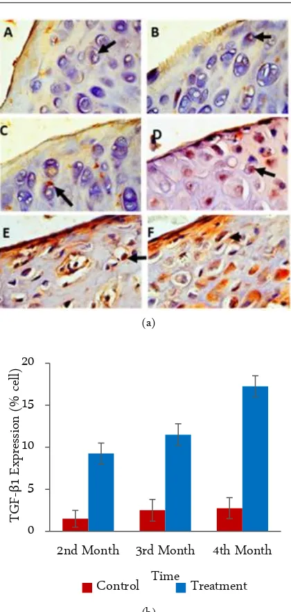
Figure 1. IHC results from the articular cartilage network joint (left panel), photomicrograph at 400×. (a) The chon-drocytes with TGFB1 expression are characterized by the brown color of the cell cytoplasm (arrows). A. Con-trol 2nd month, B. Control 3rd months, C. Control 4th
months, D. Treatment 2nd months, E. Treatment 3rd
months and F. Treatment 4th months. (b) Graphic of
the calculation analysis of all preparations of joint car-tilage tissue (right panel). The X-axis is the treatment time group and Y axis represents the mean expression of TGFB1.
groups were administered an injection of 1 cc/kg of body weight pre-condition medium 5 times over 1-week inter-val. The Wistar were observed at 2nd (P2 groups), 3rd (P3 groups), and 4th (P4 groups) months of evaluation. At this time, animals were humanely euthanized and the
cartilage was extracted. Articular cartilage tissue was placed inside a tube containing 10% formalin. Biopsy samples were sent to the Biochemistry-Biomolecular La- boratory of Medical Faculty - Brawijaya University, Ma lang.
Tissue processing (fixation and paraffin embedding) and slide preparation of 4 µm cut thickness using a ro-tary microtome was performed. An immunohistochem-istry technique was performed to observe the protein ex-pression of TGF-β1, SOX-9, Collagen type 2 and Aggre-can using specific antibodies purchased from Santa Cruz Biotech (USA) and immunohistochemistry kit using D-Bio Sys Immunostaining kit (Netherland). The sample were reacted with primary antibody of mouse anti Col-2 (COLCol-2A1 Antibody (MCol-2139) cat#: sc-5Col-2658), TGFβ1 (TGF-β1 Antibody (3C11) cat#: sc-130348), Sox-9 (Sox-9 antibody (E-(Sox-9), cat#: sc-166505) and aggrecan (aggre-can antibody (4F4), cat#: sc-33695) for 24 hours at 4°C, then cleaned with PBS pH 7.4 three times for 5 minutes. The samples were incubated using secondary antibody of biotin (rabbit anti-mouse IgG biotin labelled) for 1 h at room temperature, then washed with PBS pH 7.4 three times for 5 minutes. Strep Avidin-Horse Radin Pe-roxidase (SA-HRP) were added to the slides and incu-bated for 40 minutes then washed with PBS pH 7.4 three times for 5 minutes. Diamano Benzidine (DAB) were added to the slides and incubated for 10 minutes then washed with PBS pH 7.4 three times for 5 minutes. Counter staining the slides with Mayer Hematoxylen for 10 minutes then washed with aquadest and dried. Fi-nally, the slides were mounted with entellan and covered with cover glass. The results were evaluated using a mi-crograft photo using a Nikon E-100 microscope and Sony ICLEA7 camera with 400× magnification. Lastly, the expression was analyzed using immunoratio soft-ware (freesoft-ware from Institute of Biomedical Technology, University of Tampere).
Statistical analysis
Control and experimental groups were analyzed us-ing statistic parametric ANOVA and descriptively in mean ± SD or median, frequency is presented in per-centage. For normality or distribution, we use Kolmogo-rov-Smirnov test and two independent T-test for normal distribution data, p < 0.05 indicated a statistically signif-icant difference.
RESULTS AND DISCUSSION
To investigate the potential role of conditioned me-dium of UC-MSC in articular cartilage repair this study 0
5 10 15 20
2nd Month 3rd Month 4th Month
TGF
-β
1
E
xp
ress
ion
(%
ce
ll)
Time
(a)
(b)
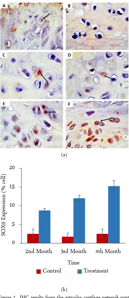
Figure 2. IHC results from the articular cartilage network joint (left panel), photomicrograph at 400×. (a) The chon-drocytes with the SOX-9 expression are characterized by the brown color of the cell nucleus (arrow). A. Con-trol 2nd month, B. Control 3rd months, C. Control 4th
months, D. Treatment 2nd months, E. Treatment 3rd
months and F. Treatment 4th months. (b) Graphic of
the calculation analysis of all preparations of joint car-tilage tissue (right panel). X axis is the treatment time group and axis Y represents the mean of SOX-9 expres-sion.
used immunohistochemical techniques, with a peroxi-dase system, to show the expression of TGF-β1, SOX-9,
type II collagen and aggrecan proteins.
The expression of TGF-β1 was evident in all study samples. Figure 1 shows that TGF-β1 is expressed in the cytoplasm of the chondrocytes of the tegmental area, as well as the transitional layer of the joint cartilage tissue. Using statistical analysis demonstrated that TGF-β1 ex-pression in the treatment group increased significantly when compared with the control group (bottom). Ex-pression of TGF-β1 (p > 0.05) between the 2nd, 3rd, and 4th observational groups in the negative control group (untreated mice). Overall, the expression of TGF-β1 in the treatment group demonstrated a gradual increase (months 2nd, 3rd, and 4th) and was significantly different in comparison with the control group (B) (p < 0.05).
Similar values were observed for the expression of SOX-9 (p > 0.05) between the 2nd, 3rd, and 4th observa-tional groups and the negative control group (untreated mice). Furthermore, there was a significant difference in the expression of SOX-9 (p < 0.05) between the 2nd, 3rd, and 4th observation groups in the treatment group (ad-ministering fasting MSC CM). SOX-9 expression demonstrated the most significant difference in the 3rd month expression (12.00 ± 0.82b) and 4th month (15.25 ± 1.5c) of the treatment group (Figure 2).
Type II collagen expression was also observed in each study sample. This result indicated that type II co- lagen is widely expressed in the cell cytoplasm in the transitional area of the articular tissue of cartilage of the joint. Using statistical analysis, the results showed that type II collagen expression was increased significantly compared to the control group (bottom). Overall, type II collagen expression in the treatment group demon-strated a gradual increase (2nd, 3rd, and 4th month) and was observed to be significantly different when com-pared to the control group (p < 0.05). Additionally, the expression of type II collagen under control conditions began in the 4th month (p < 0.05).
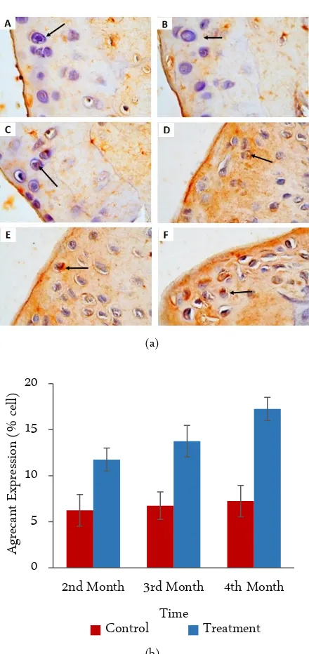
The aggrecan expression (p > 0.05) was not signifi-cantly different between the 2nd, 3rd, and 4th observa-tional groups in the negative control group (untreated mice). Based on the mean value of aggrecan expressions, the values were similar. The significant difference in ag-grecan expression (p < 0.05) was observed in the analysis between the 2nd, 3rd, and 4th month observational groups of the treatment group (MSC). Based on the mean val-ues of aggrecan expression, there was a significant in-crease in the 4th month. While in the 2nd and 3rd months aggrecan expression remained the same (Figure 3).
UC-MSC are considered medical waste and the col-lection is noninvasive. Therefore, access to UC-MSC has 0
5 10 15 20
2nd Month 3rd Month 4th Month
SOX9
E
xp
res
sion
(%
cell)
Time
(a)
(b)
Figure 3. IHC results from the articular cartilage network joint (left panel), photomicrograph at 400×. (a) Chondro-cytes with an expression of aggrecans are characterized by the brown color of the cell cytoplasm (arrows). A. Control 2nd month, B. Control 3rd months, C. Control
4th months, D. Treatment 2nd months, E. Treatment 3rd
months and F. Treatment 4th months. (b) Graphic of
the calculation analysis of all preparations of joint car-tilage tissue (right panel). The X axis is the treatment time group and the Y axis represents the average of the aggrecan expression.
not been burdened with ethical problems [10]. MSC conditioned medium (CM) could be defined as secreted factors alone that are referred to as the secretome or ex-
osome without the stem cells. In addition, CM is poten-tially less allergenic according to the selected proteins that are transplanted to some tissues. In our study, we analyzed a group of proteins that are potentially found in the medium and have a potential to be beneficial in cartilage repair. The growth, development, maintenance, and improvement of articular cartilages are strictly reg-ulated by multiple signaling pathways performed by sev-eral bioactive factors [11].
TGF-β1 is an especially significant factor in the chondrogenic differentiation of MSCs. It is considered to be a potent stimulator for proteoglycan and type II collagen synthesis [12]. Ye Li et al. reported that the ex-pression of TGF-β1 was increased in the condylar carti-lage of an injurious model of OA [7]. Injecting TGF-β1 into the periosteum of the femur stimulates chondrocyte differentiation and cartilage formation [13]. Addition-ally, we observed that the expression of TGF-β1 was slightly increased in all experimental groups, across all time points, when compared to the control groups. There are two important proteins, SOX-9 and RUNX2, which function as transcription regulators of the
TGF-β1 gene [14]. We speculate that the administration of UC MSC-CM activated one or both of the above regu-latory proteins (SOX-9 and RUNX2), resulting in in-creased expression of TGF-β1. Further, this is in line with the research of Venturin et al. who reported that TGF-β1 is expressed in all the differentiating chondro-progenitor and chondrocytes in the proliferative zone [15].
SOX-9 is expressed in all chondroprogenitor cells, predominantly in mesenchymal condensations and car-tilage. Cao et al. also found that the repair of cartilage defects can be enhanced by SOX-9 transduction [16]. They reported that the articular cartilage defects treated with MSC overexpressing SOX-9 showed the signifi-cantly better integration of the repair tissue at the inter-face with the surrounding normal articular cartilage when compared to the other groups. It is well docu-mented that TGF-β induces expression of a transcrip-tional factor SOX-9 in its signaling pathway [8]. In our study, we found that SOX-9 expression was greatly in-creased. We suggest that a high level of SOX-9 expres-sion will have beneficial effects on cartilage repair.
Type II collagen comprises more than 90% of the collagen found in adult articular (hyaline) cartilage. In this study, we observed that type II collagen was in-creased in all samples. This suggests that the CM con-tains potent components for activation of those proteins and displays promising potential for cartilage repair. It 0
5 10 15 20
2nd Month 3rd Month 4th Month
Agreca
n
t
E
xp
res
sion
(%
cell)
Time
is known that SOX-9 is a potent activator in the for-mation of collagen type II [15], which is a component of cartilage. Moreover, this is the predominant type of collagen in cartilage. The extracellular matrix is also comprised of other specific collagens such as type IX and XI [17]. Following the increase in SOX-9 mRNA expres-sion, the production of type II collagen, aggrecan, and cartilage oligomeric matrix protein (COMP) is observed [8].
Aggrecan is one of the major structural macromol-ecules of cartilage. Aggrecan function is to resist any compressive forces by maintaining the osmotic pressure balance in cartilage [18]. According to our study, we found that aggrecan expression was significantly in-creased in all samples. We speculate that aggrecan pro-duction plays a critical role in cartilage repair techniques by maintaining the collagen network. Therefore, aggre-can participates in both the demise and survival of artic-ular cartilage.
This suggests that there has been an increase in tis-sue growth according to the initial hypothesis referring to studies conducted by Mehrabani et al. [19] who ex-amined cartilage defects in 12 rabbits, in which treat-ment groups were given mesenchymal stem transplan-tation. The results of the study suggest that the nature of the predominant tissue in the transplant group had a higher score than the control group.
This study has limitations to find a solution in treat-ing cartilage defects, damaged by trauma. The outside problem is not discussed. An update of this study was the beginning of the further study for clinical experi-mental to find a solution to treat the problem of cartilage lesion in human as an effective, inexpensive, and con-venient alternative choice.
CONCLUSION
Conditioned medium from CM-UC-MSC contains proteins and tissue regenerative agents, which are se-creted by the stem cells and allow for cellular interac-tions between cell-to-cell and their microenvironment for successful chondrogenesis. Results from our study indicate that when evaluated using immunohistochem-istry, CM from UC-MSC increases the expression of protein TGF-β1, SOX-9, type II collagen and aggrecan. These increased proteins are closely related to chondro-genesis in cartilage defects in a Wistar rat.
ACKNOWLEDGMENT
The author thanks to Brawijaya University for facil-itating this research.
REFERENCES
1. Pawitan JA (2014) Prospect of stem cell conditioned me-dium in regenerative medicine. BioMed Research Interna-tional 2014: 1 – 14. doi:10.1155/2014/965849.
2. Short B, Brouard N, Occhiodoro-Scott T et al. (2003) Mes-enchymal stem cells. Archives of Medical Research 34 (6): 565 – 571. doi: 10.1016/j.arcmed.2003.09.007.
3. Chai C, Leong KW (2007) Biomaterials approach to expand and direct differentiation of stem cells. Molecular Therapy 15 (3): 467 – 480. doi:10.1038/sj.mt.6300084.
4. Kim DW, Staples M, Shinozuka K et al. (2013) Wharton’s jelly-derived mesenchymal stem cells: phenotypic character-ization and optimizing their therapeutic potential for clini-cal applications. International Journal of Molecular Sciences 14 (6): 11692 – 11712. doi:10.3390/ijms140611692. 5. Song D, Zhong Y, Qian C et al. (2016) Human umbilical
cord mesenchymal stem cells therapy in cyclophosphamide-induced premature ovarian failure rat model. BioMed Re-search International 2016: 1 – 13. doi: 10.1155/2016/25175 14.
6. Yoon HJ, Kim SB, Somaiya D et al. (2015) Type II collagen and glycosaminoglycan expression induction in primary hu-man chondrocyte by TGF-β1. BMC Musculoskelet Disor-ders 16: 141. doi:10.1186/s12891-015-0599-x.
7. Li Y, Tian AY, Ophene J et al. (2017) TGF-β stimulates endochondral differentiation after denervation. Interna-tional Journal of Medical Sciences 14 (4): 382 – 389. doi: 10.7150/ijms.17364.
8. Yu DA, Han J, Kim BS (2012) Stimulation of chondrogenic differentiation of mesenchymal stem cells. International Jo- urnal of Stem Cells 5 (1): 16 – 22. doi: 10.15283/ijsc.2012. 5.1.16.
9. Watanabe H, Tanabe N, Watanabe T, et al. (2008) Meta-bolic syndrome and risk of development or atrial fibrilla-tion: the Niigata preventive medicine study. Circulation 117 (10): 1255 – 1260. doi: 10.1161/CIRCULATIONAHA.107. 744466.
10. Nagamura-Inoue T, He H (2014) Umbilical cord-derived mesenchymal stem cells: Their advantages and potential clinical utility. World Journal of Stem Cells 6 (2): 195 – 202. doi: 10.4252/wjsc.v6.i2.195.
11. Mariani E, Pulsatelli L, Facchini A (2014) Signaling path-ways in cartilage repair. International Journal of Molecular Sciences 15 (5): 8667 – 8698. doi:10.3390/ijms15058667. 12. Yoon HJ, Kong SY, Park MH et al. (2013) Aminopropyl
carbazole analogues as potent enhancers of neurogenesis. Bioorganic and Medicinal Chemistry 21 (22): 7165 – 7174. doi: 10.1016/j.bmc.2013.08.066.
14. Goldring MB, Marcu KB (2009) Cartilage homeostasis in health and rheumatic diseases. rthritis Research and Ther-apy 11 (3): 224. doi: 10.1186/ar2592.
15. Venturin GT, Greggio S, Marinowic DR et al. (2011) Bone marrow mononuclear cells reduce seizure frequency and improve cognitive outcome in chronic epileptic rats. Life Sciences 89 (7 – 8): 229 – 234. doi: 10.1016/j.lfs.2011.06.0 06.
16. Cao L, Yang F, Liu G et al. (2011) The promotion of carti-lage defect repair using adenovirus mediated Sox9 gene transfer of rabbit bone marrow mesenchymal stem cells. Bi-omaterials 32 (16): 3910 – 3920. doi: 10.1016/j.biomateri-als.2011.02.014.
17. Ren X, Yang Z, Xu J et al. (2014) Enhanced specificity and efficiency of the CRISPR/Cas9 system with optimized sgRNA parameters in Drosophila. Cell Reports 9 (3): 1151
– 1162. doi: 1016/j.celrep.2014.09.044.