TELKOMNIKA, Vol.10, No.1, March 2012, pp. 137~146 ISSN: 1693-6930
accredited by DGHE (DIKTI), Decree No: 51/Dikti/Kep/2010 137
Classification and Numbering of Dental Radiographs
for an Automated Human Identification System
Anny Yuniarti, Anindhita Sigit Nugroho, Bilqis Amaliah, Agus Zainal Arifin Laboratory of Vision and Image Processing, Department of Informatics Faculty of Information Technology, Institut Teknologi Sepuluh Nopember Kampus ITS, Sukolilo, Surabaya 60111, Indonesia, Telp/fax 031-5939214/031-5913804
e-mail: [email protected]
Abstrak
Identifikasi manusia berbasis data gigi umum digunakan dalam forensik. Dalam kasus investigasi yang besar, proses pengidentifikasian manusia yang dilakukan secara manual memerlukan waktu yang lama. Pada makalah ini dikembangkan sebuah sistem identifikasi manusia otomatis menggunakan radiografi gigi. Sistem yang dibangun bekerja dengan 2 tahapan utama. Tahapan pertama adalah menyusun sebuah database berisi data radiografi gigi berlabel. Tahapan kedua adalah melakukan pencarian pada database untuk mendapatkan hasil identifikasi. Kedua tahapan tersebut menggunakan serangkaian proses pengolahan citra dan klasifikasi serta penomoran untuk mendapatkan pola dan fitur radiografi gigi. Pertama, dilakukan prapemrosesan yang meliputi perbaikan dan binarisasi citra, ekstraksi gigi tunggal, dan ekstraksi fitur. Selanjutnya, dilakukan proses klasifikasi gigi untuk mengklasifikasikan gigi menjadi molar dan premolar dengan menggunakan metode binary support vector machine (SVM). Setelah itu, proses penomoran pada gigi dilakukan sesuai pola molar dan premolar yang diperoleh pada tahap sebelumnya. Percobaan menggunakan 16 radiografi gigi yang terdiri dari 6 radiografi bitewing dan 10 radiografi panoramik dengan total 119 objek gigi menunjukkan nilai akurasi yang baik, yaitu 91,6% untuk proses klasifikasi gigi menjadi molar dan premolar dan 81,51% untuk proses penomoran gigi.
Kata kunci: forensik, identifikasi manusia, radiografi gigi, segmentasi, sistem penomoran gigi
Abstract
Dental based human identification is commonly used in forensic. In a case of large scale investigation, manual identification needs a large amount of time. In this paper, we developed an automated human identification system based on dental radiographs. The system developed has two main stages. The first stage is to arrange a database consisting of labeled dental radiographs. The second stage is the searching process in the database in order to retrieve the identification result. Both stages use a number of image processing techniques, classification methods, and a numbering system in order to generate dental radiograph’s features and patterns. The first technique is preprocessing which includes image enhancement and binarization, single tooth extraction, and feature extraction. Next, we performed dental classification process which aims to classify the extracted tooth into molar or premolar using the binary support vector machine method. After that, a numbering process is executed in accordance with molar and premolar pattern obtained in the previous process. Our experiments using 16 dental radiographs that consist of 6 bitewing radiographs and 10 panoramic radiographs, 119 teeth objects in total, has shown good performance of classification. The accuracy value of dental pattern classification and dental numbering system are 91.6 % and 81.5% respectively.
Keywords: forensic, human identification, dental radiographs, segmentation, dental numbering system
1. Introduction
Biometric is a tool of identification that has been broadly used in many applications. A biometric identification system is based on physical characteristics such as face [1], fingerprint, palmprint [2], eyes (iris, retina) and DNA. However, many of those characteristics are only suitable for ante mortem (AM) identification when a person to be identified is still alive. They cannot be used for postmortem (PM) identification especially in the case of decay or severe body damage caused by fire or collision [3].
On average, human h will take a long time to comple the search space by comparin matched dental and numberi identification system. tested our method not only to radiographs have wider sp radiographs, the upper and low recording phase, dental radio phase, namely preprocessing, phase are dental patterns, w
n has 32 teeth; each tooth has five surfaces, mea surfaces with various conditions. If we use dental tching of AM with PM data needs a large amount puter-aided for an identification system is needed. an automated identification system, dental featu
cted and saved in a database. During identification cted and compared to those in the database. This m plete if we do not reduce the search space. In this
ring the pattern and numbering of teeth only. This ering pattern. Therefore, we can enhance the eff
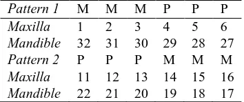
international dental numbering system which also are 32 teeth in adult people, sixteen teeth on eac
ible. Each jaw is divided into two groups, left and th comprised of two bicuspid, one cuspid, two pre research we only use molar and premolar teeth
molar teeth are usually stronger than others. ntal numbering system has teeth number from 1 to maxilla (#1), going through the maxilla to the third bering is continued to the third molar in the left ma e find the third molar in the right mandible (#32) [4]. nds of dental radiographs: bitewing, panoramic, a mages are usually used for identification. However, to bitewing radiographs, but also to panoramic radio space between upper and lower jaw, wherea lower jaw are closer.
ased identification consists of extracting dental fea this paper, the dental feature used for identifyin Premolar teeth and the numbering of each radiog have to classify each tooth in a radiograph into M ct the tooth using several image processing techn
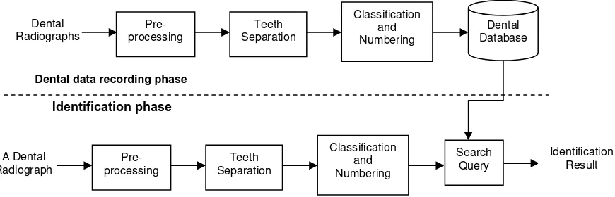
re 1. A system of dental numbering in adults
proposed system design and three main functio n, classification and numbering system, are explai implemented using Matlab 7.0.
n phases in the proposed human identification sys data recording phase and identification phase. In diographs are processed. There are three main ng, teeth separation, classification and numbering. T
TELKOMNIKA ISSN: 1693-6930
Classification and Numbering of Dental Radiographs for An Automated …. (Anny Yuniarti) 139
Figure 2. Design of the proposed human identification system
In the pre-processing step, a dental radiograph which has been digitalized are loaded from local hard disk. The dental radiograph can be bitewing or panoramic. For panoramic radiographs, we only take the molar and premolar part. Next, we perform image enhancement that aims to equalize the brightness level so that there is no pixel that has very high intensity level compare to its neighbors. This usually happens to dental fillings. Next, we perform the contrast enhancement. Generally, dental radiographs have low contrast. In order to facilitate process of teeth separation with the background, we increase the contrast using morphological operation and top-hat and bottom-hat operator [8]. After that, we perform local histogram equalization which is called Contrast-Limited Adaptive Histogram Equalization (CLAHE) [8].
After pre-processing, the grayscale digital radiographs are then converted into binary images using Otsu's thresholding method [8] followed by closing and opening operation to smooth the teeth contour and remove noises. Next, we perform horizontal integral projection [9] followed by spline method to separate the image into a maxilla image and a mandible image. Finally, we use the vertical integral projection method on each maxilla and mandible image independently to extract single tooth image.
The next process is dental feature extraction of each tooth. This step is used for classifying each tooth into molar or premolar class. The dental features are area of each tooth and ratio of each tooth's width and height. After feature extraction, we classify each tooth into molar or premolar class using binary support vector machine (SVM) method. SVM is a famous binary classification method. Given a set of training data, each marked as belonging to one of two classes, an SVM training method creates a model that predicts a new data is in one class or the other [10]. An SVM model is a representation of all data as points in space and a clear gap that separate data into two categories. This clear gap is often called as a hyperplane. This hyperplane is built as wide as possible. New testing data are then mapped into the same space and predicted as a member of a class based on which side of the hyperplane they fall on.
Otherwise, we choose Pattern 2. The next step is defining position of dental numbering based on the chosen default pattern. First, find an element of similarity matrix Okl that has maximum value, set a column index and a row index based on the element’s position, i.e. the column index = k, and the row index = l. Then, label each tooth in maxilla and mandible with number as in default pattern numbering system, i.e. pl-k+i, 1 ≤i ≤ k.
As an illustration, suppose that patterns resulted from the SVM classification results are molar-molar-molar-premolar-premolar (MMMPP) for maxilla and MMPP for mandible. Using equation (1), the similarity matrices are as shown in Figure 4. Then find the maximum value of each matrix, i.e. Smax1=5, Smax2=3, Sman1=4, Sman2=2. Thus, the score of Pattern 1 is 9,
Figure 3. Two default patterns of dental numbering
Figure 4. The similarity matrices between two default patterns and the pattern MMMPP-MMPP.
The last procedure in the proposed identification system is related to database access. Final images after the classification and numbering process are stored in the database along with dental data such as a unique serial number, name and age of the radiograph’s owner, date or recording, molar-premolar pattern, numbering pattern, area and ratio features, and file path of the original image, picture of the owner, and the classified image.
TELKOMNIKA ISSN: 1693-6930
Classification and Numbering of Dental Radiographs for An Automated …. (Anny Yuniarti) 141
3. Results and Discussion
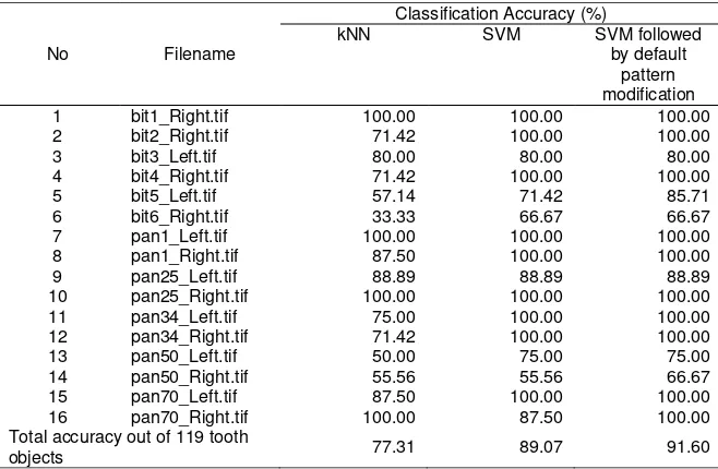
We use 16 dental radiograph images, composed of 6 bitewing radiographs and 10 panoramic radiographs. Based on an expert identification, there are 37 teeth objects identified in the 6 bitewing radiographs. Whereas in the panoramic radiographs, there are 82 teeth objects identified by the expert. Therefore, there are 119 objects of tooth in total. Three samples of the system's input image are as shown in Figure 5(a-c).
3.1. Pre-processing and Teeth Separation
In the first process, input images are successfully enhanced as shown in Figure 6. However, in the case of tooth object having too low intensity approaches background's intensity, or in the case of lower jaw bone having too high intensity approaches teeth object's intensity, this process does not perform well, as in three out of sixteen images in our experiment.
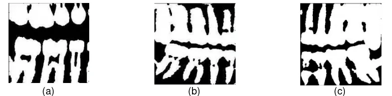
In the binarization process, the enhanced images are successfully converted into binary images. Except for the three images having the intensity problem as we explained before, all binary images have the properties as follows. The white pixels of the binary images represent teeth objects, whereas non-hole black pixels represent background. Sample outputs of the binarization process are as shown in Figure 7.
(a) (b) (c)
Figure 5. Sample input to the system: (a) a bitewing radiograph (b) a left-cropped panoramic radiograph (c) right-cropped panoramic radiograph
(a) (b) (c)
Figure 6. Results of enhancement process applied to three sample inputs as in Figure 5.
(a) (b) (c)
Figure 7. Results of binarization process applied to three enhanced images as in Figure 6(a-c).
are resulted from Figure 7(b) and (c) respectively. Here, we can successfully separate the binary image into maxilla and mandible images although the upper and lower jaws are very close in panoramic radiographs.
Each region is processed further by applying the vertical integral projection followed by spline method to separate the teeth region into single tooth region. Overall our method performs well in our experiments except for two images that have very high lower jaw bone intensity. In the case of molar tooth that has double roots in mandible, our method performs well too because we only take 3/5 upper part of mandible. Hence pixel values of tooth root are not included in the computation of teeth separation.
3.2. Classification and Numbering
In the classification process, firstly we considered a tooth object is an isolated area having more than 6000 pixels. From each tooth object, we extracted its area, ratio of height and width, and its centroid. Based on these features, we classify each tooth into molar or premolar using binary SVM method. As a comparison, we also implemented the classification using k-nearest neighbor (kNN) method, a simpler method than SVM, with k = 9.
Based on our experiments, there is significant difference between accuracy of SVM classification result and that of kNN’s result. Using the SVM method, the total accuracy value reaches 89.07% or 106 out of 119 objects were truly classified. Whereas, the average accuracy of the kNN method reaches 77.31% or 92 out of 119 objects were truly classified.
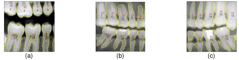
Next, we applied the numbering system by marking all teeth using a number and we also modified the class using standard numbering system in order to avoid abnormal molar and premolar pattern. As an example, if a classification process results in a pattern such as premolar-molar-premolar-premolar (P-M-P-P), then the pattern will be modified into M-M-P-P. This strategy has been able to improve the system’s accuracy to 91.60%. Hence, 109 out of 119 objects are now classified correctly. The implemented numbering system also performs well. There are 97 out of 119 objects numbered correctly. This leads to a total accuracy value of 81.51%. Details of classification and numbering accuracy value are shown in Table 1 and Table 2 respectively. Whereas, sample output images are shown in Figure 9. In Figure 9, extracted teeth are marked using a yellow line, labeled by M for molar class or P for premolar class followed by a number representing the numbering's result.
(a) (b) (c)
(d) (e) (f)
Figure 8. Results of teeth separation applied to three binary images as in Figure 7(a-c); Top row: maxilla regions. Bottom row: mandible regions.
(a) (b) (c)
Figure 9. Results of classification and numbering process applied to extracted teeth as in Figure 8(a-f).
TELKOMNIKA ISSN: 1693-6930
Classification and Numbering of Dental Radiographs for An Automated …. (Anny Yuniarti) 143 10 pan25_Right.tif 100.00 100.00 100.00 11 pan34_Left.tif 75.00 100.00 100.00 12 pan34_Right.tif 71.42 100.00 100.00 13 pan50_Left.tif 50.00 75.00 75.00 14 pan50_Right.tif 55.56 55.56 66.67 15 pan70_Left.tif 87.50 100.00 100.00 16 pan70_Right.tif 100.00 87.50 100.00 Total accuracy out of 119 tooth
objects 77.31 89.07 91.60
Table 2. The accuracy of numbering using teeth alignment No Filename Numbering Accuracy (%)
1 bit1_Right.tif 60.00 Total accuracy out of 119 tooth
objects 81.51
3.3. Identification System
The proposed automated human identification system was implemented using MySQL database server and Matlab 7.0. The system consists of four user interfaces. The first user interface is used for classification and numbering of dental radiographs. Sample input and output of the classification and numbering system is as shown in Figure 10. In the system's user interface, there are 6 buttons consisting of "Open Image" button to load an input image from local disk, "Proceed" button to perform the proposed methods, "Save" button to store the radiograph and its properties including dental pattern and numbering into the database, "Search" button to find a match of current radiograph in the database based on its properties, "Database" button to browse the database's contents, and "Exit" button to quit the application.
The second user interface aims to add a classified radiograph into the database. This interface only appears after users click the “Save” button in the first user interface. In this window, users may add additional information such as name, age, picture, and other information. Figure 11 shows an example of the action.
Figure 10. The classification and numbering system's user interface.
Figure 11. The user interface for saving new data into database.
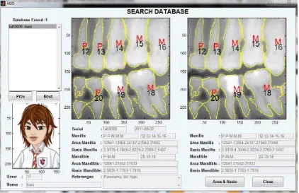
Figure 12. The user interface for searching process. Right image is the query, left image is the result.
TELKOMNIKA ISSN: 1693-6930
Classification and Numbering of Dental Radiographs for An Automated …. (Anny Yuniarti) 145
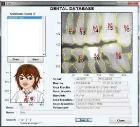
Figure 13. The user interface for querying database. Users are asked to enter the pattern or numbering in the Search textfield.
4. Conclusion
The proposed system has been successfully implemented and is able to generate dental pattern and numbering based on dental radiographs. In this paper, we have shown that our method can be applied not only to bitewing radiographs, but also to panoramic radiographs. The total accuracy value of dental pattern classification is 91.6% and the total accuracy of dental numbering system is 81.5%. However, there are some images that cannot be segmented correctly, due to low intensities of tooth objects. This error propagates into next processes and hence leads to incorrect classification and numbering. Therefore, the segmentation method still needs further research.
Acknowledgment
The authors wish to acknowledge the Institute of Research and Public Services, Institut Teknologi Sepuluh Nopember (ITS), which has financed the program through the letter of agreement implementation research: 781/I2.7/PM/2011 Date: 1 April 2011. Dental radiographs used in this research are obtained from the Hiroshima University Hospital, Hiroshima, Japan.
References
[1] Muntasa A, Sirajudin IA, Purnomo MH. Appearance Global and Local Structure Fusion for Face Image Recognition. TELKOMNIKA Indonesian Journal of Electrical Engineering. 2011; 9(1): 125-132. [2] Putra IKGD, Erdiawan. High Performance Palmprint Identification System Based On Two Dimensional
Gabor. TELKOMNIKA Indonesian Journal of Electrical Engineering. 2010; 8(3): 309-318.
[3] Lin PL, Lai YH, Huang PW. An Effective Classification and Numbering System for Dental Bitewing Radiographs using Teeth Region and Contour Information. Pattern Recognition Journal. 2010; 43: 1380-1392.
[4] Mahoor MH, Abdel-Mottaleb M. Classification and Numbering of Teeth in Dental Bitewing Images. Pattern Recognition Journal. 2005; 38: 577-586.
[6] Ammar H, Abdel-Mottaleb M, Jain A. Automated Dental Identification System (ADIS). 8th Annual International Conference on Digital Government Research: Bridging Discipline & Domains. Philadelphia, Pennsylvania, USA. 2007; 228: 248-249.
[7] Samopa F. Tooth Shape Measurement on Dental Radiographs for Forensic Personal Identification. Dissertation of Department of Information Engineering, Graduate School of Engineering, Hiroshima University. Hiroshima, Japan; 2009.
[8] Gonzalez RC, Woods RE. Digital Image Processing Using MATLAB. New Jersey, USA: Pearson Prentice-Hall, Pearson Education, Inc. 2004.
[9] Hermawati FA, Koesdijarto R. A Real-Time License Plate Detection System for Parking Access. TELKOMNIKA Indonesian Journal of Electrical Engineering. 2010; 8(2): 97-106.
[10] Duda RO, Hart PE, Stork DG. Pattern Classification Second Edition. New York, USA: Wiley-Interscience. 2001.