280
Pav'inn and TanzilBrtef
Communication
Application
of
Feulgen-Light Green
Staining Method
to
Micronuclei in Fetal Rat Blood
Jeanne Adiwinata Pawitan, Robertus Tanzil
Med J lttdones
A
micronucleusis
a
snrall spherical bodyin
thecytoplasm
of
a cell.
It
is not
associatedwith
the nucleus,but
it
contains a chromatin mass (DNA).1 Therefore, it has a similar staining quality as the main nucleus. In a nucleated cell, its diameter is smaller thanl/3
of the diameter of the main nucleus.2 Micronucleiarise when replicating cellpopulations are subjected to chromosomal breakage by clastogens (agents causing
chromosomal breakage)
or
to
chronrosome loss bymitotic
spindte dysfunction.3
Therefore,
themicronucleus tesl can be used to assess the cytogenetic damage caused by genotoxic agents, and is clainred to be as sensitive as the laborious cytogenetic analysis,
Moreover,
this test
is
now widely used
to
test themutagenicity of various agents, due
to its sinrplicity
and rapidity. The in vivo micronucleus test using fetal
mice
is
claimed
to
be
nrore sensitive
lo
:est
apremutagen that needs activation, because blood cells develop in the fetal liver, alongside hepatocytes which metabolise premutagens to the active form.4 However,
the fetal blood screened
for
micronucleus containsRNA positive young erythrocytes (reticulocytes;.3 The
micronucleus staining nrethods widely used are the
modifications
of
the Ronranowsky staining method(eg. Wright, Giemsa), which also stain the RNA. RNA
aggregates can resenrble a nricronucleus, thus causing
difficulty
in
screening.lTo ou"r"ome this
problenr, fluorescent stains which could differentiate the DNA fronl RNA were introduced.l'3'5 However, the useof
fluorescent stain needs fluorescent microscopy, andthe screening must be done quickly, due to the limited
fluorescent tinre. The Feulgen stain is a specific stain
that stains DNA, but it is usually used to stain the nuclei
in paraffin seclions, Recently, the Feulgen-Congo red
(FCR)
staining
method was developed
to
stainmicronuclei in blood smears, but the FCR stained cells were less intensively stained than the Giemsa stained
Departuent of Histoktgl,, Faculty of Medicihe, Universitl' tf In-donesia, Jakarta
Show
the
cells, and the FCR stained micronuclei were less well
pronounced.6
Con."q.,"ntly,
it
is
difficult
to
score manually, though the result is reliable. The aim of this study is to develop a staining method that only stainsthe micronuclei intensively. We used the Feulgen stain
which stains only the
DNA
(micronuclei), and lightgreen as a counter stain,
in
the expectationof
wellpronounced micronuclei.
The pregnant rats
of
the Lenrbaga Makanan Rakyat(LMR) strain were sacrificed on day lTth and 18th
of
gestation, the fetuses were severedto
get the fetalblood, and fetal blood smears were made. The slides were fixed in absolute methanol for I nrin. I and stained using the Feulgen and light green stain.T The reagents
used
in
the
Feulgen
staining
method
are
N/l
hydrochloric acid, bisulphite solution and de Tomasi
Schiff reagent,/ and 1 7o
(WV)
light green solution ascounter stain,T Staining nrethod: the slides were rinsed
in
water, andN/l
HCI (1 nrin.) consecutively,
then placed inN/l
HCI at 60 C (hydrolization), and rinsed inN/l
HCI at roonr temperature(l
min.); afler that, the slides were tranfered to Schiff'è reagent for a certaintime and rinsed 3 times consecutively
in
bisulphite solution (2 min. each);finally
the slides were rinsed well in distilled water, counterstained using light greensolution, rinsed
in
water, dehydrated through graded alcohols to xylene and mounted.T In this investigation, several hydrolization(using
N/l
hydrochloric acid)times (5, 10, 15, 25 nrin.), Schiff reagent (30, 45, 90 nrin.) and light green (2nrin., 10,2, t sec.) staining tinre were tried; each were done in duplo. The slides were examined to deternrine the shortest time which gave the best result in hydrolization, Schiff and light green staining.
The best result was obtained at hydrolization linre
of
l0
and 15 minutes, Schiff staining tinre of 45 and 90 nrinutes, and light green lime of I and 2 seconds. Usingthe Feulgen-light green stain,
the main nuclei
and micronuclei were stained pale nragenta (purplish pink)Vol 4, No 4, October - Decenber j|995
distinguishable, due to its big size, but the micronuclei
appeared
as
white dotsin
contrastwith
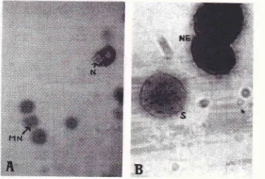
the green cytoplasm, their colour (purplish pink) were only dis-tinguishable when the microfocus was used (Fig.lA).
In this study we used fetal blood of the LMR strain rats, because fornrer results showed that the amount of cells
containing
micronuclei
washigh,
even when theanimals received
no
treatment.E Besides that, fetalblood contains
a
considerable amountof
nucleated cells of the erythrocyte lineage.e The micronuclei take the same quality of staining as the main nuclei,2 thusit
is
very easyto
detect the successof
staining, bylooking at the nuclei. The Feulgen and Rossenbeck
(1924) staining method
is a
standard technique to demonstrate the deoxyribosein
DNA.? This merhod consist of the cleavage of the purine-deoxyribose bondby mild acid hydrolysis to expose a reaclive aldehyde group, and the detection of the aldehydes by the use
of
a Schiff reagent. The reactive aldehyde combined withthe leucofuchsin in the Schiff reagent yields the forma-tion of a quinoid compound which gives a red purplish
colour."''
In
this study, several hydrolization tinreswere tried, as the correct hydrolization time suitable to the fixative used in this study (absolute methanol) was
not
available.The
bestresul
ed
usinghydrolization time
of l0
and15
ydroliza-tion time of 5 and 25 nrin.
gave
, and lesscontrast.
This finding
supports the theory rhat thehydrolysis is the critical part
of
the method, with an increasingly stronger reaction as the hydrolization timeis increased until the optim rm is reached.T Stevens and
Bancroft (1990) found that
45 min
was enough fori:,','i;.fi
stained blood smear slides, as was done in
il,:':,Ïj;:
Therefore, we tried several Schiff's reagent stainingtimes, and
we got
the same result as Stevens and Bancroft. The recommended staining time forl%
lightgreen solution is 2 min.,' but we found that 2 min. gave a very strong result, thus we shorten the staining time.
The Feulgen-light green stain does not stain the
un-evenly distributed
RNA
in
the young erythrocytes, when we use fetal blood. Therefore, no effort is neededin
differentiating the nricronucleusfrom RNA
ag-gregates, such
as
in
the
modifications
of
the Romanowsky staining merhod(Fig
lA
airdlB).
In addition, in the in vitro micronucleus test, in which thelymphocytes are screened, the Feulgen-light green
staining
method hasan
advantage.This
stainingmethod only stains the
DNA
(nricronucleus), thus noeffort is needed to differentiate the micronuclei from the aggregates
of
the azurophilic granules stained inthe
modifications
of
the
Ronranowsky stainingnrethod.12
The only
disadvantageof
Feulgen-lightStaining tlte
Micronuclei
281 [image:2.595.103.478.431.685.2]'iili:lir'illi :l'illiiii'
Figure
I'
The nicronucleus stuining. (A) IJsing rhe Fculgen-ligln greett sraittittg tttctltod, MN-
tttiuottucleus (purplistt pittk),N:ttucleus'(nagnificatiottX225.) (B)Ilsingwrighr'sstuitt,-S--stipplederyt:hrn"1r",NE:trucleutedcell .frlteeryrltrocl.te
282
Pawitan and Tanzilgreen
staining
method
is
the
need
to
use
themicrofocus,
to
differentiate white colour (RNA andother substances) from purplish pink (micronucleus).
In this study, the amount of micronuclei was not
com-pared
to
thatfound
using other staining methods.Therefore, whether there was false positive/negative results was not known, and needs further investigation.
In conclusion, the shortest time needed which gave the
best result was obtained at hydrolization time
of
10nrin., Schiff staining time
of
45 min. and light greenstaining time
of
I
second. The results showed palemicronuclei, but
whether there was false
posi-tive/negative results needs further investigation.
REFERENCES
l.
MacGregor IT, Henika PR, Whitehand L, Wehr CM. Thefetal blood erythrocyte micronucleus assay: classification of
RNA-positive erythrocytes into two age population by RNA
aggregation state. Mutagenesis 1989; 4;190-9.
2. Iskandar O. The micronucleus test [dissertation]. Iakarta :
Univ. of Indonesia, 198 1.
3. Schlegel R, MacGregor IT, Everson RB. Assesstnent of
cytogenetic damage by quantitation olmicronuclei in human
peripheral blood erythrocytes. Cancer Res 1986;
46:3717-2r.
Med J Indones
4. Cole RI, Taylor NA, Cole J, Arlett CF. Transplacental
ef-fects of chemical mutagens detected by the micronucleus
test. Nature 1979; 277 :3I1 -8.
5. Grawe
I,
Zetterberg G, Amneus H. Flow-cytometricenumeration of micronucleated polycluomatic erythrocytes
in mouse peripheral blood. Cytometry L992; l3:750-8.
6. Castelain P, van Hummelen P, Deleener A, Kirsch-Volders
M.
Automated detectionof
cytochalasin-B blockedbinucleated lymphocytes
for
scoring micronuclei.Mutagenesis 1993; 8:285- 93.
7. Stevens
A,
BancroftID,
Protein and nucleic acids.In:Bancroft fD, Stevens A, editors. Theory and practice of
histological techniques 3rd ed. Edinburgh: Churchill
Livingstone, 199 I : 143- 53.
8. Pawitan JA, Tanzil R. Pengaruh Sun-Chlorella terhadap
mikonukleasi sel darah merah pada fetus tikus. MKI 1994;
44:743-7.
9. Pawitan IA, Tanzil R. Iumlah sel seri eritrosit tak berinti
dalam darah fetus tikus putih strain Wistar pada kehamilan
hari ke 15 dan 16. In: Kumpulan abstrak pertemuan ilmiah
terbatas PAAI; 1994 lan28-29; Semarang.
10. Culling CFA. Handbook
of
histopathological andhis-tochernical techniques 3rd ed. London: Butterworths, 1975,
11. Clayden EC. Practical section cutting and staining 5th ed.
London: Churchill Livingstone, 1971.
12. Pawitan
IA.
The micronucleus test: a method used in