-Abstrak
Twnor retrobulbar nterupakan salah satu penyakit orbita. Tunor terlelakdi belakang bola nata, terkukuttg di antara tulang orbita dan berada di jaringan vital. Ganbaran klinisnya berupa eksoftaluos, dapat disertai gangguan gerak bola nnta dan penurunanr yiszs. Penbedahan cara orbitototni lateral lebih ditujukan pada tutnor orbita yang berada di daerah tenporal, apeks atau sedikit di daerah superior. Tujûan orbitototni lnteral terutama untuk nengangkat tuttror secara en toto, nrennnipulasi organ vital di orbitasedikit tnungkin untuk nenghindarkan komplikasi gatrgguan penglihatan.Peubedahan dengan cara ini nungkin nencapai tujuannya, tetapi tatnpaloya ada pengaruh jenis
tunor terhadap keberhasilan pentbedahan
arau prognosis penderita buruk Bahan penelitian sejunlah3I pasien
1'ang dilakukan penbedahan orbitototui lateral di Rwnah sakit Dr. Cipto Mangunkusunto, selanw periode Januari 1985-
1992. Kriteria prognosis yang dittilai dalan keberhasilan pengangkatan tuilror disertai dengan atau tanpa kontplikasi pada berbagai jenis twnor.Dari
keberhasilan operasi, ternlata prognosis penderita terbagidalan
3 kelonpolç yaitu :1. Hasil operasi tercapai baik dengan prognosis penderitabaih 2.Hasil operasi tercapai
bailç tetapi prognosis penderiia burulç dan 3.Hasil operasi tidak tercapai dan prognosis penderita buruk. Hasil menggaubarknn bahwa lokasi, t+,alauputt apeks sekaliputr tidak nenpengaruhi operasi orbirolotttilateral. Ketidak berhasilan operasi tanqtaknl,a dipengaruhi oleh jerus tunor.
Abstract
Retrobulbar tu,,tors are orbital disease processes situared withitt vital tissue behind the eyeball and confined v,ithin the orbital space by the orbital bones. Surgery of the retrobulbar luntors is stil! dfficult to perfonn, due to the conplex anatonl, of the orbit u,hich can deuand variatiotts in surgical approach. In the case oforbital surgerr, a surgeon will do his ut,n(rst to reach the apical area. One of the techniques v,ell knov,n
in
orbital surgerf is lateral orbitotoms,. This surgery is prinarill' ained at renovitrg zygo,naric bone cotlstituring therin of
the lateral orbital bone. The pritrcipleain of
ktteral orbitotortrf is to rennve the twnor en toto by uanipulating vital organs in the orbit as slightll,as possible in order to at'oid the conplications of visiott disorder. Thirtl'-one patients utuleru,ent lateral orbitotonD' at the Dr. Cipto Mangunkusuttto Hospital and other hospitals during the period betv,een January, I 985 to J u11' 1982. The criteriafor
successful surgery cotrsist of the evaluatiott of su.ccessful tu,,nr re,iloval v,ith or v,ithout cotttplicatiotts. Based on the saccess ofthe surgery and prognosis, the patients are divided into three groups, they 67" :1. Successfu.I su;gery', patient prognosis favorable,2.Successful surgeD:, patient prognosis dubious and 3.Utrsuccessful surgery, palienl prognosis unfavorable. Howe,ter, larcral orbitotottty did not result itt conplicatiorc in several types of tunors although they were located in the apical. This situatiott shrtu,s that even tu,nors in the apical area do not always affect the.srcce.ss of the lateral orbitotony, and thefailure of the surgery tends to be altributed nore to tuilor ry*pe.Keywords : Orbital tutnors,
Itteral
orbitototttl', T1'pes of tunors248
Moeloek et alThe Results
of
Lateral Orbitotomy
on
Orbital
Tumors
Nila
F. Moeloek, Ria Mekarwangi, Tetty A Usman, Widiarti
PRiono
INTRODUCTION
Retrobulbar tumors are orbital
diseases situatedwithin
vital
tissuebehind
theeyeball
andconfined
within
theorbit
by
theorbital bones.
Their clinical
appearance
is
often
denronstratedby
exophthalmos
that can
be
accompanied by
deterioration
of vision and limitation
of ocular
ntove-ments.Med
J Univ Indon
diag-Vol 3, No 4, October-Decenber i,994
noses, and
its
use can even be expandedto
serve as a guidancein
planning
surgeries.Surgery
of
theretrobulbar tumors
isstill difficult
to
perform,
due
to
the
complex
anatomy
of the
orbit
which
can demandvariations
insurgical
approach. Theprinciples
of
surgery, including
orbital
surgery,
presuppose
asufficient
spacefor this
undertaking
in
order
to
facilitate the removal
of
the tumor and
to
diminish
themanipulation of other
vital
tissues.In
the caseof
orbital
surgery,
a surgeonwill
dohis utmost
toreach the apical area. One
of
the techniques well
known
in
orbital surgery
is
lateral orbitotomy.
This
surgery
is
primarily
aimed
at
removing
zygomatic
boneconstituting
therim
of the lateralorbital
bÀne. l'2'3Lateral
orbitotomy
isprimarily
directed
atorbital
tumors situated at
thetemporal
andapical
spaces andto sonre
extent
at
the superior
area.
In
the
case
of
tunlors situated nasally it would
beadvisable to
reachlhe
tumor through
a
transcranial approach.
But
if
lateralorbitotomy
is toyield
the desired resultsfor this
tunror,^it should
beslighty modified to
meetspecific
needs.3The
principle aim
of lateral orbitotomy
is
torenlove
thetumor
en totoby
mani
al organsin
theorbit
aspossible
in
orclerto
omplica-tion
of
vision
disorders.
Surgery
d
is
also donefor biopsy in which
tlretumor
mass can beneither
palpated
nor clinically
diagnosed
with
suppcrting
diagnostic tools.
Though surgery
of
this
kind
nrightaccomplish its
aim, a strong mutual relationship
exists between
thetype
of tumor
and the successof
surgery,aswell
as,thepatients'
prognosis.The objective
of
this study is to perform
a re-as-sessment on the usefulnessof
lateralorbitotomy
basedon
the successof tumor
rentoval, postoperative
com_plications,
andpatient prognosis.
MATERIAL
AND
MBTHOD
This
study
includes data taken
from
the
medical
records
of
patients
who
had
lateral
or_bitotonry
at Dr. Cipto
Man
Hospital,
January 1985
to
July
1992.T
of
patier,rrs were eyetunror
patie
undergone treatntent atthe
Ocular
Tumor
ment at theDr.
Cipto
Mangunkusunto
Ho
study covers a
treat_ment
period.
Diagnosis
of
retrobulbar tumor
was
established,on the
basis
of
clinical examination
and supporting
exanrination,
of
which
one was CT-scanexamination.
Histopathological examination
still
plays
anrajor role
in
establishing diagnosis
in
these patients.
The
ktteral
Orbitotonl, on OrbitalTunrors
249majority
of patien\s underwent
lateralorbitotomy with
aWright
incision,3 while
the remainder underwent thisorbitotomy with
aslight modification,
alsocalled
the osteoplastyorbitotomy tyW
2of
Nakamura.4The criteria
for
successful surgery consist
of
rheevaluation
of successful
tumor removal
with
or
without
complications.
Patient prognosis was
determinedon
the basisof tumor
type
and the necessityfurther
treatment.The
datataken
into
accountare
:-
tumor removal : tumor
can be removed
eithcr
entoto or
in
part (excision)
-
surgical
complications
: is there any paresisof
eye muscle,orbital
hematomaor
ptosis-
visual conditions
prior
to
andpost surgery
-
tumor
types based on thehistopathologic
exanrina_tlon
-
further
treatment
on
the
basisof
tunror
types
and thepatient's prognosis
Based
on the
successof
the surgery
and
prog_nosis,
thepatients
aredivided into
three groups,
they are :l.
successfulsurgery, patient prognosis favorable
2.
successfLrlsurgery, patient prognosis
dubious3.
unsuccessful surgery,
patient
prognosis
un_favorable
The purpose
of
this three-group
division
is to
reassess the extent towhich
lateralorbitotonty
could
best serve its purpose in order that the surgerycould
be conducted moreeffectively
oncertain tuntor
types.RESULTS
Thirty-one patient
Dr. Cipto Mangun
during
theperiod
They consist
of
(58.1%);
the youngesr parienr being 4,5 yearsold,
and theoldest
70 yearsold,
the nrodebeing
3l
yearsold.
The data collected fronr those
who
underwentlateral orbitotonry
include,
the
results
of
tunror
removal; surgical conrplications; resuIting visuaI
con_ditions;
the
histopathologic
results; and the patient.s
prognosis. These results are evident
in
the
Tables1,2,3,4.
DISCUSSION
During
theperiod of
Janr.rary 1985 toJuly
1992, lateral25O
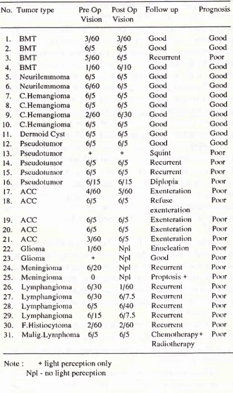
Moeloek et alTable
l.
Thesuccessof lumor rcnoval olt3l
palients undcrgoing lateral orbitotomy based on tumor tyfrc and thcir location in the orbitNo. Tumor Types
Location
Removal
To(alApical Tenrporal Sup en toto incomplote temoval
I. BMT
2. Neurilemmoma
3. C. Henrangionra 4. Dermoid Cyst 5. ACC 6. Pseudotumor
7. Glioma
8. F.Histiocytoma 9. Mal. Lynrphoma
10. Meningioma
I l. Lymphangionra
Tolal
Med
J
Utriv IndonTabcl
4.
Distribution of visual acuity bcfore to and aftcr surgcry andthc paticnt's prognosis
No. Tumor type Pre
Op
PostOp
Follow upVision
VisionPrognosis
-4-4
22
3l-4
-l-l
05-4
32-4
^a-l-l
-l-l
2-4--l
l. ., 3. 4. 5. 6. 7. 8. 9. 10. I l. t2. 13. 14. 4 2 4 I 5 5 2 I I 2 4 3t;
3 BMT BMT BMT BMT Neurilemmoma Ncurilcnrmoma C.Hcmangionra C.Hcnrangioma C.Hcmangionra C.Hcmangioma Dcrmoid Cyst Pseudolumor Pscudotumor Pscudotunror3160
31606ls
6lss/60
6lsr/60
6/106ls
6ls6160
6ls6ls
6ls6ls
6ls2160
6/306ls
6ls6ls
6ls6ls
6ls++
Gotxl Good Rccurrcnt Gotxl Good Good Goql Good Gotxl Good Good Good Squint Rccurrcnl Good Good Ptx;r Gocxl Good Good Good Good Good Good Gotxl Gcnd Ptxrr PtxrrNotè :
BMT
C. Henrangioma
ACC
F.Histicrcytonra Mal. lynrphoma
Strp 6ls 6ls 6/ls s/60 6ls 6ls 6ls
6/ r5 4l60 6ls
24 l5
: Bcnign mixcd lunror (plcinrorphic ad<:nonra) : Cavcrnous lrcnrangionra
: Adcnoid cystic carcinonra of thc lacrimal gland
: Fibrous histiocytonra
: Malignant lymphonra : Supcrior
15.
Pscudolunror16.
Pscudotunror17.
ACC18.
ACCRccurrcrrt
PtxrrDiplopia
PtxrrExcnlcralion
PtxrrRcllsc
PtxrrACC
6lsACC
6lsACC
3160Glionra
l/60Glioma
+Moningionra
6120Mcningioma
0Lyrnphangionra 6130 Lynrphangioma 6/30
Lyu.rphangionra 615 Lynrplrangioura 6l15
F.Histiocytoma 2160
Malig.Lymphoma 6/5exclltclallon
615 Exonlcration
Ptxrr615 Exr:nlcration
Poor615 Excntotation
PtxrrNpl
Enuclcation
PttorNpl
Gotxl
PoorNpl
Rccurrcnt
PtrrrNpl
Proptosis+
Ptxrrl/60 Rccurrcnt
Pttor6l'1.5 Rccurrcnt
Ptxtr6140 Rccurrcnt
Ptxrr617.5 Rccurrcnt
Ptxtr2160 Rccurrctrt
Ptxrr615
Chcnrolhcrapy+ Poor Railiothr:rapyNo(e
:
+ light pcrccption only Npl - no light pcrccptionEleven tumor types were
determined through
his-topathologic examination;
i.e.
benign
mixed
tumor,
adenoid cystic carcinoma lacrimal gland,
neurilem-moma,
dermoid
cyst, pseudotumor, cavernousheman-gioma, glioma,
meningioma,
lymphangioma,
malignant lymphoma,
andfibrous
histiocytoma.
It
is
evident
from
Table
I
that
the
lateral
or-bitotomy
procedure islimited
toorbital
tumors locatedin the temporal (or lateral), superior,
and apicalorbital
areas.
As
many as
15tumors (48.39%)
were situated
in
the temporal
area. These
figures
are
only slighty
higher than those
of
tumors located
in
the apical area;i.e.,
14patients (45.167o).
It
can also be seen
in
Table
I
that the tumors
of
24patients (77.4%) could
beremoved
entoto,
while only
7 patients
(22.6%) had tumors removed through
partial
excision.
It
is
also revealed that the
tumors situated
in
thetemporal, superior,
andapical
areas(77.4%) could
beTablc 2. Orbitotomy complications rclated to tuntor and location
19. 20. 2t. 22. 23. 24. 25. 26. 27. 28. 29. 30. 31.
No.
Tumor
Locationpaticnt lypc
in orbitEye Poslop-conrplication
Movcmcnt
Ptosis
Hcnratonra disordcr14-
Pscudolunor15.
Pscudolunror16.
Pscudotumor22.
Glioma23.
Glionra24.
Mcningionra25.
Moningioma26.
Lymphangiorna28.
Lynrplrangioma29.
Lynrphangiona Apical Apical Lalcral Apical Apical Apical Apical Apical Apical Apical No Ycs Ycs No No No Ycs Ycs Yes No + + + + +Tablc 3. Thc rolationship botwccn tunror typos and surgical rcsult and
lhc palicnl's prognosis
Gtoup Tunror lypc
Total
Succr:ssl'ul Succcssl'ulSurgcry
PnrgnosisA. Succcssl'ul
surgory
+
Strcccssl ul
ProSnosls
B. Succcssful
surgcry
+
Poorprogn()sls
C. Failc<l surgcry
+
Ptxtr
Prognosls
* BMT * Ncurilr:nrnroma * C.Hcnrangionra * Dcrnroid Cysl
* Pscudoltttnor * ACC * Glioma * F.Histitrcytonra
* Mal.lynrplronra
* Moningioma * Lynrphangionra
[image:3.595.326.561.117.513.2]Vol 3, No 4, October-Decenber 1994
removed
entoto through lateral
orbitotomy.
This
suc_cess
attests
al approach.In
thecases
isprocedure
failed
and
formed,
theHow
I
ncomplica_
tions
hough they
were
shows
thateven
tumors
in the
apical
area donot always affect
thesuccess
of
thelateral
orbitotomy,
and thefailure of
thesurgery
tendsto
beattr
The discussion
of
taken on the basis of th
Table
3.A.TUMOR TYPES OF GROUP
I
: SUCCESSFULSURGICAL
RESULTS
AND
FAVORABLE
PROGNOSIS
Benign Mixed
tumor
exists a spindle-shaped mass
clearly defined in
the areaof
the
lacrimal gland.s'7
The therapy
recommended isto remove
the tumor en toto, to reduce recurrence.Mc
NabS recommends
tumor
,"aouul
accom_periorbi
recrs
upon
.i'e
:iï:
h
this
ki
ris
f
total
success
From
Table
l,
four
patients were found with
benign
mixed tumor, that
were removed
en
toto
without any
postoperative compli.cations.
The
visual
acuity
after
surgery proved
to be
better and
thepatients'
prognoseswere
found
to
begood.
Only one
patient suffered
front
recurrence
during
treatment
Lateral Orbitotony on Orbital
Tutnors
2Sl
(Table
4, case
number 3
).
It
is
difficult
to provide
asatisfactory explanation in this
case, because when thetumor
wasremoved for
the firsttime,
thetumor
andits
rence was
associatedwith
malignant
transformation.
The
patient's
prognosis worsened and eventually
shepasseci away.
Neurilemmoma
Neurilemmoma
is
a primary peripheral nerve tumor
consisting mainly
of
the proliferation
of
Schwann.s;iï:,ii".Ti
ffJ'ï:i*:
tween 20
and
sexis found.
From Table
l,
two adult patients
werefound
with
neurilemmoma located
in the
superior
orbit. The
tons
I
on.
Cavernous hemangioma
Cavernous
hemangioma is
avascular
tumor
most
fre_quently
encounteredtumor
isgenerally
unrelatively slow
yet
pr
lated,
possessing
noregressive nature. Proptosis is
generally axial,
becausetumor locations are mostly
at
the apical
area.s
OnCT-scan examination
the tumor upp"u., as a
spindle
shaped mass
wirh definite
boundaiies and
is situated
within
the muscle cone.252
Moeloek et althe
vision
after
surgery was good,
andeven became
increasingly better (Table
4, casesnumber
9).Dermoid Cyst
The cyst walls consist
of
squamous
epithelium
anddermis
andthe lumen contains keratin
andhair.
Der-moid
cysts
are usually in thetemporal
or superonasal
areas
of
the
orbit. Their clinical
symptoms vary
depending
on thelocation
and the size of the cyst.The
appearances
of
dermoid cysts on
CT-scan are quite
characteristic; i.e, lesions
appearto
beclearly
defined
with thin
walls
and the lumen
is of
low
density."
Dermoid
cysts located
anteriorly
can cause defectsof
the bone
to
where they are
attached,
usually
in
thezygomatic
frontal
region.
Table I
shows apatient
with
atemporally
situateddermoid cyst
which was
removed
entoto
without any
post-operative
colnplications
(Table
2). Visual
acuity
after
surgery
remained
good (Table
4),
and
the
patient's prognosis
wasequally
good.It
isevident from
the datacollected
that the lateralorbitotomy
performed
in
the
first
group was
success-ful;
i.e,
tumors were removed en toto without
postsurgery complications. Even visual acuity
showed
definite improvement
in
those cases
and
in all
thepatient. Prognoses were favorable except
for
onepatient whose benign
mixed tumor
recurred.
In
order
to
avoid
recurrence,Mc
Nab emphasizes the necessityof removing tumor along with
the
periorbital
fascia.oIt is sufficient
to saythat
lateral orbitotomyperformed
in
the
first
group
of
tumor types
succeeded
in
ac-conrplishing
the purposesof the treatment.
B.
THE
SECOND GROUP
OFTUMOR
TYPBS
:SUCCESSFUL SURGERY
WITII
DUBIOUS
PROGNOSIS
Adenoid Cystic Carcinoma
Adenoid
cystic
carcinoma
is afrequently encountered
epithelial
carcinonra
of
the
lacrimal
gland.
This
kind
of
tumor is
one
of
the malignant
tumors
of
lacrima!
gland
that
can
causedeath.e Generally this tumor is
found
in
young adults
with
symptoms
of
unilateral
proptosis
with downward and medial displacement
of
the eye and is accompanied by pain. The pain
indicates
the
spread
of
the tumor
to the periorbit
and
netves.Through CT-scan,
it is
revealed
that the
mass is
spindle-shaped
with
irregular
boundaries.
The bone
erosion at the
area ofthe superotemporal
orbit is not
always distinct except when the
mass
is
relatively
large. The prognosis
of
the patient
with
malignant
tunror
of
lacrinral gland
is
usually poor. Recurrence
Med
J
Uttiv Indonrate is reasonably
high, and it
frequently
invadesorbi-tal
bones.As
seen in Table 1,five
patients were
diagnosedhistopathologically as
having
adenoid
cystic
car-cinoma.
All
thetumors were removed
entoto
without
any
postsurgery
complication
(Table 2). The patients
visual acuity both
prior to
and
after the
surgery
remained
good.However,
because
of
the poor histologic
charac-teristics of
thetumor,
and itsfurther growth
to stageIII
and
IV,
toprolong
thepatients lives further surgery in
the form
of
orbital
exenteration
coupled
with
radio-therapy
was necessary.
Visual acuity
of
the patients
waslost.
Pseudotunror
Pseudotumor
is not a true
neoplasm,
but
consists
of
inflammatory
cells
forming
urnur. in the
orbit.s'6
This tumor usually
affects young adults
but
can
alsoaffect children. There
is
no
sex prevalence, and
thetumor
canoccur
unilaterally
or
bilaterally.
The clinical
appearanceis
of
palpebral edema,
con-junctival
chemosis,ocular
motility disturbances,
prop-tosis, and pain. Occassionally
visual
reduction
or
diplopia
may
occur.The
inflammatory process can involve the
whole
orbit
or
certain
tissues
only,
such as the lacrimal
gland,
extraocular
muscles,orbital
fat,
or
vascular
tissue.CT-scan
appearances arevaried.
It
could
appearto
beretrobulbar
edema,optic
nerve
inflammation,
muscleenlargement, or
an infiltrative*orr.'0
The
patient's
prognoses ishopeful,
yet thepreser-vation
of
visual acuity largely depends
on
the
extentand the
severity
of
the
inflanrnatory
process and theeffective
response tocorticosteroids
andradiotherapy.
From
Table
l, it is
seen
that there wcre five
patientswith
pseudolumors
locatcd at the temporal andapical
areas.
Tunrors located
at
the temporal
areaoriginated from
the
lacrinral gland
and
their
ren.rovalen toto was relatively easy
to
perform.
On
the othcr
hand,
the
en toto removal in theapical
area wasrela-tively
more
difficult,
and great care
hr',Jto
be
taken. Onepatient
was found to have chronic'nyositis
of the
lateral rectus muscle
which on
CT-scan examination
appeared
as
a
mass.
Only
an
extirpation biopsy
wasperformed via
lateralorbitotomy.
Further
treatmentby
radiotherapy proved
to have
no effect,
and thepatient
still
complained
of diplopia (Table 4, casenumber
l6).
In four
patients
whosetumors were
removed
entoto,
post surgical complications
occurred
consisting
of
limited
eye movement
in
one
patient
and ptosis
Vol 3, No 4, October-Deceuber 1994
Visual acuity
bothprior
to and after the surgerydid not
change
significantly overall. During further
treatmentit
wasfound
that onepatient suffered
from
reccurrenceand
another
from
strabismus,
while two others
hadcomplaints
of
diplopia
(Table
4,
cases number
13,14,15,16).
It
seems
that the prognosis
for
goodvisual
acuity
is
not
very encouraging,
while
theprog-nosis
for survival
remains favorable.
Glioma
Glioma
is aprimary
tumor
of theoptic
nerveglial
cell.s
This
tumor
is
most
commonly found
amongchildren,
and
comprises l-27o
of all orbital
tumors.
Nearly
50%of
thetumors
arediagnosed
in
children
aged under 5.It
appearswith
unilateral proptosis. Bilateral proptosis
is a
pathognomonic
sign
of
glioma
accompained by
neurofibroma, and cafe-au-lait spots
may be
en-countered.5'6Management
of
thetumor varies according
to the size andlocation
of
thetumor,
aswell
as thevisual
condi-tion of
thepatient.
It
is
difficult
to
assess thepatient's
prognosis
dueto
thevariation
of
the disease'shistory.
Generally,
thevisual prognosis is discouraging,
while
the
life
prognosis
dependson
the extension
of
tumor
invasion intracranially.
On CT-scan the
optic
nerveappears
enlarged
and intracranial extension
of
thetunror through
theoptic
foramen
can be seen.Table
I
reveals
two
five
year
old girls
with
glionra.
Their tuntors
were renroved
in toto, but
postsurgical conrplication
occurred
consisting
of
limited
eye htovement and ptosis.
Visualacuity definitely
wor-sened.
It
wasalso not possible to maintain
the eyesin
good
condition, which
finally
neccessitatedenuclea-tion of
the eye andrepair
of
the ptosis.Fibrous Histiocytoma
Fibrous histiocytonra, frequently
referred
to
as
fibro
xanthonta, is a tuntor
of histiocytes
usually interntixing
with
fibroblasts
or fibrocytes.)
Its
clinical
appearanceis
generally unilateral
proptosis,
with
the
tumor
situated
at
the superior
or
nasal area
of
orbit.
This
tumor lends
to
cause reduction
of
vision on
con-junctival
chemosis andparalysis
ofocular
muscles. OnCT-scan
it
appears as a round-shaped nrasswith
either
clear, conjunctival
chentosis and paralysis
of
ocular
muscles
definite or irregular
boundaries,
andit
is
dif-ficult
to differentiate
it
from
neurilemmoma,
cavern-ous
hemangioma,
or
hemangiopericytoma.
Bone
erosion is
rarely
encountered, except
in
the
advanced stages.)The
surest
way to
deal
with this kind
of
tumor is
to renroveall
thetumor
tissue. Should a recurrenceoccur,
lnteral
Orbitototttl, on OrbitalTunnrs
253further excision
mustcover
awider
area, andif
neces-sary anorbital
exenterâtion
must be resortedto
in
this
case.
During
therecurrence,
thetumor
will tend
to
bemore
aggressive
and
malignant.
Generally,
radio-therapy carried out on
this
tumor
doesnot
yield
goodresults, and
its
recurrency
rate
is
high. Death
could
follow
dueto
adirect
metastasesintracranially.
Table
I shows one
patient who underwent lateral
orbitotomy
due tofibrous histiocytoma.
Thetumor
wassituated at the
apexand was removed
in toto without
post surgery complications. The visual acuity
both
prior
to and after surgery remainsrelatively
unchanged(Table 4).
In view of
thepossibility of
the recurrenceof this tumor,
thepatient's prognosis
remainsdubious.
Malignant Lymphoma
Lymphoid
tumorsgenerally originate in lymph
glands,yet they
canalso develop outside
lymph
glands,
suchas those found
in
the orbit.5'o
Clinically
lymphoid
tumors usually
present as progressive
proptosis,
yetthis kind of tumor
is
not
acconrpaniedby pain,
disor-ders
in ocular
motility,
reduction
in
vision, or
enlarge-mentof
the lacrimal gland.
It
isdifficult
todifferentiate malignant lymphoma
originating
in
the
lacrimal gland-from
epithelial
primary
tumor
of
thelacrimal
gland.5On
CT-scan thetumor
appearsoval or
elongated
andgrows along
the rectus muscle.Most
tumor
of
this kind
areconfined
tosoft
tissueof
theorbit
and do not cause bone erosion.5Table
I
reveals onepatient
whounderwent lateral
orbitotomy
for
malignant lymphoma located
at
thelacrimal gland. The tumor
was
removed
conrpletely
without
post surgery complications. The visual acuity
of
the patient
remained
unaffected, yet
because thepatient's condition
necessitated
cytostatic
treatment andradiotherapy, his prognosis
depends on theexlent
of
the disease.The
secondgroup
of
tunlors
exhibit
reasonablygood surgical results
in
which the
tumors
were
renoved
entoto. However
onfollow-up it
was shownthat the
patients had
poor
prognoses. performing
lateral
orbitotomy
on these casesshould
belimited. In
the
caseof
adenoid
cystic
carcinoma,
with
the tumor
at an early
stage
and
of
small size, when
it is
still
possible to remove
theorbital
tissue around thetumor
without
resorting to exenteration,
alateral orbitotonry
could be utilized.
If
the
diseasehas
reachedand
ad-vanced
stage,
an exenteration
is
nrore
appropriate.Appropriate evaluation
would
include biopsy
todeter-mine diagnosis
first. Fine
needleaspiration biopsy
isrecomnrended
in
an
effort
to
avoid
surgery. This
254
Moeloek et alwith
corticosteroid
or radiotherapy. Lateralorbitotomy
can
be utilized
if
necessary,yet
it
must be
done by
taking
into
account any possible complications.In
thecases
of
glioma, fibrous histiocytoma,
and malignant
lymphoma, lateral orbitotomy can be carried out
to
remove the tumor.
It
should be borne in mind
thatultimately in
the patients prognosis may be afunction
of
the natureof
the tumor.C. THE TIIIRD
GROUP OF
TUMORS
:THE
RESULTS OF
SURGERY
ARE NOT
ACCOMPLISHED,
AND
TIIE
PATIENTS
PROGNOSIS
UNFAVORABLE
Lymphangioma
Lymphangioma is
abenign tumor
pres-ent sincebirth.
It
is
slowly
progressive,
sothat
clinical
signs areonly
apparent
after
several months
or
several years after
birth.
Proptosis is foundunilaterally.
Blood
filled
mul-tilocular
cysts are frequently encountered.Hemorrhage
into the
orbit
results
in a
mass usually
known
as
a "brown cyst".
Pressureon
the eye
will
causesecondary glaucoma,
edemaof
the optic
disc,and loss
of
vision. On
CT-scan, a
multilocular
cystic
massis
seenwithin orbital
tissue
with
undefined
ir-regular boundaries,
of high
density
without
contrast,
and
without
changes in density after contrastinjection,
Four
patients
with
lymphangioma
underwent
lateral
orbitotomy,
as can be seenin
the TableL Only
one tumor was completely
removed,
and
complica-tions occurred
in
three, such as
restriction
of
ocular
movement
in two
andptosis
in
another
two.
Extirpa-tion of this tumor
isdiffic'ult,
evenwith
gradual,partby
part excision it appearsimpossible
to avoid postopera-tive complications. No improvement of vision occured
after
surgery, and prognosis
is doubtful
in
respectto
the progressive natureof
the tumor.It
is
difficult
to
determine
the
best
way
to
dealwith
this type
of
tumor
becausetreatment
will
dependon
such
factors
as
visual acuity,
and size as
well
aslocation of
the
tumor.
If
the
tumor is
confined
to
theoptic nerve and
does
not disturb vision,
immediate
ro.g".y is
not
neces.ury.s
On the other
hand,
if
thetumor
showsany
progressivity
and sharp reductionin
vision
occurs,
surgery
is called
for,
and
the type
of
surgery recommended is lateralorbitotomy.
The prog-nosisfor visual acuity of
thepatient
with
tumor
located at the apical area is usually discouraging.Meningioma
Meningioma
is a tumor originating
fro_mthe
menin-gothelial cells
of
the
arachnoid
layers.rJ The
arach-Med
J
Univ Indonnoid can
befound in the
orbit
as the outer layersof the
optic nerve.
Primary meningioma
arisesfrom
theoptic
nerve arachnoidal
layer
within
the
orbit
of
optic
foramen.
Secondary
meningioma
arises
from the
in-tracranial arachnoid layer contiguous to the
orbit.
Primary
meningioma occurs
in
the 30
-
50 yearsage
group,
and secondarymeningioma usually
presentin
thefifth
decade.This tumor is
most often
found in
women, and
usually
Caucasion.5'6Meningioma
is
a
slowly
progressive
gro_wingtumor
of local malignancy,without
metastases.tThe
clinical characteristic
of
meningioma
is
slow
but
progressive
unilateral
lossof
vision.
CT-scan examinations
areimportant to
evaluatethe
extent
of the tumor. On
CT-scan
a high
density
lesion can be seen whose density increases uponinjec-tion
of
contrast.
The tumors may
have
regular or
ir-regular
boundaries.Table
1 reveals two patientwith
meningioma who
underwent lateral orbitotomy.
The tumors were
not
removed en
toto,
and even
excision
was
difficult
be-cause
of
the hard
consistency
of
the tumor and its
tendency to bleed. Complication occuring aftersurgeryin
two
patients
were restricted eye movement
andptosis.
Visual
acuity after
surgery progressively
deteriorated.
Recurrences
during
follow up
led
to
semi-exenteration
of
the orbit.The above data shclw that
surgical
results
in
thethird
group on lymphangioma and meningioma are notfavorable.
Tumors could
not
beconpletely
removed
and
surgical cornplications frequently occur
such
asretricted
eye movement and ptosis.Lateral orbitoton-ry
on these
cases
should
be carefully
considered in
respect
to
the
difficulty of
the operation, the surgical
complications that may occur, and the poor prognosis
of
the patient.In
the caseof lymphangioma
andmenin-gioma, the lateral
orbitotomy is
not adequate
to yield
the desired results. There are
still
numerousdifficulties
to be faced
in this
group
if
lateral
orbitotomy js
to
beperformed. Recently
analternative
methodusing
CO2laser, has been devised.
Chardll
has already used COz laser therapy on a caseof
meningioma
of
the sphenoid boneby
first
exposing
thefield of
operation
by
meansof
lateral orbitotonry.
The
tumor
was destroyed
by
COz laser
burning.
The
major
advantageof this
therapy lies
in its
capacity to
eliminate
the possible hemorrhagefrequently
interfer-ing in
the removalof
the tumor.CONCLUSION
In
the
view
of
the results
of
this review
of
lateralorbitotomy
conducted on patients
with
tumors
of
theVol 3, No 4, October-De':entber 1994
prognoses,
it
is
this kind of
surgery
begiven careful
c
efore
it
is
attempted
in
order
toensure
ults,In
thelight of
all
thesepossibilities, it
seems thatlateral
orbitotomy
still
has aplace
in
dealing
with
this group
of
tumors, yet
it would
surely
be moreeffective
if
this
kind
of
surgery is coupled
with
other therapy,
such asCO2 laser.
REFERENCES
1. Kronlein RU. Zur Pathologie und operativen behandlung der
dermoidcysts der Orbita. bruns
klin
Chirl9g2;4:
149. 2. Henderson JW. orbital turnors. Brian C Decker, a Divisionof Thieme-Stratton Inc. New
york,
l9g03. Bonavolonta G. Evoluzione dell'orbitotomia temporale. Att
610 Congr. Societa Oftalrnologica Italiana, Rorne,
l9gl.
lnteral
Orbitotomy on OrbitalTunors
2554. Nakamura
y.
Osteoplastic Orbitotomy for Orbital tumor surgery, 5th International Symposium on Orbital Disorders, September 1985, Amsterdam. Orbitl9g6;5
:235 _7. 5. Shield JA. Diagnosis and Management of Orbital Ttimors.Philadelphia : WB Saunders, 1989.
6. Henderson fW, Farrow GM, Devine KD, Miller RH. Orbital Tumors. Philadelphia: WB Saunders, 1973.
7. Kincaid
MC,
Green WR. Diagnosric Merhodsin
Orbital Disease.Ophthalmology 1984; 9l : 7 19-25.8. McNab
AA.
lacrimal fossa lesions clinical features of over400 consecutive cases. Satellite Symposium on Intraocular and Adnexal Tumorc, and Orbital Diseases, Bali 1990
9. Grove
AS.
Giant Dermoid Cyst ofthe Orbit.
Ophthal_mologica 1919; 86: 15 13-20.
10. Ensmann D, Donaldson Dermoid SS, Marshall WH, Kriss
IP. Computed Tomografi
in
Orbital pseudotumor. Radiol_ ogy 1976;l2O:597-û1.
ll. Char
cesin
orbital
Tumors Diagnosis andïrera
gress of Asia pasific AcadeÀy of Oph_