168
The Effect of Yoghurt Supplementation on Rats (
Rattus Norvegicus
)
that Formaldehyde Exposure on Oxidative Damages and Protease
Enzymatic Activities of Gastrointestinal
Chanif Mahdi* and Aulaniam*
Departement of Chemistry, Faculty of Mathematics and Natural Sciences, Brawijaya University,Jln Veteran, Malang 65145
Abstract
Formaldehyde is a simplest organic compound of aldehyde or alkanal group. Formaldehyde is a toxic and carcinogenic substance. Formaldehyde contamination through food or continuous feeding diet is very dangerous for the body, especially for organ like hepar and kidney, because formaldehyde is sources of reactive oxygen species (ROS) and free radicals substances for the body. Purpose of the study is to know the effect of formaldehyde exposure and yoghurt supplementation on profile and characteristics of rats (Rattus norvegicus) protein gastrointestinal tissues and protease enzyme activities. The research methods is laboratory method. The protease enzyme activities were determined by spectrophotometry method. The result showed that formaldehyde exposure through the feeding diet of rats affect on oxidative damages, histopathology damage, and protease activity of gastrointestinal protein tissue. Yoghurt supplementation affect on decreasing on oxidative damages, histopathology damages, and increasing protease activity of gastrointestinal tissues.
Key words : Formaldehyde exposure; yogurt; oxidative damages, histodamages, profile protein; protease activity
INTRODUCTION
Formaldehyde or formalin is a simplest organic compound of aldehid or alkana group. Formaldehyde is a toxic and carcinoogenic substance. Formaldehyde contamination through the food and feeding diet with high doses level is very danger for the body. Yoghurt or yogurt is a fermented milk product of milk pasteurized that rich on vitamins and amino acids, as a potential source of antioxidant, that be expected as a protective agent of toxic substances.
Formaldehyde is a reactive com-pound, it’s easy to react to nucleophilic groups, especially to NH2 group of protein
(Enzyme system), that can effect on decreasing specific activity of enzyme, especially of oxidative phosporilation System of cytochrome P450 disturbed. Overall of these will affect to acidosis condition of the cells and tissue, and
tendency to over production of reactive oxygen species (ROS) and free radical substance, as a result of disturbed of beta oxidation Cycle. All of them will affect to decrease ATP production and tendency to dead of cells necrosis (Lee, 2003; Bray, 2005).
Over production of reactive oxygen reactive and free radical substances will affect of both to cell membrane damages, ion Ca++ accumulation in cytosol, and stimulated inflammation of the cells and tissue of gastrointestinal, that characterized by generation of nitrooxide radicals (NO.), and if it reacts with radical superoxide
malondialdehyde (MDA), nitro-oxide
MATERIALS AND METHODS Material
Chemicals. Chemicals and
instrumentation: Formaldehyde, yoghurt, tyrosine, hematoxylen, eosine, ethanol 98 %, SDS, acrilamid, bisacrilamid, glycerol, aquadest, xillol, paraffin, bromophenol blue, commasive, brilliant blue.R. 350, tris bas, B- marcaptoetanol, ammonium persul-phate, NaCl, KCl, Na2PO4, KH2PO4, TCA,
TBA, HCl, aquabidest, MDA Kit.
Instruments. Analytic Balance AE
50; Spectrophotometer UV-Vis; electro-phoresis; Vortex Gua Hug; Spuit 1 mL; Gavage; Volumetric Glaases; Water Bath, Micro pipette; Micri Tip; Eppendof.
Animal experiment. Twenty five of
8-10 week old male rats (Rattus
norvegicus), with the body weigh 100-120
g, were devided into 5 groups, each of group contain 5 rats. Group A was the control, Group B, C, D and E were administrated with formalin treatment of each were 25 ppm; 50 ppm; 75 ppm and 100 ppm. All of each group without and supplementation before and after formalin treatment.
Methods
Hispathologies observation.
Gastrointestinal tissues (Jejunum) were fixed with 4 % of buffer formalin two times for 24 hours. Hydrated tissue using alcohol rise, and then inserted into xyllen twice. Incorporated gastrointestinal tissue into soft paraffin, then into hard paraffin, then on the block paraffin and allowed to stand at room temperature. Cutting the tissue is done by using a rotary microtome with thickness of 10 µM. demounting on the glass slide using mayer albumin. The preparation is then stained with Eosin hematoxylen. Staining and preparation were incubated into mayer hematoxilene. Further preparation were washed with water and be dehydrated with alcohol rise. The preparation was washed with water and dried amounted with entelen.
Measurement of Malondialdehyde
(MDA). As much as 1.8 g of rats jejunm
mortar, placed on cold block of ice. Then add 1 mL of NaCl 9.0 percent. Homogenate transferred into micro tubes and centrifuge at a speed of 8000 rpm for 20 minutes. 100 µL of supernatant was added 550 µL distilled water. Then 100 µLof Na- thiobarbituric acid. At each reagent homogenized with vortex. Then centrifused at 500 rpm for 10 minutes.
Furthermore, the supernatant
absorbance was measured with a
spectrophotometer at maximum wave length (530 nm), and plotted on standard curve that has been made to calculate the concentration of MDA (Farmakology Laboratory ).
Measurement enzyme protease activity (using Folin reagent). 250 µL of casein substrate into sample and blank reaction tube. Then incubated for 5 minute at 370C, and then added 50 µL enzyme solutions (Crude enzyme). Incubated at 370C for 10 minutes. Then centrifuged at 6000 rpm for 5 minutes. Supernatant was taken and used for colour reaction.
In the Colour reaction supernatant 250 µL samples and blank solution were taken then added 625 µL sodium carbonate and 125 µL Folin solution. Then further incubated at 370C for 30 minutes. Then measured the absorbance in maximum wave length (660 nm),
The effect of formaldehyde exposure through the feeding diet of rats on
jejunum histopathology MDA
production.
170
number cells of jejunum. The control treatment showed that the number jejunum cells were full. And It will be decreasing continuously related with increasing dosis of formaldehyde treatment, that be characterized by increasing of level of holes of jejunum tissues. Also table 1 and table 2 showed that with the increasing level of formaldehyde exposure through the feeding diet could lead to increase
malonyldialdehyde as a parameter of membrane cell damages, and decreasing of proteolytic activity of jejunum enzyme.
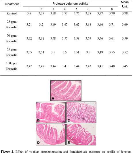
From Figure 1 and Table 1 and 2 showed that formaldehyde exposure with the dosis of 0 ppm; 25 ppm; 50 ppm; 75 ppm and 100 ppm affect on decreasing of number cells of jejunum. The control treatment showed that the number jejunum cells were full.
Figure 1. Effect of formaldehyde exposure on histopathology of rat’s jejunum. A. Jejunum control; B. Jejunum 25 ppm; C. Jejunum 50 ppm; D. Jejunum 75 ppm; E. Jejunum 100 ppm.
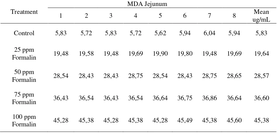
Table 1. Effect of formaldehyde exposure through the feeding diet on MDA production on jejunum tissue.
Treatment
MDA Jejunum
1 2 3 4 5 6 7 8 Mean
ug/mL
Control 5,83 5,72 5,83 5,72 5,62 5,94 6,04 5,94 5,83
25 ppm
Formalin 19,48 19,58 19,48 19,69 19,90 19,80 19,48 19,69 19,64
50 ppm
Formalin 28,54 28,43 28,43 28,75 28,54 28,43 28,75 28,65 28,57
75 ppm
Formalin 36,43 36,54 36,43 36,54 36,64 36,75 36,86 36,64 36,60
100 ppm
It will also be decreasing conti-nuously related with increasing dosis of formaldehyde treatment, that be charac-terized by increasing of level of holes of jejunum tissues. Also, table 1 and table 2 showed that with the increasing level of
formaldehyde exposure through the
feeding diet could lead to increase malonyldialdehyde as a parameter of membrane cell damages, and decreasing of proteolytic activity of jejunum enzyme.
Table 2. Effect of formaldehyde exposure through the feeding diet on proteolitic activity
Treatment Protease Jejunum activity Mean
Unit
1 2 3 4 5 6 7 8
Kontrol 3,8 3,79 3,78 3,77 3,76 3,78 3,77 3,79 3,78
25 ppm
Formalin 3,71 3,7 3,69 3,67 3,67 3,68 3,66 3,71 3,69
50 ppm
Formalin 3,62 3,61 3,58 3,57 3,58 3,59 3,56 3,61 3,59
75 ppm
Formalin 3,55 3,54 3,5 3,5 3,51 3,5 3,49 3,55 3,52
100 ppm
Formalin 3,47 3,47 3,44 3,43 3,44 3,43 3,41 3,48 3,45
172
Table 3. Effect of yoghurt supplementation and formaldehyde exposure on level of MDA production. (ug/mL).
Treatment 1 2 3 4 5 6 7 8 Mean
25 ppm
Formalin +
Yogurt
6,15 6,15 6,04 5,94 6,36 6,15 6,36 6,26 6,18
50 ppm
Formalin +
Yogurt
15,43 15,43 15,32 15,21 15,11 15,32 15,43 15,53 15,35
75 ppm
Formalin +
Yogurt
17,56 17,56 17,45 17,88 17,99 17,67 17,77 17,88 17,72
100 ppm
Formalin +
Yogurt
19,37 19,26 19,26 18,94 18,84 19,05 18,94 19,16 19,10
Table 3. Effect of yoghurt supplementation and formaldehyde exposure on jejunum proteolytic activity (Unit)
Treatment 1 2 3 4 5 6 7 8 Mean
25 ppm Formalin +
Yogurt
3,75 3,74 3,72 3,7 3,71 3,7 3,72 3,75 3,72
50 ppm Formalin +
Yogurt
3,68 3,66 3,64 3,64 3,63 3,65 3,66 3,67 3,65
75 ppm Formalin +
Yogurt
3,6 3,59 3,55 3,56 3,57 3,57 3,55 3,6 3,57
100 ppm Formalin +
Yogurt
3,55 3,54 3,53 3,52 3,51 3,5 3,48 3,54 3,52
It suggest that formaldehyde expo-sure can lead to oxidative stress, and will
be followed by oxidative damage,
inflammation, increasing MDA product-ion, de-creasing of protein content or decreasing enzymatic activities and cell
death, either necrosis or apatosis ( Dassagayam, et al 2004; Murray, 2005; Gulec, 2006).
histopathology profile, MDA production and Proteolytic enzyme activity
Based on figure 2 and table 2 and table 3 showed that yoghurt supple-mentation treatment of formalin exposure rats, induced decreasing of cells dead, decreasing of MDA production and increasing proteo-lytic activity of jejunum. This is according to Eltean (2005) and Bray (2006) Which states that yoghurt is rich on some vitamins and amino Acids , especially A, B, C, D and E vitamins, that have potent as antioxidant and detoxicant potent.
CONCLUSION
Formaldehyde as potent as a source of ROS and free radical substances could lead histopathology damages and cells dead, and cold lead increasing of MDA as parameter of cells damage, and lead to decrease proteolyditic activity of jejunum. Yoghurt supplementation could lead to decrease histopathology damages and de-creasing cells dead, and dede-creasing MDA production, and increasing proteolytic activity of jejunum.
ACKNOWLEDGMENT
The Authors are thankful to the head and all staff and assisten student of Biochemistry Laboratory of Chemistry Departement Faculty of Mathematic and Natural sciences, Brawijaya University for their kind help in preparation animal and reagent, during investigation analysis datas.
Dassagayam, TPA., Tilac, JC., Bolor, KK., Ghoshodifi, S., And Dile, RD. 2004. Free radical and in human health. Current status and future prospect. Radiation biology and health science division. JAPI Vol 52 : 2- 7.
Kumar, V., Cotarns, RS., Robin, SL. 2003. Robbins basic pathology. 7rd ed. Arrangement with elsiver Inc. New York USA. P. 3-31 ; 113- 150.
Laboratorium Farmakology. 2007. Intro of
oxidant-antioxidant measurement.
Fakultas Kedokteran Universitas
Brawijaya, Malang.
Lee, WM., 2003. Drug induced
hepatology. N Engl J Med. 349 (5) : 474- 484.
Miller, K. 2002. Immunocytochemical techniques. Theory and practice of histological technique. Fith edition. Churchill livingstode. London. P.421- 464.
Teng, S. Beard, K., Pourahman, J., Mondem, M., Easson, E., Poon, R., Obrean, PJ. 2001. The formaldehyde
metabolic detocfication enzyme