I ter atio al Sy posiu o Resear h i I ovatio a d Sustai a ility ISoRIS ’ -16 October 2014, Malacca, Malaysia Special Issue
Sci.Int.(Lahore),26(5),1741-1744,2014 ISSN 1013-5316; CODEN: SINTE 8 1741
A BRIEF REVIEW OF SURFACE MESHING IN MEDICAL IMAGES FOR
BIOMEDICAL COMPUTING AND VISUALIZATION
Shazwani Alias1, Zuraida Abal Abas2, A. Samad Shibghatullah3, A. F. Nizam Abdul Rahman4 Optimization, Modelling, Analysis, Simulation and Scheduling (OptiMASS) Research Group
Fakulti Teknologi Maklumat & Komunikasi, Universiti Teknikal Malaysia Melaka, Hang Tuah Jaya, 76100 Durian Tunggal, Melaka, Malaysia.
Email: [email protected], {2zuraidaa, 3samad, 4fadzli}@utem.edu.my
ABSTRACT:A visual representation of the interior of a body is important for clinical analysis and medical intervention.
The technique, process and art of creating this visual representation are called medical imaging. The images produced from medical imaging need to be analyses by using Finite Element Method (FEM) especially for intraoperative registration and biomechanical modeling of the tissues. This medical model ranges from the smallest vascular to bones and the complex brain. In order to use FEM, the images need to go through surface meshing generator. Although numerous mesh generation methods have been described to date, there is a few which can deal with medical data input. In this paper, a briefing review of surface meshing that can deal in medical images is presented especially in biomedical computing and visualization. Some automatic mesh generators software used in medical imaging is also discussed such as ScanIP, MIMICS, TETGEN, NetGen, BioMesh3D,CUBITMesh and Gmsh.
KEYWORD –surface mesh, biomedical image, Finite Element Method,visualization 1.0 INTRODUCTION
Finite element method (FEM) is a numerical method for approximating solutions to boundary value problem. The governing equations for most of physical phenomena are often described by partial differential equation. FEM with mesh generation has also been applied to solve problems in biomedicine, material sciences and engineering. However, mesh generation in biomedicine are still facing major obstacle where the time taken for the analysis is about 80%. It can simulate the physical phenomena and are more commonly applied in engineering sector such as heat transfer, mechanical deformation, fluid flow and electromagnetic wave propagation [1,2].
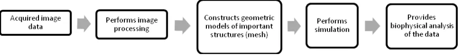
Biomedical image analysis has a pipeline that guides researchers in meshing the image as illustrated in Figure 1. The first step in generating mesh is to acquire the image data. The image can be in the form of CT, MRI scans or X-Ray. The next step is to perform image processing. Then we have to construct the geometric models of important mesh, starting from meshing the surface to transforming it into 3D images. The output from the previous step is then used to perform simulation and the result provides biophysical analysis of the data [3].
The purpose of this review paper is to introduce the readers to the existing technique in meshing especially in biomedical field. Apart from that, it is also to give an overview to newcomers on the field on the existing biomedical meshing techniques. Surface mesh is the process of discretizing a domain into smaller elements in order to conduct numerical simulation. As we are well aware, the technology has seen a
rapid advancement lately. With these technologies being applied into medical field, we can assist the medical team in decision making process. Meshing has played a very huge role in rendering images in medical imaging. With the increment of illnesses, most of it requires medical imaging machines such as MRI, CT scans, X-ray and many more in order to determine what or where the source of illness is in the human body. It is hard to study in-vivo of biological structures [4]. Computer-aided planning of surgeries employs mesh processing. These tools has proven to be vital in preparing surgeon to plan on how to conduct the operation of difficult and complex area of human body such as the spine, face or brain. In biomechanics, mesh generation has helped the orthopedic surgeon in modelling human gait to better understand the complexity of neurological and developmental issues of multiple-sclerosis patients. Computational models are also used to perform electrophysical simulations of ablation, ischemia, and arrhythmia. Other than that, mesh generation are mostly used in visualization of the human body [3]. Mesh generation or grid generation is often used interchangeably. It refers to the process of generating polygonal or polyhedral meshes that approximates a geometric domain. Meshes can be categorized into two; structured and unstructured. The basic difference between the two lies in the form of the data structures [5]. Structured mesh generates uniform shapes and the common 2D shape is quadrilateral and hexahedral in 3D
I ter atio al Sy posiu o Resear h i I ovatio a d Sustai a ility ISoRIS ’ -16 October 2014, Malacca, Malaysia Special Issue
ISSN 1013-5316; CODEN: SINTE 8 Sci.Int.(Lahore),26(5),1741-1744,2014 1742
Amesh is categorized as structured if the internal points are connected independently to their neighbors from their position. Unstructured mesh is the opposite of structured mesh. The common shapes for this type of mesh are triangular in 2D and tetrahedral in 3D [6]. There are three main techniques that are popular in triangle and tetrahedral meshing; Octree, Delaunay and Advancing Front. In order to mesh the surface of the medical images, there are criteria that need to be followed in order to get the best quality meshes that also preserve the structure of the image. Surface mesh or 2D mesh is the first step in creating a 3D mesh. However, in this paper surface mesh will be the topic of discussion and how it is applied to medical image and the criteria that needed to be followed in preserving the images.
2.0 CRITERIA FOR BIOMEDICAL SURFACE MESH GENERATION
Although unstructured mesh generation algorithm has been developed many years before and some are offered as commercial packages, they are often restricted to mechanical engineering application only [7-10]. With the evolving domain of biomedical analysis and simulation, the generation of unstructured meshes for human anatomical domains needs more flexible and effective techniques in order to tackle their topological and geometrical complexities [9]. Human body is different from engineering field in terms of complexity. We need to carefully follow the contour of complex human organs such as brains and heart while in engineering the surfaces are usually smooth and are easy to work with.
Certain criteria are required when generating surface mesh in biomedical images. Prior knowledge regarding the subject that we want to mesh is required. When dealing with human anatomy, we should have knowledge on the part that we want to take as subject so that we can project the mesh more accurately. When dealing with vascular geometries, it is important to take note that the model must represent the thin wall line of the blood vessel [11]. In order to provide a better guidance to artificial human femur, it is important that we understand the physiology of the femur and also response in a fracture[12].
Mesh has also been used to model in human prosthesis. First we need to understand the mechanics of the prosthetic and how it is used on human limbs. It is important to take note how the residual limb surface contacts the inner part of the prosthetic socket [13]. Another important criteria in doing surface meshing is the image quality.
CT scan and MR images have different approach in producing the image and the image produced depicts different view of the same object. [14] state that the parameter that needs to be take note is when using CT scan are the voltage and current intensity of the X-ray lamp, voxel dimensions, slice thickness, image dimensions and radiation exposure.
3.0 SURFACE GENERATION
Surface meshing is the process to discretize a domain into smaller elements in order to conduct numerical simulation. Is it also known as the 2D mesh. The surface meshing between anatomical features extracted from images have
made it possible for medical applications in confirming types of diagnoses, surgery simulation and planning, and therapy evaluation [3]. However, we need to take note of the difference between the medical image data with CT scan and MRI. CT scan are better in representing bony structure but it emits radiation while MRI detects soft tissue more accurately [4]. Isocontouring is a very useful tool in constructing surface data and a popular technique in producing isosurfaces is marching-cube algorithm. The active and challenging topic in constructing geometric surfaces to be used as the input boundaries in generating mesh include surface feature (contour) extraction and matching, mesh warping and deformation, conformal or harmonic mapping between surfaces, surface evolution, dynamic modeling, and motion tracking [3].
2.1 Technique/method
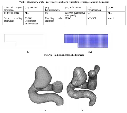
In the paper [11], MRI was used to capture the vascular structure image. Deformable surface model or 3DAO is used for image segmentation which is a collection of connected non-planar simplex faces with connectivity number 2, which means that every 3DAO simplex node have exactly 3 neighbor vertices. The simplex surface is first transform into triangulation before it can be used as an input for 3D tetrahedral mesh generation algorithm. Since the technique is to be used on hemodynamical simulation in vascular segmentation process. Laplacian smoothing and mesh simplification techniques such as decimation and quadric clustering were applied in order to improve the mesh quality and subsequent computational efficiency. The initial mesh was first smooth and then simplified before being smoothed again.
In the paper written by [15],they focused in Sub-cellular structures : transverse tubules (T-tubules), surrounding junctional sarcoplasmic reticulum (jsr) in ventricular muscle cells (or myocytes). They capture the image using electron microscopic tomography. The 2D surface generation was reconstructed or tiled by using the modules in IMOD. However, the outcome of the process results in noisy and extremely low-quality angle. A modified marching method was used to improve the quality of the mesh by re-meshing the surface again in order to get a quality tetrahedral. The next paper focuses on human femur and written by [12]. In this paper, they used already existing software, MIMICS, to generate the surface mesh and CT scan image was used as the image data. In order to have better understanding of mechanism failure and providing guidance for the design and operation of femur replacement, Finite Element Analysis of femur under physiologic condition is important. Surface mesh are created using FEA Remeshing in MIMICS
after the 3D models have been created. It’s a tool that allows
I ter atio al Sy posiu o Resear h i I ovatio a d Sustai a ility ISoRIS ’ -16 October 2014, Malacca, Malaysia Special Issue
Sci.Int.(Lahore),26(5),1741-1744,2014 ISSN 1013-5316; CODEN: SINTE 8 1743
.
Table 1 : Summary of the image sources and surface meshing techniques used in the papers
Type of subject (anatomy)
[11] Vascular [14]
Femur(animals)
[15] Sub-cellular [12]
Femur(human)
[4] IVD
Source of image MRI CT Electron microscopic
tomography
CT MRI
Surface meshing techniques
3DAO/ Deformable surface model
Marching cube algorithm
IMOD MIMICS Voxel
(a)
(b)
Figure 2: (a) domain (b) meshed domain
Figure 3: Mesh image of Intra-cranial aneurysm [11]
Intervertebral disc (IVD) is the focus in this paper. The author [4] uses voxel-based geometry to generate the mesh. FORTRAN code was developed in order to generate the segmented 2D medical image and it will be used to carry out the 3D reconstruction of the voxel-based geometries that will be used to generate the FE mesh.
There are many techniques in generating the surface mesh either using software/tools or manual/traditional way. Some existing software or tools that can generate mesh automatically such as ScanIP; a software from Simplewire that provides an image processing environment by rapidly converting 3D image data (taken from MRI, CT, micro-CT) into computational models [16]. MIMICS have been widely used for mesh generation. It is 3D medical imaging software that can do segmentation, surface meshing to produce the 3D model from the imaging data [17]. TetGen [18], CUBIT
Mesh [19], Gmsh [20] and NetGen are some of the other popular automatic mesh generation software. BioMesh3D is a software that focus in generating mesh of biomedical images developed by University of Utah [21].
4.0 CONCLUSION
I ter atio al Sy posiu o Resear h i I ovatio a d Sustai a ility ISoRIS ’ -16 October 2014, Malacca, Malaysia Special Issue
ISSN 1013-5316; CODEN: SINTE 8 Sci.Int.(Lahore),26(5),1741-1744,2014 1744
ACKNOWLEDGEMENT
This research is funded by the Ministry of Education Malaysia and Universiti Teknikal Malaysia Melaka under
the Fundamental Research Grant Scheme
FRGS/2/2013/ICT07/FTMK/02/7/F00190.
REFERENCE
[1] J. Zhang, Image-based Geometric Modeling and Mesh Generation, Lecture No. Springer, 2013.
[2] J. Shewchuk, “Unstructured mesh generation,” Comb. Sci. Comput., pp. 259–299, 2011.
[3] J. A. Levine, R. R. Paulsen, and Y. Zhang, “Mesh processing in medical-image analysis--a tutorial.,”
IEEE Comput. Graph. Appl., vol. 32, no. 5, pp. 22–8, Jan. 2012.
[4] S. Cortez, J. Claro, and J. Alves, “3D reconstruction of a spinal motion segment from 2D medical images: Objective quantification of the geometric accuracy of
the FE mesh generation procedure,” Bioeng.
(ENBENG), 2013 IEEE 3rd Port. Meet., no. February,
pp. 1–6, 2013.
[5] J. Thompson, B. Soni, and N. Weatherill, “Handbook
of Grid Generations,” Vasa, 1999.
[6] S. Rebay, “Efficient unstructured mesh generation by means of Delaunay triangulation and Bowyer-Watson
algorithm,” J. Comput. Phys., 1993.
[7] C. Lederman, A. Joshi, I. Dinov, L. Vese, A. Toga, and
J. D. Van Horn, “The generation of tetrahedral mesh models for neuroanatomical MRI.,” Neuroimage, vol. 55, no. 1, pp. 153–64, Mar. 2011.
[8] J. Bronson, J. A. Levine, and R. Whitaker, “Lattice cleaving: A multimaterial tetrahedral meshing
algorithm with guarantees,” IEEE Trans. Vis. Comput.
Graph., vol. 20, pp. 223–237, 2014.
[9] D. Szczerba, E. Neufeld, M. Zefferer, G. Szekely, and
N. Kuster, “Unstructured mesh generation from the
Virtual Family models for whole body biomedical
simulations,” in Procedia Computer Science, 2010, vol. 1, pp. 837–844.
[10] A. Corrigan, F. F. Camelli, R. Löhner, and J. Wallin,
“Running unstructured grid-based CFD solvers on
modern graphics hardware,” Int. J. Numer. Methods Fluids, vol. 66, pp. 221–229, 2011.
[11] S. De Putter, F. N. Van De Vosse, F. a. Gerritsen, F.
Laffargue, and M. Breeuwer, “Computational Mesh
Generation for Vascular Structures with Deformable
Surfaces,” Int. J. Comput. Assist. Radiol. Surg., vol. 1, no. 1, pp. 39–49, Mar. 2006.
[12] A. Francis and V. Kumar, “Computational Modeling of Human Femur using CT Data for Finite Element
Analysis,” Int. J. Eng. Res. Technol., vol. 1, no. 6, pp. 1–7, 2012.
[13] S. Zachariah and J. Sanders, “Automated hexahedral mesh generation from biomedical image data:
applications in limb prosthetics,” IEEE Trans. Rehabil. Eng., vol. 4, no. 2, pp. 91–102, Jun. 1996.
[14] D. Lopes, J. Martins, E. Pires, L. Rodrigues, E. de Las
Casas, and R. Faleiros, “A geometric modeling
pipeline for bone structures based on computed
tomography data: a veterinary study,” in Conference,
Computational Vision and Mewdical Image
Processing; VipIMAGE 2007 VipIMAGE ECCOMAS,
2007, pp. 217–222.
[15] Z. Yu, J. Wang, Z. Gao, M. Xu, and M. Hoshijima,
“New software developments for quality mesh generation and optimization from biomedical imaging
data.,” Comput. Methods Programs Biomed., vol. 113,
no. 1, pp. 226–40, Jan. 2014.
[16] “3D image processing software from Simpleware.”
[Online]. Available:
http://www.simpleware.com/software/scanip/. [Accessed: 10-Jul-2014].
[17] “Mimics®.” [Online]. Available:
http://biomedical.materialise.com/mimics. [Accessed: 10-Jul-2014].
[18] H. Si, “TetGen: A Quality Tetrahedral Mesh
Generator.” [Online]. Available: http://wia
s-berlin.de/software/tetgen/. [Accessed: 10-Jul-2014]. [19] “Sandia National Laboratories: CUBIT Geometry and
Mesh Generation Toolkit.” [Online]. Available:
https://cubit.sandia.gov/. [Accessed: 10-Jul-2014]. [20] “Gmsh: a three-dimensional finite element mesh
generator with built-in pre- and post-processing
facilities.” [Online]. Available: http://geuz.org/gmsh/.
[Accessed: 10-Jul-2014].