Cytoglobin expression in oxidative stressed liver during
systemic chronic normobaric hypoxia and relation with HIF-1α
Abstrak
Latar belakang: Hati merupakan organ yang sensitif tehadap hipoksia dan hipoksia dapat menstabilkan
HIF-1α. Saat yang bersamaan, hipoksia juga menghasilkan
senyawa oksigen reaktif (reactive oxygen species, ROS)
yang dapat ditangkal oleh Cygb. Tujuan penelitian
ini untuk mengetahui apakah hipoksia sistemik kronik normobarik menginduksi ekspresi Cygb dan apakah
proses ini berhubungan dengan stabilisasi HIF-1α. Metode: Penelitian ini menggunakan 28 tikus jantan Sprague-Dawley, BB 150-200 g. Tikus dibagi menjadi
7 kelompok: kelompok kontrol dan kelompok perlakuan yang ditempatkan dalam sungkup hipoksia (O2 10%: N2
90%) selama 6 jam, 1, 2, 3, 7 dan 14 hari. Semua tikus dieutanasia setelah perlakuan dan jaringan hati diisolasi,
dihomogenisasi dan dianalisis untuk penetapan stres
oksidatif, ekspresi Cygb dan HIF-1α.
Hasil: Ekspresi mRNA dan protein meningkat pada
hari-1 perlakuan dan mencapai maksimum Cygb pada hari-2
perlakuan hipoksia. Ekspresi menurun pada hari-3 dan sedikit meningkat pada hari-14 hipoksia. Korelasi antara ekspresi Cygb dan parameter stres oksidatif menunjukkan hubungan kuat. mRNA Cygb, demikian pula proteinnya, menunjukkan kinetik yang sama dengan HIF-1, meningkat
pada hari-1 dan hari-2.
Kesimpulan: Hipoksia normobarik sistemik kronik dan/atau
stres oksidatif menyebabkan pasang naik (up-regulation) mRNA HIF-1α yang berkorelasi dengan ekspresi mRNA dan protein Cygb. mRNA dan protein Cygb memperlihatkan
kinetik yang sama dengan HIF-1, semuanya meningkat pada hari-1 dan hari-2 yang mungkin disebabkan karena Cygb
diatur oleh HIF-1, tetapi kemungkinan juga dikendalikan
oleh faktor lain selain HIF-1.
Abstract
Background: Liver is sensitive against hypoxia and hypoxia will stabilize HIF-1α. At the same time, hypoxia will produce reactive oxygen species (ROS) which can be scavenged by Cygb. The purpose of our study is to know, if normobaric hypoxia can induce Cygb expression and its association with HIF-1α stabilization.
Methods: This is an experimental study using 28 male Sprague-Dawley rats, 150-200 g weight. Rats are divided into 7 groups: control group and treatment groups that are kept in hypoxic chamber (10% O2: 90% N2) for 6 hours, 1, 2, 3, 7 and 14 days. All rats are euthanized after treatment and liver tissue are isolated, homogenized and analyzed for oxidative stress parameter, expression of Cygb and HIF-1α.
Results: Expression of Cygb mRNA and protein was increased on the day-1 after treatment and reach the maximum expression on the day-2 of hypoxia treatment. But, the expression was decreased after the day-3 and slightly increased at the day-14 of hypoxia. The correlation between expression of Cygb and oxidative stress parameter was strongly correlated. Cygb mRNA, as well as protein, showed the same kinetic as the HIF-1, all increased about day-1 and day-2.
Conclusion: Systemic chronic hypoxia and/or oxidative stress up-regulated HIF-1α mRNA which is correlated with the Cygb mRNA and protein expression. Cygb mRNA as well as Cygb protein showed the same kinetic as the HIF-1, all increased about day-1 and day-2 suggesting that Cygb could be under the regulation of HIF-1, but could be controlled also by other factor than HIF-1.
Keywords: cytoglobin, HIF-1α, liver, oxidative stress, systemic chronic normobaric hypoxia
pISSN: 0853-1773 • eISSN: 2252-8083 • http://dx.doi.org/10.13181/mji.v23i3.1025 • Med J Indones. 2014;23:133-8 Correspondence author: Sri W.A. Jusman, [email protected]
B a s i c M e d i c a l R e s e a r c h
Copyright @ 2014 Authors. This is an open access article distributed under the terms of the Creative Commons Attribution-NonCommercial-ShareAlike 4.0 International License (http://creativecommons.org/licenses/by-nc-sa/4.0/), which permits unrestricted non-commercial use, distribution, and reproduction in any medium, provided the original author and source are properly cited.
Sri W.A. Jusman,1 Febriana C. Iswanti,1 Franciscus D. Suyatna,2 Frans Ferdinal,3 Septelia I. Wanandi,1
Mohamad Sadikin1
1 Center of Hypoxia and Oxidative Stress Studies (CHOSS), Department of Biochemistry & Molecular Biology, Faculty of Medicine,
Universitas Indonesia, Jakarta, Indonesia
2 Department of Pharmacology, Faculty of Medicine, Universitas Indonesia, Jakarta, Indonesia
Cytoglobin (Cygb) and neuroglobin (Ngb) are two new globins which play role in intracellular respiration in human and other vertebrates. Both, Cygb and Ngb are structurally similar to myoglobin (Mb). Kinetic and structural studies show that Cygb and Ngb belong to the class of hexa-coordinated globins, distinct from the other globins, hemoglobin
(Hb) and Mb which are classiied as
penta-coordinated globins. The physiological functions of hexa-coordinated globins are still unclear, but biochemical studies showed that they can scavenge toxic species, such as nitric oxide, peroxynitrite, hydrogen peroxide and supposed to be involved in oxygen storage and delivery.1-4 Studies showed
the important role of Ngb in neuronal oxygen
homeostasis and hypoxia protection.5 Cytoglobin
is expressed in ibroblasts and related cell type
from different tissues and in distinct cell types of brain and retina. It might be involved in the oxygen-consuming maturation of collagen
proteins. Although its structure is well deined, its
physiological function is still unclear. It has been suggested that Cygb has a role in protecting cells against oxidative stress.6,7
In the liver, overexpression of Cygb reduces extracellular matrix deposition in toxic and cholestatic models of liver injury and promotes recovery from previously initiated damage-induced
ibrogenesis. By inhibiting free radical-induced
activation of hepatic stellate cells, Cygb plays an
important role in controlling tissue ibrosis.8 Liver
is known as an organ which is sensitive against hypoxia. Hypoxia itself will stabilize
hypoxia-inducible factor-1α (HIF-1α). At the same time, as
a form of oxidative stress, hypoxia will produce the reactive oxygen species (ROS). As mentioned before, ROS can be scavenged by Cygb. It should be important to know, if normobaric hypoxia will induce Cygb expression in associated with
HIF-1α stabilization. For this reason, we performed
an experiment in which rats were exposed to continuous normobaric hypoxia for several periods.
We examined the relative transcriptional changes of Cygb in a situation of systemic chronic normobaric hypoxia using real-time quantitative PCR and changes of expression of Cygb protein
using enzyme-linked immunosorbent assay
(ELISA) technique. We also measured the
expression of HIF-1α mRNA using real time
RT-PCR.
METHODS
Animals
Twenty eight male Sprague-Dawley rats, weighing 150-200 g used in this study were kept under constant condition (light to dark cycle, 12 hours and 12 hours, 22-24°C room temperature, with food and water
ad libitum. The animals were kept in the animal house of Department of Biochemistry & Molecular Biology, Faculty of Medicine Universitas Indonesia. The protocol was approved by Ethical Committee of Center Research and Health Development, Ministry of Health Republic of Indonesia (Balitbangkes RI) No LB.032.02/KE/4783/08. The treatment groups
were placed in hypoxic chamber (10% O2: 90%
N2) for 6 hours and 1, 2, 3, 7 and 14 days. The rats
were euthanized by ether anesthesia. The liver were immediately removed, weighed and stored in deep frozen refrigerator (-86°C).
Isolation of total RNAs
Total RNA from rat liver was isolated and extracted from liver tissue using TriPure Reagent Isolation Kit (Roche). RNA concentration was determined using spectrophotometer.
Relative expression of cytoglobin and HIF-1α
mRNA using real time RT-PCR.
Real time RT-PCR was performed using Mini-Opticon (BioRad). An amount of 500 ng of total RNA isolate per 50 µL of RT reaction were used and reverse-transcribed
and ampliied to cDNA using real time RT-PCR with
iScript One-Step RT-PCR with SYBR Green(BioRad,
USA). Beta-actin gene was used as internal control.
The primer for cytoglobin, HIF-1α and β-actin are
designed with Primer3 based on GeneBank (NM_
024359 for HIF-1α; NM_130744.2 for Cygb and
NM 031144 for β-actin).
The primers of HIF-1α are: forward 5’-CGA AGA ACT CTC AGC CAC AG -3’ and Reverse 5’-AGC TCG TGT CCT CAG ATT CC-3’ with end-product
174 bp; the primers of Cygb are: forward 5’- AGA
ACC TGC ATG ACC CAG AC -3’and Reverse
5’-GGA AGT CAT TGG CAA ACT CC–3’ with
Real time RT-PCR reaction mixture consist of 25 µL
SYBR Green RT-PCR reaction Mix; 1.5 µL of each forward and reverse primer; 1 µL RNA template;
20 µL nuclease-free water and 1 µL iScript Reverse Transcriptase. The protocol for real-time RT-PCR
were: synthesis cDNA at 50°C for 10 minutes;
inactivation of iScript Reverse Trancriptase at 95°C
for 5 minutes; 39 cycles at 95°C for 10 seconds,
60°C for 30 seconds and 72°C for 30 seconds. A melting curve was performed to verify the presence of a single amplicon. Non-template control (NTC) was used as a negative control. Real-time RT PCR data was calculated according to Livak method.
Expression ratio:
2ΔCt target (calibrator – test)
2ΔCt ref (calibrator – test)
= 2-[(Ct target (tes) – Ct target (calibrator)]-[(Ct ref(test) – Ct ref (calibrator)] = 2-ΔΔCt
Quantiication was performed by dividing the mean
expression value of the hypoxic samples by the the normoxic sample. The result represents the factor of higher or lower expression in hypoxic samples. The data were analyzed by ANOVA.
ELISA of cytoglobin protein
ELISA plates were coated with liver homogenate (1/40 dilution factor), overnight, 4°C in coating buffer (carbonate: bicarbonate buffer pH 9.6). The plates were blocked with 5 % BSA (bovine serum albumin) for 90 minutes in room temperature with shaker and subsequently washed three times with PBST (PBS supplemented with 0.05% Tween 20).
The irst antibody (rabbit polyclonal antibody raised
against amino acids 1-190, Santa Cruz, sc-66855) 1/500 dilution, was incubated for 90 minutes in room temperature with shaker and subsequently washed three times with PBST. The second antibody, goat anti rabbit IgG (Sigma), 1/1000 dilution in PBST, were added to each well and incubated 60 minutes in room temperature with shaker. After additional
washing, the bound antibody was quantiied by
addition of ABTS solution A: ABTS solution B (1:1). The intensity of the production color was read with ELISA reader at l405 nm.
RESULTS
The expression level of HIF-1α mRNA and
cytoglobin in rat liver tissue under systemic chronic normobaric hypoxia (10% O2 for 6 hours, 1, 2, 3, 7
and 14 days) was determined using real time RT-PCR (MiniOpticon, BioRad with CFX manager software)
and quantiication of relative expression was made
using Livak method.
Expression of HIF-1α mRNA in liver tissue of rat
exposed to systemic chronic normobaric hypoxia.
Quantitative real time RT-PCR revealed an
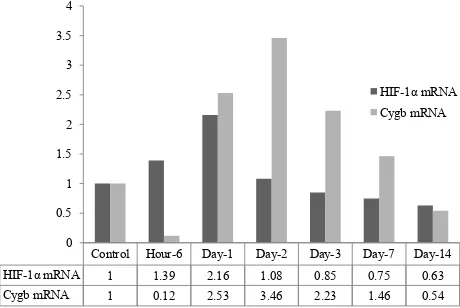
up-regulation of HIF-1α mRNA 1.39 fold after 6 hours
induction of systemic chronic hypoxia, reach its peak
after day-1 of exposure and down-regulated into 1.08; 0.85; 0.75 and 0.63 fold after 2; 3; 7 and day-14 of
systemic hypoxic condition respectively (Figure 1).
Cygb mRNA during systemic chronic normobaric hypoxia.
Relative expression of Cygb mRNA using quantitative real time RT-PCR did not showed an up-regulation of
Figure 1. Relative expression of HIF-1α mRNA in liver of rat under systemic chronic normobaric hypoxia (10% O2) for hour-6, 1, 2, 3, 7 and day-14. Beta-actin gene was used as control gene and
quantiication was made by using Livak method
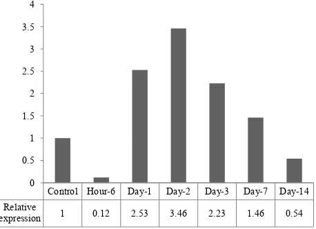
Figure 2. Relative expression of Cygb mRNA in liver of rat un-der systemic chronic normobaric hypoxia (10% O2) for 6 hrs, 1, 2, 3, 7 and 14 days. Beta-actin gene was used as control gene and
quantiication was made by using Livak method
Control Hour-6 Day-1 Day-2 Day-3 Day-7 Day-14
Relative
expression 1 1.39 2.16 1.08 0.85 0.75 0.63 0
0.5 1 1.5 2 2.5
Control Hour-6 Day-1 Day-2 Day-3 Day-7 Day-14
Relative
expression 1 0.12 2.53 3.46 2.23 1.46 0.54 0
Figure 3. Comparison of relative expression of HIF-1α & Cygb mRNA in liver of rat tissue under systemic chronic nor-mobaric hypoxia
Cygb mRNA after 6 hours induction of systemic chronic hypoxia, but showed an up-regulation on the day-1 by a factor 2.53 times and reach its peak by 3.46 factor at day-2 and down-regulated 2.23 fold at the day-3, and decreased after day-7 and day-14 (Figure 2, 3).
Correlation of relative expression of HIF-1α &
Cygb mRNA
Statistical analysis of the correlation between
HIF-1α mRNA and Cygb in liver of rat tissue
under systemic chronic hypoxic condition showed positive weak correlation (Pearson = + 0.26, p < 0.05) (Figure 4).
Concentration of Cygb protein under systemic chronic normobaric hypoxia.
Detection of cytoglobin protein in liver homogenate of rat using specific antibody
Figure 4. Correlation of Cygb and HIF-1α mRNA expression showed weak positive correlation (Pearson, r = +0.26, p < 0.05)
Figure 5. Concentration of Cygb protein in liver tissue of rat exposed to systemic chronic normobaric hypoxia (10% O2) (ELISA method). Data are represented as ratio of dilution factor (titer) of liver homogenate
Group Mean log
titer Mean titer Ratio
Control 2.66 457 1
Hour-6 2.2 158 0.35
Day-1 3.03 1072 2.34
Day-2 3.26 1820 3.98
Day-3 2.66 457 1.00
Day-7 1.9 79 0.17
Day-14 2.43 269 0.59
Table 1. Cygb protein concentration in liver of rat under sys-temic chronic hypoxia
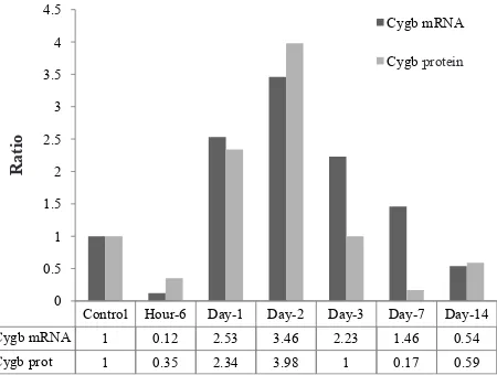
antiCygb polyclonal (full length 190 amino acids) raised in rabbit showed that the concentration, represented as log dilution factor of liver homogenate was increased since day-1 (2.34 fold) after exposure to hypoxic condition and reach the peak (3.98 fold) at 2 of hypoxia. After day-3 of exposure to systemic hypoxic condition, the Cygb protein was decreased and slightly increased on the day-14 at the end of observation, as shown in Table 1 and Figure 5.
Correlation of Cygb mRNA and Cygb protein
Figure 6 represent the correlation of Cygb mRNA and Cygb protein in liver tissue of rat exposed to systemic chronic normobaric hypoxia. The correlation between the Cygb mRNA and Cygb protein is strong positive correlation (Pearson = 0.85, p < 0.05) (Figure 7).
Control Hour-6 Day-1 Day-2 Day-3 Day-7 Day-14 HIF-1α mRNA 1 1.39 2.16 1.08 0.85 0.75 0.63 Cygb mRNA 1 0.12 2.53 3.46 2.23 1.46 0.54
0 0.5 1 1.5 2 2.5 3 3.5 4
HIF-1α mRNA Cygb mRNA
y = 0.596x + 0.950
0 0.5 1 1.5 2 2.5 3 3.5 4
0 0.5 1 1.5 2 2.5
Cy
g
b
m
RNA
HIF-1αmRNA
Control Hour-6 Day-1 Day-2 Day-3 Day-7 Day-14
Cygb
protein 1 0.35 2.34 3.98 1 0.17 0.59
0 0.5 1 1.5 2 2.5 3 3.5 4 4.5
Figure 6. Comparison of Cygb mRNA and Cygb protein in liver of rat under systemic chronic normobaric hypoxia (10% O2)
Figure 7. Correlation of Cygb mRNA and Cygb protein
DISCUSSION
In our previous study, we have demonstrated that systemic chronic hypoxia lead to oxidative stress in liver tissue since the early phase of hypoxic condition.9 It had been reported that administration of
H2O2 to pulmonary artery smooth muscle cell as such
or as a product of NADPH-oxidase addition provoke
the activation of HIF-1α.10 But in a pathological
condition, the reactive oxygen spesies does not come
from external H2O2 and even does not necessarily in
the form of this compound. Often the pathological condition appears as a result of a hypoxia condition,
which, in turn caused by insuficient perfusion
of the related tissue or organ. Though at a glance it is paradoxical, in all cases hypoxia occurs and produces a phenomenon named as “reperfusion
injury”. It is explained by the modiication of xanthin
dehydrogenase (XDH) into xanthin oxydase (XO) through a proteolytic activity of a protease which
becomes active in hypoxia. XO produces H2O2. We
have proved also that chronic normobaric hypoxia produces oxidative stress as indicated by TBARS value.9
Whether the oxidative stress, induced by systemic
chronic hypoxia and the up-regulation of HIF-1α
mRNA is correlated with Cygb expression is still
unclear. HIF-1α mRNA increases within 6 hours
in hypoxia and arrives at the maximum value after one day in hypoxia, after which it decreases gradually and even attaints the lower values than the control (Figure1). This phenomenon was observed also with another organ in the same condition, for example in heart muscle and in
stomach.11,12 Though observations were limited to
1α mRNA, apparently it is igured the
HIF-1 response. It could be supposed that as HIF-HIF-1 control an important number of genes needed for survival in early hypoxic condition and the adaptation succeeded, hence there would no need to maintain the HIF-1 at measurable level anymore. In another experiment, we observed that rats can survived in such hypoxic condition up to 14 days (data not presented).
In this study we found that expression of Cygb mRNA and Cygb protein in increased after 6 hours of induction of hypoxia, continued in the day-1, reached a peak on the 2 and decreased on the 3, day-7 and day-14. This suggested that hypoxia and/or oxidative stress would induce the expression of Cygb mRNA and the synthesis of Cygb protein. Cygb is a member of globin protein family, whose function is considered to be in relation with intracellular oxygen or oxidative state homeostasis. This is well showed by the other globin protein, namely myoglobin, another member of globin protein widely distributed in striated muscle. There are a number types of
globin protein in Caenorhabditis elegans. It was
reported that in such a tiny worm, there are 33 different globins whose expression are under the control of HIF-1. However, ten of them apparently
are not under sole HIF-1 control.13
Cytoglobin gene is a hypoxia-induced gene, which is upregulated during chronic hypoxia in a hippocampal neuronal cell line and in multiple metabolically active
tissues of murine.14 The mechanism of induction
of Cygb is HIF-1α dependent. HIF-1 is unique
among mammalian transcription factors regarding
the speciicity and sensitivity of its induction by
hypoxia.14 We found that Cygb, mRNA as well as
protein, showed in general the same kinetic as the
Control Hour-6 Day-1 Day-2 Day-3 Day-7 Day-14
Cygb mRNA 1 0.12 2.53 3.46 2.23 1.46 0.54
HIF-1, all increased about day 1 and day 2. It may lead to suggest that Cygb could be under the regulation of HIF-1. Cygb can bind O2 and it is assumed that it could have the same function as intracellular O2 reserve as do myoglobin in striated myocytes. However, statistical analysis showed that Cygb expression
was weakly, but signiicantly correlated with the
HIF-1. The data should be regarded carefully. The
correlation study was made between HIF-1α mRNA
and Cygb mRNA. As known, the merely presence of mRNA of any protein does not necessarily means the presence of corresponding coded protein. On the other hand, Cygb is assumed can scavenging free radicals which is formed even under hypoxia condition. In this regard, it is possible that free radicals itself could act as a signal for intracellular communication as indicated by nitric oxide. Cygb, therefore could be needed for quenching the action of free radical, after it completed the signal function. From this perspective, the synthesis of Cygb could be controlled also by other factor than HIF-1. If this is the case, then correlation of HIF-1 and Cygb is not as strong as if Cygb is controlled solely by HIF-1. The same phenomenon is found in myoglobin, other extraerythrocyte haemoglobin. This globin, widely distributed in striated muscle, is considered also as O2 buffer for the metabolically very active organ as striated muscle. This globin has also free radical scavenging activity. Cytoglobin like myoglobin, while under control of HIF-1, is also under control of another factor, calcineurin.15
Systemic chronic hypoxia and/or oxidative stress
would induce up-regulation of HIF-1α mRNA which
is correlated with the Cygb mRNA and protein expression. Cygb, mRNA as well as protein, showed in general the same kinetic as the HIF-1, all increased about day 1 and day 2. It may lead to suggest that Cygb could be under the regulation of HIF-1, but could be controlled also by other factor than HIF-1.
Conlicts of interest
All authors have nothing to disclose.
REFERENCES
1. Pesce A, Bolognesi M, Bocedi A, Ascenzi P, Dewilde S, Moens L et al. Neuroglobin and cytoglobin. Fresh
blood for the vertebrate globin family. EMBO Rept.
2002;3(12):1146-51.
2. Hamdane D, Kiger L, Dewilde S, Green BN, Pesce A, Uzan J, et al. The redox state of the cell regulates the ligand
binding afinity of human neuroglobin and cytoglobin. J Biol Chem. 2003;278(51):51713-21.
3. Ebner B, Panopolou G, Vinogradov SN, Kiger L, Marden MC, Burmester T, et al. The globin gene family of the cephalochordate amphioxus: implications for chordate
globin evolution. BMC Evol Biol. 2010;10:370.
4. Blank M, Wollberg J, Gerlach F, Reimann K, Roesner A, Hankeln T, et al. A membrane-bound vertebrate globin.
PLoS ONE. 2011;6(9):e25292.
5. Schmidt M, Gerlach F, Avivi A, Laufs T, Wystub S, Simpson JC, et al. Cytoglobin is a respiratory protein in connective tissue and neurons, which is up-regulated by
hypoxia. J Biol Chem. 2004;279(9):8063-9.
6. Hankeln T, Ebner B, Fuchs C, Gerlach F, Haberkamp M, Laufs TL et al. Neuroglobin and cytoglobin in search of their role in the vertebrate globin family. J Inorg Biochem.
2005;99(1):110-9.
7. Fordel E, Thijs L, Moens L, Dewilde S. Neuroglobin and cytoglobin expression in mice. Evidence for a correlation with with reactive species scavenging. FEBS J.
2007;274(5):1312-7.
8. Xu R, Harrison PM, Chen M, Li L, Tsui L, Fung PCW, et al. Cytoglobin overexpression protects against
damage-induced ibrosis. Mol Ther. 2006;13(6):1093-9.
9. Jusman SW, Halim A, Wanandi SI, Sadikin M.
Expression of hypoxia-inducible factor-1α (HIF-1α) related to oxidative stress in liver of rat-induced
by systemic chronic normobaric hypoxia. Acta med
Indones. 2010;42(1):17-23.
10. Bonello S, Zahringer C, BelAiba RS, Djordjevic T, Hess J, Michiels C, et al. Reactive oxygen species activate the
HIF-1α promoter via a functional NFκB site. Arterioscler Thromb Vasc Biol. 2007;27(4):755-61.
11. Ferdinal F, Suyatna FD, Wanandi SI, Sadikin M. Expression of B-type natriuretic peptide-45 (BNP-45) gene in the ventricular myocardial induced by systemic chronic
hypoxia. Acta Med Indones. 2009;41(3):136-43.
12. Syam AF, Simadibrata M, Wanandi SI, Hernowo BS, Sadikin M, Rani AA. Gastric ulcers induced by systemic
hypoxia. Acta Med Indones. 2011;43(4):243-8.
13. Hoogewijs D, Geuens E, Dewilde S, Vierstraete A, Moens L, Vinogradov S, et al. Wide diversity in structure and
expression proiles among members of Caenorhabditis
elegans globin protein family. BMC Genomics.
2007;8:356.
14. Fordel E, Geuens E, Dewilde S, Rottiers P, Carmeliet P, Grooten J, et al. Cytoglobin expression is upregulated in all tissues upon hypoxia: An in vitro and in vivo study by quantitative real-time PCR. Biochem Biophys Res Commun. 2004;319(2):342-8.
15. Singh S, Manda SM, Sikder D, Birrer MJ, Rothermel BA, Garry J, et al. Calcineurin activates cytoglobin transcription in hypoxic myocytes. J Biol Chem.