Autopsy indings in severe malaria – a case report
Dedi Afandi1, Budi Sampurna2,Inge Sutanto3, J. Wirasmi Marwoto4, Nurjati Chairani4, Sutisna Himawan4, Rawina W3, Ivan Riyanto5
Abstract
Severe malaria, caused by Plasmodium falciparum infection, has a high mortality rate and is the main cause of death in malaria. Since clinical autopsy is unpopular in Indonesia, autopsy examination in malaria cases is rarely done. We reported a forty three year old woman from non endemic area that was dead because of severe malaria. Diagnosis was concluded from autopsy, histopathology, and toxicology. (Med J Indones 2008; 17: 210-5)
Abstrak
Malaria berat yang disebabkan oleh infeksi Plasmodium falciparum memiliki angka mortalitas yang tinggi dan merupakan penyebab utama kematian pada malaria. Pada kematian akibat malaria jarang dilakukan pemeriksaan otopsi karena belum populernya otopsi klinis di Indonesia. Dilaporkan seorang perempuan berusia 43 tahun dari daerah non endemik dengan penyebab kematian malaria berat. Diagnosis ditegakkan melalui otopsi, pemeriksaan histopatologi, dan toksikologi. (Med J Indones 2008; 17: 210-5)
Keywords: Plasmodium falciparum, cerebral malaria, black water fever
Malaria has caused over 1.2 million deaths all over the world in year 2002, it’s the fourth among deaths caused by infectious and parasitic diseases. There are approximately 400 million new malaria cases all over the world and 7% of those numbers were found in Asia (World Health Organization, 2002).More than 45% of world population is contacted with malaria. In Indonesia, Malaria spreads all over the island particularly in the eastern islands.1,2,3
Diagnosis of severe malaria is based on the appearance of asexual forms of Plasmodium falciparum in a patient with a potentially fatal manifestation or com plication
1 Department of Forensic Medicine and Medicolegal, Faculty of Medicine, University of Riau, Pekanbaru, Indonesia. 2 Department of Forensic Medicine and Medicolegal,
Faculty of Medicine, University of Indonesia/Dr. Cipto Mangunkusumo Hospital, Jakarta, Indonesia
3 Department of Parasithology, Faculty of Medicine, University of Indonesia, Jakarta, Indonesia.
4 Department of Anatomical Pathology, Faculty of Medicine, University of Indonesia/Dr. Cipto Mangunkusumo Hospital, Jakarta, Indonesia
5 Sixth year medical student, Faculty of Medicine, University of Indonesia, Jakarta, Indonesia.
of malaria, in whom other diagnosis have been excluded (WHO, 1990). The potentially fatal manifestations and complications are: (1) cerebral malaria, (2) severe anemia, (3) renal failure, (4) hypo glycemia, (5) Adult Respiratory Distress Syndrome (ARDS)/pulmonary edema, (6) circulatory collapse, (7) bleeding and blood clothing disturbance, (8) convulsion, (9) acid-base disturbance, (10) hemo globinuria (black water fever) in acute malaria.2-5
Due to rarity in complete clinical autopsy examination in Indonesia, particularly in malaria case, we reported a complete clinical autopsy in a case of severe malaria.,4,5,6
CASE
later, she went to a hospital and got hospitalized by the recommendation of the doctor there. Five hours after hospitalized, she started to have convulsions and her general condition was deteriorating. She then died 3 hours later. Her family was suspicious that her death was due to improper medical treatment, so they reported the case to the police for some investigation.
We did external examination on the corpse. Further, standard procedure for toxicologicy and histo pathology was done. All organ samples were taken including brain, liver, and blood. Further, urine, gastric content and bladder content were withdrawn.
RESULTS
External examination
External examination was done 10 hours post mortem. The corpse was an oriental female, well nourished, 43 years old, white to yellow skin color, with black hair, 154 cm in height. She was naked. The lower left second molar was missing, no denture was observed. The irises were brown in color, the pupils were measuring 5 mm in diameter; the conjunctives were white in color. Rigor mortis had fully developed; Livor mortis was found on the back, red-purple in color and bleached by pressure. Putrefaction signs were not observed. There was no sign of injuries. Cyanosis was found on inger nails. The skin was not icteric. Needle-marks were found on both of cubital regions.
Autopsy indings
Autopsy was done 30 minutes after external examination.
Head:
Brain and skull were intact. The brain weighed 1250 grams. Venous congestion on the surface of the brain was observed. No injury was found.
Neck, trachea and esophagus:
The hyoid bone, thyroid and cricoid cartilages were intact. The esophagus and trachea contained red mucous and food remnant. The tracheal mucous was hyperemia. No signs of injuries on the esophagus and trachea walls.
Thorax:
There was dark red luid in the thoracic cavities, 80 cc in the right and 15 cc in the left one. There were no signs of injury on the thoracic wall.
Lungs: Upper lobes of both lungs were purple red in color and spongy; many petechial hemorrhages were found on their surfaces. Lower lobes of both lungs were dark red in color; and hematomas were found on their back surface. The right lung weighed 300 grams and the left one 330 grams.
The heart was brownish red in color. The circum ference of tricuspid valve was 10 cm, and the mitral, pulmonal, and aortic valve circumference were 9 cm, 5.5 cm, and 5 cm respectively. The right ventricle wall was 2 mm thick and the left was 9 mm. The coronary vessels were not sclerotic or narrowed. The inter-ventricular septum, the left wall and the right wall of ventricles showed no remarkable changes. The heart weighed 300 grams. There were many petechial hemorrhages on the back surface of the heart.
Abdomen and pelvis:
Stomach contained 50 ml of food remnant. Spleen was grey-purple in color, showed no remarkable change and weighed 120 grams.
The liver was extremely big, far from normal, yellowish brown in color, soft and brittle (fragile), with rather blunt edges, and weighed 1420 grams. Further, the cut surface structure was unclear.
Duodenum, intestines, colon and adrenal glands showed no remarkable changes. Pancreas was yellow brown in color, showed no remarkable change, and weighed 80 grams.
Renal capsule was easily detached. It contained red dark luid and longitudinal section appearance was changed. Petechia was found in renal pelvis.
Both of ovarium showed no emarkable changes. Uterus measures 10 cm x 7 cm x 3.5 cm and contained a small amount of blood.
Histopathological indings
Brain:
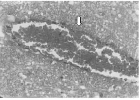
The brain was edematous and hyperemic, especially in the white matter. The blood vessels were congested and clogged by erythrocytes cluster. There were ine black pigments in the erythrocytes and inside the capillary lumens (Figure 1), which did not disappear by bleaching technique, was not blue on Yzer staining method, and negative on Fontana-Masson staining method. It was conirmed that the pigments were malaria pigments.
Heart:
The heart was edematous and hyperemic. The morphology of the cardiac muscle ibers showed no remarkable changes. There were no signs of bleeding or inlammation, but the parenchyma contained pigments inside and outside the capillary lumen.
Lungs:
The anterior parts of the lungs were slightly emphysematous and hyperemic, but the posterior ones were severely congested and edematous (Figure 2). There was no leucocyte proliferation (signs of inlammation).
Liver:
The liver showed varying appearance, especially at R3 area which was diffusely necrotic and bleeding. There were degenerative and fatty liver processes. Pigments were found every where in the parenchyma (Figure 3).
Pancreas, intestines and spleen:
The pancreas contained moderate necrotic tissue. Intestines contained early necrotic tissue and pigments especially at the mucous membrane. Spleen were congested and contained pigments.
Kidneys:
The kidneys were severely congested. There were pigments inside the glomerulus. Some tubular epithelia were necrotic and sloughed off into the lumen on some places. These indings are consistent with early acute tubular necrosis (Figure 4).
Uterus:
The uterus showed pigments in the erythrocytes inside the capillaries, and there were occlusion of capillaries by erythrocyte clusters, but there was no stromal breakdown except at the upper endometrium that showed necrosis and sloughing (Figure 5).
Toxicological examination
Toxicological examination was done at Forensic Laboratory Center, Police Department of the Republic of Indonesia, Jakarta. The examination indings: There were no poison found in the blood, stomach content, urine, brain, and liver.
Figure 1. Malaria pigments in liver (H&E after bleaching X 100)
Figure 2. Renal microphotograph showing acute tubular Figure 3. Malaria pigments in uterus with spontaneous necrosis with malaria pigments (H&E after bleaching x 100) bleeding (H&E after bleaching x 40)
DISCUSSION
There were 4 types of malaria found in human based on the types of parasite. Mortality of malaria is often caused by complication of severe malaria due to P. falciparum infection. The complication happened because the parasite had the capability of sequestrating into organ capillaries which did not happen in the other types of parasite. The effects of sequestration could cause function disturbance of the organs and lead to organ failure.2,3,6-8
After external examination, we still couldn’t determine the cause of death and had to wait for histopathological and toxicological examination indings. At this time we
were thinking that her dead was caused by some kinds of disease while keeping the probability that she was poisoned.
Although statistically cardiovascular disease is the most common cause of silent dead (more than 50% is caused by ischemic heart disease),9 we could not ind any heart abnormality in this case. Macroscopically there was no thickening or plug in the coronary artery and no sign of old or new infarct. Microscopically, though there was no sign of inlammation or even bleeding in the myocardium, malaria pigments were found.
was very important at this time. To make the inding deinite, we did 4 staining methods for all organ samples: (1) Hematoxylin and Eosin to show malaria pigment that was molasses-yellow to black in color.10 (2) Hematoxylin and Eosin staining after bleaching to exclude formalin pigment, (3) Yzer staining to exclude hemosiderin pigment, and (4) Fontana-Masson staining to exclude melanin pigment.
Malaria pigments were found in the brain, heart, liver, intestines, spleen and kidney. Multiple organ failures were found on heart, lungs and kidneys and spontaneous bleeding was showed inside the uterus. All data were consistent with severe malaria.2-5,8
The brain was edematous and hyperemic especially in the white matter which differ cerebral malaria from viral encephalitis where the hyperemia was found mostly in substansia grisea.7,10 Brain blood vessels was congested and clogged by erythrocyte clusters. Fine black pigments were found in the erythrocytes and inside the capillary lumen. These indings it the signs of cerebral malaria where the malarial pigment was only found inside the capillary lumen and never been in the brain tissue.11
The liver showed blunt edges; the surface was yellowish brown and smooth but fragile, while the cut surface structure was unclear. Necrosis was found under microscope especially in R3 area, the closest part to the center of the lobule. These indings showed liver failure which also it with malaria falciparum case theoretically which show degeneration and central lobular necrosis.2,6 Histopathological examination showed malaria pigments at the parenchyma, that was diffusely necrotic and showed fatty liver processes. Study in Jodphur,12 reported that histopathological feature in liver due to P. falciparum showed malaria pigments (90%), liver cell necrosis (18,3%) and fatty change (15%).
The spleen was normal in size, no splenomegaly was found. This inding is in line with the theory stating that splenomegaly is usually found in chronic malaria, children and semi-immune people, but not in severe malaria. No abnormality was found microscopically. This fact may happen when the disease is acute due to the difference in the ability to sequester in different organ. From a prospective study,13 Chotovanich et al reported that ring-infected erythrocyte surface antigen (RESA) was not found in red blood cells (RBCs) in spleenic blood smears from 5 patients with acute falciparum malaria.
In this case, macrocopic appearance of the kidneys and microscopic indings revealing early acute tubular necrosis and malaria pigment in the glomerulus indicated an acute attack of falciparum malaria.4 Newman et al,14 reported that renal failure and cerebral malaria was common clinical complication among U.S. travellers (43.9% and 48% respectively), and malaria pigments were found in some organs in autopsy indings (in 18 out of 50 cases).
Disseminated intravascular coagulation was supposed to happen in this patient, based on the many petechiaes that were found in the heart and lung, conirmed microscopic indings in some organs, and uterus spontaneous bleeding.4,11 The negative inding of thrombus in blood vessels of the organs might be due to increasing post mortem ibrinolysin activity.
Post mortem diagnosis of severe malaria was determined by the appearance of the parasite and or malaria pigment in the organs. The most accurate prove for the presence of the parasite involve poly merase chain reaction tests for plasmodium DNA and TaqMan real-time PCR methodology. Laser light depolarization analysis for malarial pigment (hemozoin) in white blood cells is also speciic for malarial infection and has a sensitivity of 80%.5 Sometimes, the parasite can be found in peripheral blood smear by using malaria detection method (thin and thick ilm).5,6,10
According to WHO criteria, diagnosing cerebral malaria as the cause of death should be carefully considered, and should be based on the inding of malaria parasite or pigment in the brain, that is usually hard to ind in post mortem examination. In this case, we found malaria pigments in the brain, but we diagnosed the case as severe malaria, since the inding of malaria parasite or pigment in the brain might give various symptoms depending on the sequestration density, while death could be due to other organ failure.
In conclusion, the cause of death for this patient was multiple organ failure due to severe malaria.
REFERENCES
Murray CJ, Lopez AD. Global burden of disease and injury 1.
series. Geneva: World Health Organization; 2004.
Supargiono. Immunopathology and clinical manifestations 2.
of severe malaria with special references to Plasmodium
falciparum. Med J Indones. 1998;7:8-15.
Widodo D, Pribadi MJ, Zulkarnain I. Malaria serebral. Maj 3.
Nand N, Aggarwal H, Sharma M, Singh M. Systemic 4.
manifestation of malaria. JIACM. 2001;2(3): 189-94. Rudman MS. Diagnostic dilemma: is it severe malaria ? 5.
Med J Therapeut Africa. 2007;1:41-4.
Despommier DD, Gwadz RW, Hotez PJ, Knirsch CA. 6.
Parasitic diseases. 4th ed. New York : Apple Trees Prod;
2000.
Clark IA, Alleva LM, Mills AC, Cowden WB. Patho genesis 7.
of malaria and clinically similar conditions. Clin Microbiol Rev. 2004; 17(3):509-39.
Dondorp AM. Pathophysiology, clinical presentation 8.
and treatment of cerebral malaria. Neurology Asia. 2005; 10:67-77.
Huikuri HV, Castellanos A, Myerburg RJ. Sudden death 9.
due to cardiac arrhythmias. N Engl J Med. 2001; 345(20): 1473-82.
Cross SS. Diagnostic pathology in clinical practice. In: 10.
Underwood JCE, editor. General and systematic pathology.
3rd ed. London: Tottenham Court Road; 2000.p.57-69.
Pongponratn E, Turner GDH, Day NPJ, Phu NH, Simpson 11.
JA, Stepniewska K, et al. An ultrastructural study of the
brain in fatal Plasmodium falciparum malaria. Am J Trop
Med Hyg. 2003; 69(4):345-59.
Baheti R. Laddha P, Gehlot RS. Liver involvement in 12.
falciparum malaria – a histo-pathological analysis. JIACM. 2003;4(1):34-8.
Chotivanich K, Udomsangpetch R, McGready R, Proux S, 13.
Newton P, Pukrittayakamee S, et al. Central role of the spleen in malaria parasite clearance. JID.2002;185:1538-41. Newman RD, Parise ME, Barber AM, Steketee RW. 14.