Treatment Options of Lemmel’s Syndrome: A Case
of Benign Obstructive Jaundice in the Elderly
Abigail Prasetyaningtyas*, Aru Wisaksono Sudoyo**, Perdana Aditya Rahman*
*Department of Internal Medicine, Faculty of MedicineUniversity of Indonesia/Dr. Cipto Mangunkusumo General National Hospital, Jakarta **Division of Hematology and Medical Oncology, Department of Internal Medicine
Faculty of Medicine, University of Indonesia/Dr. Cipto Mangunkusumo General National Hospital, Jakarta
Corresponding author:
$UX:LVDNVRQR6XGR\R'LYLVLRQRI+HPDWRORJ\DQG0HGLFDO2QFRORJ\'HSDUWPHQWRI,QWHUQDO0HGLFLQH
University of Indonesia/ Dr. Cipto Mangunkusumo General National Hospital. Jl. Diponegoro No. 71 Jakarta Indonesia. Phone: +62-21-3162497/3919680; Facsimile: +62-21-3926286. E-mail: arusudoyo @yahoo.com
ABSTRACT
Lemmel’s syndrome, also known as duodenal diverticulum obstructive jaundice, is a rare cause of benign obstructive jaundice that should be included in the differential diagnosis of biliary obstruction when PAD is present, in the absence of cholelithiasis or other detectable obstacle. Diagnosing Lemmel’s syndrome could be challenging, but being aware of this condition is important to avoid mismanagement and it begins with
LGHQWL¿FDWLRQRISHULDPSXOODU\GLYHUWLFXOD3$'ZKLOHLQWHUSUHWLQJDQ\ELOHGXFWLPDJLQJ,WFDQEHPLVLQWHUSUHWHG
as periampullary tumors, biliary stones, or pancreatic pseudocyst. Symptomatic patients can be successfully managed endoscopically in many cases but surgical management would be necessary in selected cases.
We present a patient with benign obstructive jaundice caused by Lemmel’s syndrome who was successfully treated with endoscopic sphicterectomy. A 67 years old female presented to the emergency department with chief complaint of jaundice. The patient was assesed to have obstructive jaundice cause by a duodenal mass, elevation of transaminase enzime supected caused by drug induced liver injury, hypertension (controlled), and
DQWHULRUH[WHQVLYHFRURQDU\LVFKHPLD(QGRVFRSLFUHWURJUDGHFKRODQJLRSDQFUHDWRJUD¿(5&3VKRZLQJPXWLSOH
giant diverticle in second part of duodenum, stenosis of the distal common bile duct (CBD) with compression of diverticular extra luminal as a differential diagnosis. Endoscopic ultrasound (EUS) was performed to exlude a periampullary tumor, resulting distal CBD stenosis due to compression of multiple PAD. We performed an endoscopic sphinterectomy (EST) and the stent was removed. A further evaluation of the tuberculous lymphadenitis
ZDVSODQQHGDVRXWSDWLHQWVHWWLQJ2QHPRQWKIROORZXSQRUHFXUHQFHRIMDXQGLFHZDVREVHUYHG
Keywords: Lemmel’s syndrome, obstructive, jaundice, endoscopic sphicterectomy
ABSTRAK
Sindrom Lemmel, yang juga dikenal sebagai divertikulum duodenum ikterus obstruktif, merupakan penyebab yang jarang dari ikterus obstruktif jinak yang harus dipikirkan dalam diagnosis diferensial obstruksi bilier dengan divertikula periampula, saat tidak ditemukannya cholelithiasis atau obstruksi lainnya. Mendiagnosis sindrom Lemmel merupakan suatu tantangan, tetapi menemukan kondisi ini penting untuk menghindari salah dalam
SHQDWDODNVDQDDQSDVLHQGDQKDOLQLGLPXODLGHQJDQLGHQWL¿NDVLGLYHUWLNXODSHULDPSXODNHWLNDPHPEDFDVHWLDS
Dalam laporan kasus ini, kami melaporkan seorang pasien dengan ikterus obstruktif jinak yang disebabkan oleh sindrom Lemmel, yang berhasil diterapi dengan dengan sphincterotomi endoskopi. Seorang wanita 67 tahun datang ke gawat darurat dengan keluhan utama ikterus. Pasien ini didiagnosa memiliki ikterus obstruktif yang disebabkan oleh massa duodenum, kenaikan enzim transaminase yang dicurigai disebabkan oleh kerusakan hati akibat obat, hipertensi (terkontrol), dan iskemia koroner anterior yang luas. Endoskopi retrograde
FKRODQJLRSDQFUHDWRJUD¿(5&3PHQXQMXNNDQGLYHUWLNXOXPUDNVDVDPXOWLSHOSDGDEDJLDQNHGXDGDULGXRGHQXP
stenosis saluran empedu umum distal dengan kompresi divertikulum luminal tambahan sebagai diagnosis
GLIHUHQVLDO(QGRVNRSLXOWUDVRQRJUD¿GLODNXNDQXQWXNPHQJHNVOXVLWXPRUSHULDPSXOD\DQJPHQ\HEDENDQVWHQRVLV
distal CBD akibat kompresi dari beberapa divertikula periampula. Sphinterektomi endoskopi dilakukan dan stent diangkat. Evaluasi lebih lanjut dari limfadenitis TB direncanakan dalam rawat jalan. Setelah satu bulan follow-up, tidak ada jaundice yang muncul kembali.
Kata kunci: sindrom Lemmel, obstruktif, ikterus, sphinterektomi endoskopi
INTRODUCTION
Lemmel’s syndrome, also known as duodenal diverticulum obstructive jaundice syndrome was ¿UVWGHVFULEHGLQE\/HPPHOFKDUDFWHUL]HGE\ obstructive jaundice due to periampullary diverticula (PAD), in the absence of cholelithiasis or other detectable obstacle. Very few cases of Lemmel’s syndrome have been published and fully investigated.1-3 Duodenal
diverticulum is a well known entity since the early HLJKWHHQWKFHQWXU\ZKHQLWZDV¿UVWUHSRUWHGE\D)UHQFK pathologist, Pierre Jean Baptiste Chomel, in 1710.4 The
duodenum is the second most common site of diverticula LQWKHVPDOOERZHOIROORZLQJWKHMHMXQXP,WLVGLI¿FXOW to ascertain the exact prevalence of duodenal diverticula; they are seen in 1-6% of upper gastrointestinal contrast studies, 12-27% of endoscopic studies and in 15-22% of autopsies.4,5 Diverticula usually found in people above 40
and has a tendency to increase with age.5–9 PAD occurred
in up to 65% of elderly patients in some studies.10
Diverticula occur at locus minoris in the duodenal wall such as the site of entry of the common bile duct, pancreatic duct and perivascular connective tissue sheath. The etiology is not clear, it might be the end result of duodenal motility disorder, advancing age, progressive weakening of intestinal smooth muscles and increase of intraduodenal pressure may all encourage the outpouching of the duodenum.11 About
70-75% of duodenal diverticula are periampullary. 3$'ZHUHGH¿QHGDVH[WUDOXPLQDORXWSRXFKLQJVRI the duodenum adjacent to or containing the ampulla of vater or intraluminal component of the common bile duct (CBD). Diverticula arising within 2-3 cm radius of the ampulla but not containing it are referred to as juxtapapillary diverticula (JPD). However, if the papilla arises within a diverticulum it is called an intradiverticular papilla (IDP).5,11
In the majority of cases, diverticula arise on the inner or pancreatic border of the duodenum. The possibly of PAD should be kept in mind while LQWHUSUHWLQJDQ\ELOHGXFWLPDJLQJ,WFDQFUHDWHD¿OOLQJ defect in biliary passage; hence, can be mistaken for periampullary tumors or biliary stones. It can also be misinterpreted as pancreatic pseudocyst when it is large DQGÀXLG¿OOHG11 We report a case of an elderly patient
with osbtructive jaundice due to Lemmel’s syndrome that was succesfully managed endoscopically.
CASE ILLUSTRATION
A 67 years old female presented to the emergency department with chief complaint of jaundice. Three weeks before admission, she noticed yellow-colored sclera and brown discoloration of urine. The patient also complained of nausea and decrease in appetite. Weight loss and fever was denied. Five week before admission, the patient was diagnosed with tuberculous lymphadenitis and was taking anti tuberculosis drugs (rifampicin, pyrazinamide, and isoniazid), and took the drugs for two weeks. She complained nausea and stopped consuming the drugs. She went to a hospital in Purwakarta then referred to Jakarta for further investigation. Abdominal computerized tomography (CT) performed with result of distal bile duct blockage. She was advised to undergo endoscopic UHWURJUDGHFKRODQJLRSDQFUHDWRJUD¿(5&3DW&LSWR Mangunkusumo Hospital.
icteric with no tenderness in abdomen. Laboratory tests showed hemoglobin 12.4 g/dL, hematocrit 35.4%, leukocytes 6300/mm3, and platelets 171,000/mm3.
There was an elevation of liver function test AST of 447 U/L and ALT of 204 U/L, total bilirirubin of 12.92 mg/dL, direct bilirubin of 9.82 mg/dL and indirect bilirubin of 3.1 mg/dL, gama glutamyl traspeptidase of 57 mg/dL, alkaline phosphatase of 137 mg/dL, and non-reactive for HBsAg, anti-HCV and IgM anti HAV.
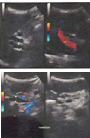
ECG examination showed sinus rhythm, QRS rate of 62 times per minute, normoaksis, PR interval 0.12 seconds, QRS interval 0.8 seconds, no changes in the ST waveform, T inversion in I, AVL, V1-V6, and no hypertrophy. The X-ray examination in 22 January UHYHDOHGDRUWDFDOVL¿FDWLRQHORQJDWLRQDQGQR abnormalities of the lungs. Abdominal CT scan with contrast in 13 January 2015 show thickening wall of the pylorus (1.28 cm) and duodenal wall at second part and third part (approximately: 2.32 cm) suspected was an intraluminar mass, widening CBD (13.6 mm) and pancreatic duct (2,8mm); simple cyst in the upper pole of the right kidney (8 mm), multiple cysts in the lower pole of the left kidney (the largest size of 1.44 cm), DQGDRUWLFFDOFL¿FDWLRQ)LJXUH
The patient was assesed to have obstructive jaundice cause by a duodenal mass, elevation of transaminase enzime supected caused by drug induced liver injury, hypertension (controlled), and anterior extensive coronary ischemia. Prophylactic cefoperazone sulbactam 2 gram per day was administered for prevention of cholangitis, total bilirubin decreased to 6,89 mg/
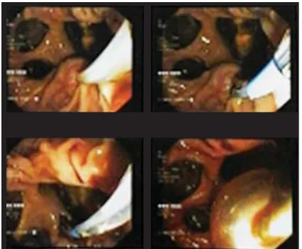
G/ (QGRVFRSLF UHWURJUDGH FKRODQJLRSDQFUHDWRJUD¿ (ERCP) and CBD stenting performed. The ERCP showing mutiple giant diverticle in second part of duodenum, stenosis of the distal CBD with compression of diverticular extra luminal as a differential diagnosis (Figure 3). Endoscopic ultrasound (EUS) was performed to exlude a periampullary tumor, resulting distal CBD stenosis due to compression of multiple periampullary diverticula (PAD).
)LJXUH$EGRPLQDO&7VFDQVKRZHGWKLFNHQLQJZDOORIWKHS\ORUXVFPDQG
duodenal wall at second part and third part (approximately: 2.32 cm) suspected was an intraluminar mass, widening CBD (13.6 mm) and pancreatic duct (2,8mm); simple
F\VWLQWKHXSSHUSROHRIWKHULJKWNLGQH\PPPXOWLSOHF\VWVLQWKHORZHUSROHRI WKHOHIWNLGQH\WKHODUJHVWVL]HRIFPDQGDRUWLFFDOFL¿FDWLRQ
Figure 3. ERCP showed periampullary diverticula and stenosis of the distal CBD
We performed an endoscopic sphinterectomy (EST) and the stent was removed (Figure 4). Patient was discharged with billirubin further decreased to 2,1 mg/dL. A further evaluation of the tuberculous lymphadenitis was planned as outpatient setting. One month follow-up, no recurence of jaundice was observed.
Figure 4. Endoscopic Sphincterectomy showed the stent was removed
DISCUSSION
Lemmel’s syndrome is characterized by obstructive jaundice due to periampullary diverticula (PAD), in the absence of cholelithiasis or other detectable obstacle.1-3 Most patients with Lemmel’s syndrome present with jaundice, abdominal pain or acute cholangitis, mimicking periampulary tumors.2,12,13
Diverticula of the gastrointestinal tract are outpouchings of all or part of the intestinal wall which can occur anywhere throughout the gastrointestinal tract. The duodenum is second most common site of diverticula in the gastrointestinal tract after colon, followed by jejunum, ileum and stomach. Duodenal GLYHUWLFXODZDV¿UVWUHSRUWHGE\WKH)UHQFKSDWKRORJLVW , Pierre Jean Baptiste&KRPHOLQDQG¿UVWZHOO
documented report was made by Morgagni in 1762 and
it was regarded an anatomic curiosity until 1913 when radiological demonstration was done by JT Case, who displayed roentgenograms of 4 cases.1,14,15
PAD were
GH¿QHGDVH[WUDOXPLQDORXWSRXFKLQJVof the duodenum adjacent to or containing the ampulla of vater or intraluminal component of the CBD. Diverticula arising within 2-3 cm radius of the ampulla but not containing it are referred to as juxtapapillary diverticula (JPD). However, if the papilla arises within a diverticulum it is called an intradiverticular papilla (IDP).5,11
It is difficult to ascertain the true prevalence of duodenal diverticula; they are seen in 1-6% of upper gastrointestinal radiologic exam, 12-27% of endoscopic studies and in 15-22% of autopsies.5,13
Duodenal diverticula are rare below age 40 years and has a tendency to increase with age.5–9,11 They
FDQEHFODVVL¿HGDVHLWKHUFRQJHQLWDORUDFTXLUHGDQG intraluminal or extraluminal. They typically occur in the periampullary region, along the medial aspect of the second and third part of the duodenum.16,17
Incidence of Lemmel’s syndrome seems higher in patients with intradiverticular papilla than in patients with a juxtapapillary diverticulum, possibly because of their larger size and closer relation to the ampulla.13
Among duodenal diverticula, PAD is the most common type comprising about 70% to 75% of all duodenal diverticula. Most PAD are asymptomatic but complications can occur in about 5% of cases and they include bleeding, perforation, diverticulitis, pancreatitis, choledocholithiasis, cholangitis, jaundice, enterolith or bezoar formation, intestinal obstruction, etc. Among these complications, hepatocholangiopancreatic disease seldomly occurs in the absence of choledocholithiasis and is termed Lemmel’s syndrome.1,2
Pathologic mechanisms through which Lemmel’s syndrome is thought to occur include the following. First, diverticulitis or direct mechanical irritation of 3$'PD\FDXVHFKURQLFLQÀDPPDWLRQRIDPSXOODDQG OHDGWRFKURQLF¿EURVLVRISDSLOODSDSLOOLWLVFKURQLFD ¿EURVD6HFRQG3$'PD\FDXVHG\VIXQFWLRQLQWKH VSKLQFWHURI2GGL7KLUGIRRGGHEULVÀRZLQJLQWRD diverticulum cause distal CBD or ampulla directly compressed mechanically by PAD. 1–3,6,20,21 In our case,
3$'¿UVWPDNHGDFKURQLF¿EURVLVRISDSLOODWKDWOHDG to papillary stenosis. Second PAD directly compressed the distal CBD, PAD seems to have expanded with resultant extrinsic compression of distal CBD . CBD ZDVH[SORUHGLQRXUSDWLHQWE\(5&3DQGFRQ¿UPHGE\ endoscopic ultrasound, no other etiology of obstructive MDXQGLFHFRXOGEHLGHQWL¿HGRWKHUWKDQH[WUDOXPLQDO compression by PAD.
Prior to the 1970s, PAD was diagnosed coincidentally during barium meal or surgery, and the discovery rate was low (estimated at less than 1%). After the 1970s, the widespread use of ERCP led to increase diagnosis of PAD.22 Diagnosing Lemmel’s syndrome could
be challenging, but being aware of this condition is important to avoid mismanagement and it begins with LGHQWL¿FDWLRQ RI 3$' 3$' DUH EHVW GHPRQVWUDWHG using a side-viewing endoscope during ERCP. On CT scan or MRCP, PAD appear as thin-walled cavitary lesions situated on the medial wall of the duodenum second part that typically contain gas. However, PAD are sometimes filled with fluid and frequently be misinterpreted with pancreatic pseudocyst, pancreatic abscess, cystic neoplasm in the pancreas head or even metastatic lymph node.1,23–25 Therefore, high index of
suspicion is necessary to establish right diagnosis in such cases. In our case, the common bile duct dilatation ZDVDW¿UVWFRQVLGHUHGFDXVHE\PDVVKRZHYHUDIWHU (5&3DQGWKHQFRQ¿UPHGE\HQGRVFRSLFXOWUDVRXQG WKHGLODWDWLRQZDVFRQ¿UPHGFDXVHE\WKHFRPSUHVVLRQ of PAD. Currently, the diagnosis of Lemmel’s syndrome is mostly made by EUS and ERCP. These examinations FRQ¿UPWKHGLDJQRVLVH[FOXGHRWKHUSRVVLEOHFDXVHV such as choledocolithiasis and tumors, and allow to perform treatment by endoscopic sphincterotomy. 13
The therapeutic options for Lemmel syndrome are surgical resection, endoscopic intervention and conservative treatment.3 The most simple treatment of
Lemmel’s syndrome is endoscopic sphincterotomy to release CBD obstruction.13 But until now, there are no
guidelines of the management of Lemmel’s syndrome. Earlier this century, surgical diverticulectomy was IUHTXHQWO\ FDUULHG RXW IRU QRQVSHFL¿F V\PSWRPV
There is now consensus that elective surgical treatment of asymptomatic or minimally symptomatic diverticulum is not justified. Surgical procedures for diverticula in the second part of duodenum are SDUWLFXODUO\GLI¿FXOWVLQFHRIWHQLWUHTXLUHVPRELOL]DWLRQ of the duodenum which is retroperitoneal. Surgical or endoscopic interventions should only be reserved for symptomatic diverticulum.11 Diverticulectomy
for abdominal discomfort and indefinite pain is dangerous and unrewarding; it carries a high morbidity and mortality. Only 50% of patients treated with diverticulectomy were relieved of their symptoms.11,26
The patient in our case was successfully treated by endoscopic sphincteroctomy. Generally, the length of EST is shorter in patients with PAD than in those without PAD due to the weakness of the sphincter of choledochus and risk of perforation in patients with PAD.10,29 The complication of EST is pancreatitis (5.4
%) and hemorrhage (2.0 %).32 Until now, guidelines
regarding the therapeutic indication of Lemmel’s syndrome have not been established, so we must select a suitable therapeutic strategy for each patient. We should consider the patient’s quality of life and comorbidity, because Lemmel’s syndrome is a benign disease and is usualy found in the elderly.
REFERENCES
1. Kang HS, Hyun JJ, Kim SY, Jung SW. Lemmel ’ s syndrome, an unusual cause of abdominal pain and jaundice by impacted intradiverticular enterolith? J Korean Med 2014:874-8. 2. Rouet J, Gaujoux S, Ronot M. Lemmel’s syndrome as a rare
cause of obstructive jaundice. Clin Res Hepatol Gastroenterol 2012;36:628-31.
3. Takakura K, Koido S, Kajihara M. Repetitive acute cholangitis and pancreatitis due to Lemmel syndrome: A case report. Int J Diagnostic Imaging 2014;1:88-91.
4. Thorson CM, Ruiz PS, Roeder RA, Sleeman D, Casillas VJ. The perforated duodenal diverticulum. Arch Surg 2012;147:81-8.
5. Lobo DN, Balfour TW, Iftikhar SY. Periampullary diverticula: consequences of failed ERCP. Ann R Coll Surg Engl 1998;80:326-31.
6. Zoepf T, Zoepf D, Arnold JC, Benz C, Riemann JF. The relationship between juxtapapillary duodenal diverticula and disorders of the biliopancreatic system: analysis of 350 patients. Jpn J Gastroenterol 2001;1:54.
7. Boix J, Lorenzo-Zúñiga V, Añaños F, Domènech E, Morillas RM, Gassull MA. Impact of periampullary duodenal diverticula at endoscopic retrograde cholangiopancreatography: a
SURSRVHGFODVVL¿FDWLRQRISHULDPSXOODU\GXRGHQDOGLYHUWLFXOD
Surg Laparosc Endosc Percutan Tech 2006;16:208-11. 8. Rajnakova A, Goh PM, Ngoi SS, Lim SG. ERCP in patients
with periampullary diverticulum. Hepatogastroenterology 2003;50:625-8.
diverticula with bile duct stones and with technical success of endoscopic retrograde cholangiopancreatography. Endoscopy 2004;36:1050-3.
10. Kim HW, Kang DH, Choi CW, et al. Limited endoscopic s p h i n c t e r o t o m y p l u s l a rg e b a l l o o n d i l a t i o n f o r choledocholithiasis with periampullary diverticula. World J Gastroenterol 2010;16:4335-40.
11. 5L]ZDQ 00 6LQJK + &KDQGDU9 =XO¿TDU 0 6LQJK9 Duodenal diverticulum and associated pancreatitis: case report with brief review of literature. World J Gastrointest Endosc 2011;3:62-3.
12. Yoneyama F, Miyata K, Ohta H, Takeuchi E, Yamada T, Kobayashi Y. Excision of a juxtapapillary duodenal diverticulum causing biliary obstruction: report of three cases. J Hepatobiliary Pancreat Surg 2004;11:69-72.
13. Chiang TH, Lee YC, Chiu HM, Huang SP, Lin JT, Wang HP. Endoscopic therapeutics for patients with cholangitis caused by the juxtapapillary duodenal diverticulum. Hepatogastroenterology 2006;53:501-87.
14. Teven CM, Grossman E, Roggin KK, Matthews JB. Surgical management of pancreaticobiliary disease associated with juxtapapillary duodenal diverticula: case series and review of the literature. J Gastrointest Surg 2012;1:1436-41.
15. Mahajan SK, Kashyap R, Chandel UK, Mokta J, Minhas SS. Duodenal diverticulum: review of literature. Indian J Surg 2004;66:10-12.
16. Kua JE, Seah A, So JB. Periampullary diverticulum: a case of bleeding from a periampullary diverticula. Ann Acad Med Singapore 2005;34:636-8.
17. Harford, William. Duodenal diverticula. Sleisenger and Fordtran’s Gastrointestinal and Liver Disease 2010;337-8. 18. Egawa N, Anjiki H, Takuma K, Kamisawa T. Juxtapapillary
duodenal diverticula and pancreatobiliary disease. Dig Surg 2010;27:105-9.
19. Ko KS, Kim SH, Kim HC, Kim IH, Lee SO. Juxtapapillary duodenal diverticula risk development and recurrence of biliary stone. J Korean Med Sci 2012;27:772-6.
20. Qi C, Zhaodong L, Shengwei L. Diagnosis and treatment of juxta-ampullary duodenal diverticulum. Clin Investig Med 2010;33:298-303.
21. Macari M. Original report cystic neoplasms of the pancreas:
&7DQG05LPDJLQJ¿QGLQJVLQVHYHQ$-5$P-5RHQWJHQRO
2003:195-9.
22. Ono M, Kamisawa T, Tu Y, Egawa N. Clinical imaging MRCP and ERCP in Lemmel Syndrome. J Pancreas 2005;6:277-8. 23. Sherlock, Sheila. Imaging of the biliary tract: interventional
radiology and endoscopy disease of liver and billiary system. Blackwell Science 2002;562-78.
24. Mathis KL, Farley DR. Operative management of symptomatic duodenal diverticula. Am J Surg 2007;193:305-9.
25. Martins PN, Benckert C, Vetzke-Schlieker W, Pratschke J, Tullius SG, Neuhaus P. Intraduodenal diverticulum associated with a double common bile duct causing recurrent pancreatitis and cholangitis: Report of a case. Surg Today 2007;37:320-4. 26. Abu Dayyeh BK, Baron TH. Endoscopic sphincterotomy: