240
Istiantoro et al. Med J IndonesSix-month follow-up
of laser
in-situ
keratomileusis
for
myopia
Istiantoro,
Tjahjono D.
Gondhowihardjo,
JohanHutauruk
Abstrak
Penelitian retrospektif untuk menilai hasil refraksi dari laser in-situ keratomileusis
(lnsik) menggunakan excimer
Laser untuk menentukan ketepatan, prediktabilitas, keamanan dan stabilitas koreksi miopia. lnsik dilakukan pada 475 mata pada 302 penderita miopia dengan ekwivalen sferis (ES) dari450 sampai
-27diopti
(D). Diba7i dalam grup A (ES kurang dari4.00D),
B (SE4.00
sampai-l
1.99) dan C (ES -12.00D atau lebih). Pengamatan dilakuknn sampai 6 bulan. Hasil: pada semua grup hanya 122 mata
mendapat pengamatan 6 bulan. Rerata pra-bedah ES -8.45D
t4.66
dan rerata pra-bedah silinder -1.10D + 1.07 (dari plano sampai4.00
D). Pada 6 bulnn, hasil koreksi dalam kisaran 2.00 D dari koreksi seharusnya tercapai pada 95.6 Vo grup A, 97.7 Vo grup B dan 78. I7a grup C. Rerata regesi kurang dari 1.00 D terdapat pada semua grup pada pengamatan 6 buLan. Komplikasi terdapat pada 29 mata (6 Vo) untuk seluruh grup. Kesimpulan: Lasik sangat efektif dan tepat untuk koreksi miopia rendah dan sedang. Untuk miopia tinggi (>-12,00 D) efektivitas danprediktabilitas cukup baik. Stabilitas refraksisampai6 bulantampaknyabaik, namundiperlukanpengamatan Lebih Lama lagi.Abstract
Retrospective study to evaluate the refractive results of laser in-situ keratomileusis (LASIK) using excimer laser performed on
myopic
eyes;
correct myopia. I'ASIK wasperformed
on
ranging from4.50
to -27.00diopters
(D).
to -11.99 D), or C (SE -12.00D or
higher).
2 eyes at 6 months. The meanpreoperative spherical equivalent was -8.45D 4.66 [SD], and the mean preoperative cylinder was
-l.I1D
!
1.07[SD]
(range: plano to -6.00D). At 6 months, 95.6Vo of the eyesin Group
A, 97.7Vo in Group B, and 78.lVo in Group C were within 2.00D
of intended correction. The mean regression at 6 months was less than 1.00 D in all Broups. Complications were observed in 29 eyes (6Vo) ofall
groups. Conclusion: LASIK was found to be very effective and predictable in the coruection of low and moderate myopia. For high myopia (> - l 2.00D|
the effectiveness and predictability of LASIK werefairly
good. Results after 6 months tend to suggest the stability of postoperative refraction, but longlerm follow-upwill
be required to make further conclusions.Keywords: Insik, excimer laser, comealflap technique
The use of
ultraviolet
193-nmexcimer laser
for corneal
surgery has been suggested
in
several studies since thefirst
study
by Trokel
et
all in
1983.Radiation emitted
by
theargon
fluoride
excimer laser
has demonstrateda
unique
ability
to
ablate corneal tissue
with
sub-accuracy, le
Its
appticat
1989* with
ogy known
as photorefractive keratectomy(PRK)
hasevolved,
there are2
major problems
encountered. Thefirst problem
is
subepithelial
hazein
the
visual
axis,Department
of
Ophthalmology, Facultyof
Medicine,Universitt
of
Indonesia, Jakarta, Indonesiaand the second
is
thepredictability
andstability of
therefractive results
in
high myopia.
Since
PRK
did not prove
its
safety, effectivene_ss, andpredictabilig/
in
myopia higher
ihan 6.00
D.5'6
Pal-likaris
et
al' suggested
the corneal
flap
technique
for
"laser
in
situ
keratomileusis" (LASIK)
for high
levels
of
myopia.
LASIK is
a refractive surgery
technique
that
usesmicrokeratome
to
raise a corneal
flap
andfollowed
by an excimer
laser to ablate thestromal
bed. 8,9Vol 8, No 4, October - December I999
METHODS
Patients
One
hundred twenty-nine
males andl23
females (475 eyes)underwent
LASIK
proceduresfor
the correctior.rof myopia.
Eyes were treated betweenMarch
25,1997
and September27,
1997.Their
age rangedfrom
17to
62
years (mean: 30.5 years), and the
preoperative
refraction
(spherical equivalent)
rangedfrom -0.50
to
J1
.OOdiopters
(D)
(mean*
standarddeviation,
-8.45+
4.66
D).
The
meanpreoperative
cylinder
was-1.10
+
1.07D
(range,
0.00 to-6.00
D).
Stability
of
refraction was
documented
by
previous
clinical
records, and anypatient
exhibiting a
refractive
change
of
at
least 0.50
D
during the last year
wasexcluded. Exclusion criteria
for LASIK
proceduresincluded
keratoconus,
flat cornea,
dry
eye,
or
other
corneal inflammatory
diseases.No
subject had
any signsof
systemic
disease.The
eyeswere
divided into
three groups basedon
theamount
of
preoperative spherical equivalent
(SE).
Group
A
included
134 eyeswith
SE
lessthan
-6.00
D.
Group
B
included
219 eyeswith
SE between-6.00
D
and-11.99 D;
andGroup
Cincluded
122 eyeswith
SE-
I 2.00D
orhigher. Informed
consent was obtainedfrom
all patients after
adetailed description of
LASIK
procedures and
athorough
review of
theprocedure's
known risk.
Preoperative examination
Preoperative evaluations were performed
on
all
patients
included
external,
slit-lamp,
airddilated
fun-dus examination.
Visual acuity (Snellen) was
evaluatedwithout
correction
andwith
manifest
refrac-tion.
Other measurements taken werekeratometry
andultrasonic pachymetry. Topographical
analysisof
the cornea was attempted on eachpatient.
If
thepupil
wasdilated, 2
(two)
dropsof
pilocarpine
2Vo were used toconstrict the
pupil
making centration
of
the
suction
ring
easier.Surgical procedure
The ocular surface was
anesthetizedwith
atotal
of
3drops lidocaine
hydrochloride
20
mg/ml
(Xylocaine@2Vo) at
30, l5
and 5minutes before surgery.
Onedrop
of
Chloramphenicol O.5
Vo(Cendo
Fenicol@)
wasgiven every 5 minutes
for
15minutes before
surgery.The eyelids were
separatedwith
an
eyelid
speculum and the eye was cleanedwith
normal
saline.Six months follow up Lasikfor
myopia
241A landmark
was made on the corneawith
gentianviolet
using
a corneal
marker
with
3.0 and
10.5
mm
rings
linked
by
a pararadial
line.
The
automated corneal
shaper, automated
microkeratome (Chiron
Vision)
and suctionring
were assembledby
the surgeonwith
a 160pm
or
I 30 pm plate, depended on the corneal thicknessand the degree of intended ablation. Then a suction
ring
was applied, centered
on the
previous marks.
In-traocular pressure(IOP)
wasverified
to be greater than65 mm
Hg with Barraquer
tonometer. Theoptical
zoneof
theflap
cut was measured to be greater thanT .2mm
using
a 7.2mm applanation
lens.The corneal
surface
was irrigated
with
balance
saltsolution
and
the
microkeratome head
was
placed
in
position to
produce
acorneal
flap. After the
cornealflap was formed, the microkeratome
and
its
suction
ring
wereremoved
and theflap
wasreflected nasally.
Both
the
internal
side
of
the
flap
andthe corneal
bedwere irrigated
with
normal saline
toremove
epithelial
waste.
The
exposed stromal
bed was then
ready
for
laserablation.
An ArF
excimer laser
system(Chiron 217)
was
used to correctrefractive
errors. Theexcimer
laserproduced
193-nmultraviolet light with
afluence
of
160 mJ/cm2 and a pulse rateof
5Hz.
The laser system's computer
program
was also used torecord
parameters
such
aspatient identification
andablation
depth, rate and diameter.
The
LASIK
algo-rithm
program
was usedwith Maria
Clara's
modifica-tion (Table
I ).After
excimer
laserablation,
the ablated bed wasirrigated with
balance salt
solution.
The
cor-neal bed
andthe
inner
surface
of
the
flap
were dried
with
absorptive
sponge, and
the
flap
was
realigned
with
themarks to its
original position.
Striae
test wasperformed
to
determine
that
the
flap
was
properly
seated,by
pressing
gently
atthe
edgeof
therecipient
with
closed
tying
forceps and watching
for
striaeradiating
up
into
the flap.
At
the end
of surgery,
Chloramphenicol
0.5Va (Cendo-Fenicol@,pT.
CendoIndonesia)
eyedrops
wereinstilled into
the
eye. Eyes wereprotected
with
aclear shield after
surgery.Table
l.
Maria Clara's modification of myopic normogram for LASIKSpherical Equivalent (D) Correction added (D)
0.00 to - 2.00 -2.50 to - 4.00 - 4.25 to - 6.00
> -6.00
242 Istiantoro et aL.
Postoperative
examination
Follow-up
examinations were
scheduledafter
surgeryat
the
lst
day,
lst
week, and
lst,
3rd and
6th
month
respectively. In addition
tovisual
acuity,refraction
andslit
lamp examination
of the cornea,
flap and
anterior
segment
were performed
at each visit.Corneal
topog-raphy was
only
performed
if
visual acuity
on
theoperated eyes
were not better than the
preoperative
best
corrected visual acuity. Chloramphenicol
0.5Vo (Cendo Fenicol@ PT.Cendolndonesia)
èye drops wereinstilled three
times
a dayfor
thefirst
7 days. Eyes werealso
randomly divided
into
two
groups, treated
with
eithercorticosteroid
ornonsteroidal anti-infl ammatory
drug
(NSAID)
eye drops. Patients were
advised to
avoid direct
pressureto the
eye
for
12 weeks.Table 2. Patients characteristics
Med J Indones
RESULTS
For all
groups,
meanpreoperative uncorrected visual
acuity
(UCVA) was 0.05
+
0.09D; mean
preoperative
spherical
equivalent (SE) was
-8.45
+
4.66
D;
and mean preoperativecylinder
was-1.10 +
1.07D
(range,0.00
to -6.00 D).
The number
of
eyes
in
the
threegroups
(A,
B
and
C)
which
was followed-up
wasshown
in
Table
2.Table
3 summarizes
the
refractive results
of
eachgroup.
Thepostoperative
predictability
after
6 monthsin
group
A
demonstrated
that 79.lVo
of
eyes were
within +
0.50 D,93.5Vo were
within
t
1.00
D,
and95,6Vo
were
within + 2.00
D.
Attempted
versus
achieved
correction
at 6months
is
shown
in Figure
1.Preoperative Number of eyes at tbllow-up Group
Mean UCVA Mean spherical equivalent (D) Mean cylinder (D)
t
DayIM
3M 6MA B C 134
2t9
t22
0.106 0.033 0.017-
3.04-
8.02 -14.76 -0.69 -1.15 -1.57 1342t9
t22ll3
87
46180 t4l
44104 8t
32Total 475 0.05 + 0.09 -8.45 + 4.66 -1.10
r
1.07 475 397 309 122n = Number of eyes, M = month(s)
Table 3. Refractive results
UCVA Spherical Equivalent (D) Spherical correction (D)
Mean + SD Mean + SD Measurements
Mean + SD
Group A (< -6.00)* Preoperative
I day
I
month 3 months 6 monthsGroup B (-6.00 ro -l 1.59) Preoperative
I
dayI
month 3 months 6 monthsGroup C (-12.00 or higher)* Preoperative
I
dayI
month 3 months 6 months0.106
r
0.15 0.677!
0.2'70.833
+
022 0.867+
0.20 0.879+
0.210.033
+
0.04 0.582+
0.24 0.761+
0.22 0.783+
0.23 0.821t
0.240.017
+
0.01 0.453+
0.26 0.462X
0.29 0.459+
0.27 0.481+
0.26-3.40
+
1.39 0.49+
0.76 0.33+
0.74 o.22+
0.72 0.19r
0.73-8.02
+
1.67 0.71t
0.91O.il
r
0.72 0.04r
0.65 -0.11+
0.71-14.76
+
3.04 -0.15t
1.68 -1.23+
1.72 -1.59+
1.88 -1.37+
t.633.91
t
1.753.75
+
2.02 3.60+
2.08 3.03+
1.788.68
+ 2.0',7
8.29+
t.76 8.ll
+
t.75 7.93+
1.42t4.52
+ 13.25 + 12.53 + t2.24 +2.82 2.67 2.45 2.75
Vol 8, No 4, October - December 1999 Six months follow up
lnsikfor
myopia
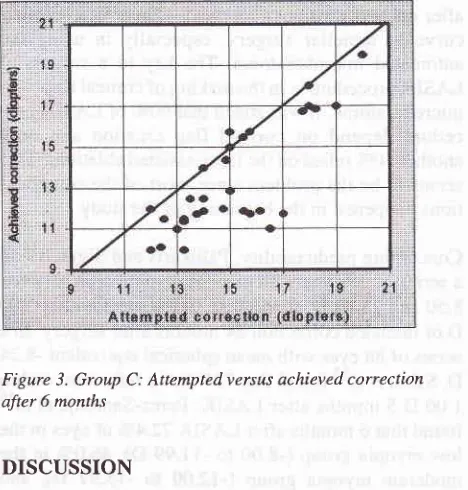
243Figure 3. Group C: Attemptedversus achieved correction after 6 months
DISCUSSION
The goal
of
this study
wasto determine the safety
andefficacy of excimer
laserkeratomileusis
as atreatment
to correct myopla.
Corneal clarity
is
the main factor
in
evaluating
thesafety
of
the
procedures.
Several
studieslo-I2
iave
found varying
degreesof subepithelialhaze
afterPRK.
The corneal
opacification correlated
with
the presenceof subepithelial
activatedfibroblasts
and the presumed svnthesisof
newcollasen
and other comDonentsof
theextracellular
matrix.F3 Marshall
and'
cowork"rsg
reported thatalthough
mostcellular activity
afterPRK
stopped at three months
postoperatively, the
healing
process
continued
for
upto
8months postoperatively.
By
8months,
themorphology of
the
cornea was nearnormal with
the exception that
Bowman's
membranewas
absent andthere was
still
a degreeof
disorder
in
theimmediate subepithelial stromal fibers. In
LASIK,
haze occurs
in
different
anatomical
location than in
PRK.
By
applying
laser energydirectly
to thestromal
bed,
LASIK'spares
Bowman's membrane and
theepithelium,
thusavoiding
theproblems
associatedwith
deepsurface ablation.
In
this
study,
all
corneas
wereclear before surgery. The
degreeof
hazeafter
LASIK
was
minimal
in
all
eyes.
This finding
indicates that
preservation
of Bowman's layer
may be
important in
corneal
healing.Complications
werefound
in
6Voof
eyesof
all
groups, [image:4.595.78.309.84.288.2] [image:4.595.77.305.527.720.2]although none was
vision
threatening.
The most
fre-quentcomplication
of LASIK
proceduresis
epithelial
ingrowth,
possibly
from
debrisin
theinterface.
In
this
study,
interstitial
debris was
found
in
8 eyes,
secondFigure
L
Group A: Attempted versus achieved correction after 6 monthsIn group
B,
the predictability after 6
months
demonstrated that the SE
of
81.87o of eyes werewithin
+ 0.50D,
90.9Vo werewithin +
I .00D
and 97,7 Vo w erewithin +
2.00
D.
Figure 2
shows the attempted versusachieved
correction
of
group
B
after
6
months. In
group C thepostoperative
predictability
after 6 months demonstrated that theSE
o140.6Vo of eyes werewithin
0.50
D,
53.lVo were
within
1.00D,
and78.l%o were
within
2.00D.
Attempted
versus achievedcorrection
in group
C after
6months is shown
in
Figure
3.Complications
ofLASIK
procedures were found in 67oof
eyes(29
of
475
eyes). Thesecomplications,
which
were associatedwith
thecreation of
cornealflap,
wereas
follows: epitheliopathy/erosion (16
eyes),
intersti-tial
debris (8
eyes),incomplete flap (2
eyes),free flap
(2
eyes), andslipped
flap
(l
eye). In
caseof
free
flap
and slipped
flap,
the
flap
waselevated, repositioned,
and secured
with
l0/0 nylon
sutures.Other
complica-tions were
leave unmanaged.244 Istiantoro et al.
after
corneal erosions
(16 eyes).
There
is
alearning
curve
to
lamellar
surgery,
especially
in
using
theautomated
microkeratome. The key
to
a successful
LASIK
procedure isin
themaking of
cornealflap
with
microkeratome.
It was stated
that
90Voof LASIK
pro-cedure depend
on
corneal
flap
creation and^only
another lOVo
reliedon
the laser-assistedablation.' This
seems
to
be the
problem
sincemost
of
the
complica-tions
happenedin
thebeginning
of
the
study.Concerning
predictability, Pallikaris
andSiganosla
in
a series
of
39
eyeswith
preoperative myopia
between8.50 and
25.87D, found
that
79.2Vowere
within
2.00D of
intended correction
24months after
surgery.In
aseries
of
88 eyeswith
meanspherical equivalent -8.24
D,
Salah etall5 found
that 72Vo had arefraction
within
1.00
D
5 months after
LASIK. Perez-Santonja
et all6
fbund that
6months
afterLASIK
72.4Voof
eyesin
thelow myopia group (-8.00 to
-11.99
D),46.0Vo
in
the
moderate myopia group (-12.00
to
-15.99
D),
and50.}Vo
in
thehigh myopia
group
(-16.00 to -20.00D),
were
within
1.00D of
emmetropia.
In
this study,
at6
months
after
surgery, the
achievedcorrection
was
within
2.00
D of
attemptedcorrection
in
44
eyes (95.65Eo)in
group
A,
43
eyes (97.73Vo)in
group
B,
and25
eyes (78.13Vo)in
group C. The total
number
of
eyes at6-month
follow-up were
122 eyes.Refiactive
results
after 6
months
in
group
A
showedthat
therewas
atendency toward overcorrection
(Fig-ure
l).
This
is
in
contrast to refractive
resultsin
group
C,
which
showed that mostof
the cases wereundercor-rected
(Figure
3). Thesefindings
may indicate
that the laserablation normogram
usedin
this study
(Table
l,
Maria Clara's modification)
needmore
adjustment.The
meanpostoperative spherical equivalent
seemsto
stabilize early
in
the postoperative period.
The
mean regression between the1tt
and 6thmonth
was less than 1.00D. Stability of
the spherical equivalent
with
time
may have to do
with initial
site of tissueremoval below
Bowman's
layer in
LASIK, which
seems toprovoke
aless intense
healing
response.However,
in view of
therelatively
short
follow-up
at the
current
stage ofthis
study,
it
is
too
early
to
conclude that the stability
suggested
by
the meanrefractive
results after 6months
postoperatively
is
predictive
of
future
refractive
stability.
Statistical
analysis wasnot
donein
this
study, because the numbersof
eyesduring
follow-up
wereconstantly
changing.
Med J Indones
CONCLUSION
The
results
of
this
study
show
that
laser in-situ
keratomileusis
with
the
193-nm argon
fluoride
ex-cimer
laser can correctmyopia
with
good accuracy andpredictability.
Results
also tendto
suggeststability
of
refraction. Further investigations
should
be
made to
obtain an improved ablation normogram
to
correct
extremely high myopia.
REFERENCES
l. Trokel SL, Srinivasan
R, Braren B. Excimer laser surgeryof
the comea. Am J Ophthalmol 1983; 96:710-5.2. Marshal J, Trokel S, Rothery S, Krueger RR. A comparative study
of
corneal incisions inducedby
diamond and steel knives and two ultraviolet radiations from an excimer laser.Br J Ophthalmol 1986; 70:482-501.
3. Kerr-Muir MG, Tokel SL, Marshal J, Rothery S.
Ultrastruc-tural comparison of conventional surgical and argon fluoride excimerlaserkeratectomy. Am JOphthalmol 1987;l
03:448-53.
4. McDonald
MB,
KaufmannHE,
FrantzJM,
Schofner S, Shofner S, Salmeron B, et al. Excimer laser ablationin
ahuman eye. Arch Ophthalmol 1989;10'l:641-2.
5. Hamberg-Nystrôm H, Fagerholm P, Sjôholm C, Tengroth B. Photorefractive keratectomy for 1.5 to 18 diopters
of
myopia. J Refract Surg 1995; 11:52261-7.
6. Seiler T, Holschbach A, Derse M, Jean B, Genth U. Com-plications of myopic photorefractive keratectomy with the
excimer laser. Ophthalmology 1994; 101:153-60.
7.
Pallikaris
IF,
Siganos
DS. Excimer laser
in
situ
keratomileusis and photorefractive keratectomy for correc-tion of high myopia. J Refract Corneal Surg 1994;
l0:498-510.
8. Casebeer JC, Slade SG, editors. LASIK, Lamellar refractive
surgery: technique, technology and complications. Boston: Chiron Vision, 1996.
9. Slade SG.
LASIK
and lamellar refractive surgery. Boston: Ophthalmology Interactive CD-ROM, 1995.10. Waring GO
III,
Maloney RK, HagenKB
et al. Refractive and visual resultsof
a multicentertrial
of
excimer laserphotorefractive keratectomy. Ophthalmology
1992; 99(9S):106ll.
Gartry DS, Kerr
Muir MG,
MarshallJ.
Excimer laser photorefractive keratectomy: I 8-month follow-up. Ophthal-mology 1992; 99 :1 209 -1912. Seiler T, Wollensak J. Myopic photorefractive keratectomy with the excimer laser: one-year follow-up. Ophthalmology
l99l;98:1156-63
13. Del Pero
RA,
Gigstad JE, RobertsAD,
Klintworth GK'Martin CA,
L'EsperanceFA
Jr, et al.
A refractive
andhistopathologic study
of excimer laser keratectomy in
primates. Am J Ophthalmol 1990; lO9:419-2914. Pallikaris IG, Siganos DS. Laser
in
situ keratomileusis to treat myopia: Early experience.J
Cataract Refract SurgVol 8, No 4, October - December 1999 Six months follow up Lasikfor
rnyopia
245
15. Salah T, Waring GO III, El-Maghraby A, Moadel K,
Grimm
16. Perez-Santonja JJ, Bellot J, Claramonte P, Ismail MM,Allo
SA. Excimer laser in situ keratomi leusis under a comeal
flap
JL. Laserin
situ keratomileusis to conect high myopia. J