Urinary stone characteristics of patients treated with
extracorporeal shock wave lithotripsy in Cipto Mangunkusumo
Hospital Jakarta, 2008–2014: a gender analysis
Keywords: ESWL, female, male, urinary stone, urolithiasis
pISSN: 0853-1773 • eISSN: 2252-8083 • http://dx.doi.org/10.13181/mji.v24i4.1258 • Med J Indones. 2015;24:234–8
• Received 25 Jun 2015 • Accepted 30 Dec 2015
Correspondence author: Endrika Noviandrini, [email protected]
Copyright @ 2015 Authors. This is an open access article distributed under the terms of the Creative Commons Attribution-NonCommercial 4.0 International License (http://creativecommons.org/licenses/by-nc/4.0/), which permits unrestricted non-commercial use, distribution, and reproduction in any medium, provided the original author and source are properly cited.
Endrika Noviandrini, Ponco Birowo, Nur Rasyid
Department of Urology, Faculty of Medicine, Universitas Indonesia, Cipto Mangunkusumo Hospital, Jakarta, Indonesia
C l i n i c a l Re s e a rc h
ABSTRAK
Latar belakang: Pasien dengan batu saluran kemih di Indonesia terus meningkat setiap tahunnya di kedua gender. Data menunjukkan insiden penyakit batu saluran kemih umumnya di temukan lebih tinggi pada pria dibandingkan wanita. Tujuan dari penelitian ini untuk mengetahui karakteristik dari Pasien dengan batu saluran kemih, baik pria maupun wanita yang menjalani extracorporeal shock wave lithotripsy (ESWL) di Rumah Sakit Cipto Mangunkusumo (RSCM), Jakarta tahun 2008–2014.
Metode: Penelitian ini menggunakan data dari rekam medis pasien ESWL tahun 2008–2014 di RSCM Jakarta. Kami mengambil 5.174 dari 6.020 data, dikarenakan ketidaklengkapan status. Data kemudian di sortir berdasarkan jenis kelamin, usia, lokasi batu, opasitas batu, ukuran batu, riwayat ESWL pada pasien dan dianalisis menggunakan SPSS v.20 untuk Mac.
Hasil: Dari 5.174 data, didapatkan insiden Pasien dengan batu saluran kemih pada pria dua kali lebih besar dibanding wanita (66,3%:33,6%) dan terjadi pada usia produktif (65,2% pria dan 65.9% wanita). Batu ginjal unilateral menjadi batu yang paling banyak ditemukan di kedua gender (50,2% pria dan 57,2% wanita) dengan lokasi yang terbanyak yaitu kaliks inferior (24,8% pria dan 28,9% wanita). Sebanyak 72,9% merupakan batu radioopak (73,7% pria dan 71,5% wanita). Rerata ukuran panjang batu pada pasien pria dan wanita adalah 11,34±7,15 mm dan 11,90±7,54 mm, secara berurutan. Penelitian ini juga menunjukkan bahwa 79,3% pasien datang untuk terapi ESWL pertama.
Kesimpulan: Batu saluran kemih ditemukan dua kali lebih tinggi pada pria dibandingkan wanita dan terjadi pada usia produktif. Batu ginjal unilateral yang berlokasi di kaliks inferior menjadi batu yang paling banyak di temukan di kedua gender. Sebagian besar batu merupakan batu radio opak.
ABSTRACT
Background: The incidence of urinary stone patient in Indonesia has increased every year in both genders. Data showed that urolithiasis was higher in male rather than female. The aimed of this study was to describe the characteristics of urinary stone found in patient who underwent extracorporeal shock wave lithotripsy (ESWL) at Cipto Mangunkusumo Hospital, Jakarta from 2008–2014.
Methods: Data obtained from ESWL medical record Cipto Mangunkusumo Hospital, Jakarta from 2008–2014. We obtained 5,174 out of 6,020 data due to incompleteness data record. We sorted data records by gender, age, stone location, stone opacity, size of the stone, and history of ESWL, and analyzed by statistic tools (SPSS v 20 for Mac).
Results: From 5,174 records, we found that the incidence of urinary stones was two times higher in male rather than female (66.3%:33.64%), occurred mostly in productive age (65.2% male, 65.9% female). Unilateral kidney stone was most common location found for both gender (50.2% male, 57.2% female), and most frequent site located in calyx inferior (24.8% male, 28.9% female). About 72.9% stone was radiopaque (73.7% male and 71.5% female). The mean size of the stone in male and female was 11.34±7.15 mm and 11.90±7.54 mm, respectively. This study also showed that 79.3% patients came for first ESWL.
Urinary stone disease has become one of the most common urologic diseases found in Indonesia. In Cipto Mangunkusumo Hospital (CMH), the numbers of urinary stone incidences has been increased every year. During 1997–2002, the numbers of patients who underwent extracorporeal shock
wave lithotripsy (ESWL) was increased five times
than before.1 Research shows that urinary stone disease occurs mostly in man, rather than woman. And it usually happens in their productive ages.1
There are some procedures that can be used as the treatment of urinary stone disease. One of the most common procedures used among urologist is ESWL. ESWL not only known as the non-invasive procedures, but also for its good in stone free rate outcomes.2 The successful rate of ESWL as the treatment of all urinary tract stone was 96%.3 Another supporting research showed, about 87% stone free rate was achieved after three months follow up in urethral stone treatment by using ESWL and no major complications found.4
The aimed of this study was to describe the urinary stone characteristic in male and female patient who underwent ESWL at CMH, Jakarta, during 2008–2014.
METHODS
This research used secondary data taken from medical record of urinary stone patient who treated with ESWL at CMH, Jakarta from 2008–2014. There were 6,020 patients who underwent ESWL during
those specific years, only 5,174 with complete
medical records included in this research. The inclusion criteria of this research were female and male patient who had urolithiasis and underwent ESWL during 2008–2014, in Cipto Mangunkusumo Hospital, Jakarta. We exclude patients with incomplete data, and patients with CBD stone. The variables were age, gender, and location of stone, stone site, stone opacity, stone size, and history of ESWL. We divided the stone location into unilateral kidney stone, bilateral kidney stone, unilateral urethral stone, bilateral urethral stone and bladder stone. The stone site consisted of calyx inferior stone, calyx media stone, calyx superior stone, distal urethral stone, proximal urethral stone, and
bladder stone. We also classified patient age
into three categories: under 19 years old, 20–50 years old, and more than 51 years old. This criteria
of age was made based on literature which stated that the highest incidences of urolithiasis was in patient around 20–50 years old.5 Confidentiality of subjects identity were guaranteed.
After collecting the data, we sorted it by gender, and analyzed it with SPSS v.20 for Mac. The frequency distribution of stone characteristic in both gender and the mean size of the stone were analyzed by using this statistic tools.
RESULTS
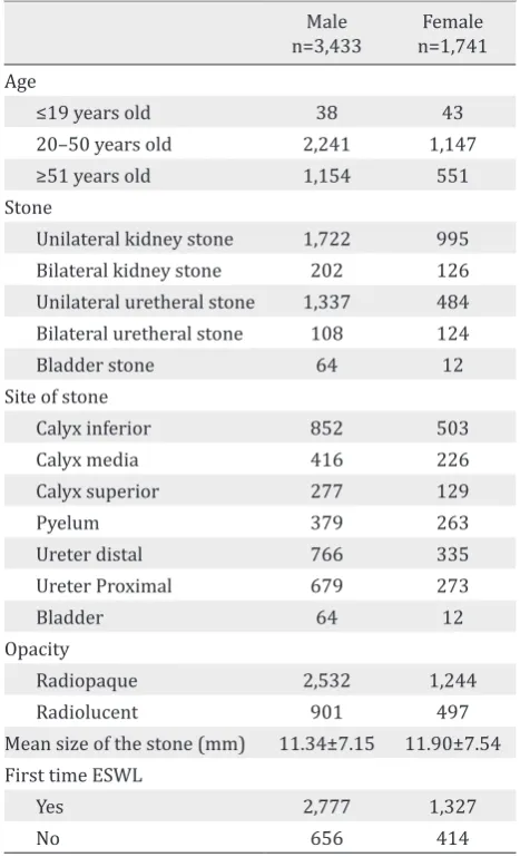
Table 1 describes the urinary stone characteristics in patients related with gender. Urinary stone found mostly in male in their productive ages. Unilateral kidney stone, which located in the calyx inferior of the kidney was mostly found with radioopacity.
Male n=3,433
Female n=1,741 Age
≤19 years old 38 43
20–50 years old 2,241 1,147 ≥51 years old 1,154 551 Stone
Unilateral kidney stone 1,722 995 Bilateral kidney stone 202 126 Unilateral uretheral stone 1,337 484 Bilateral uretheral stone 108 124
Bladder stone 64 12
Site of stone
Calyx inferior 852 503
Calyx media 416 226
Calyx superior 277 129
Pyelum 379 263
Ureter distal 766 335
Ureter Proximal 679 273
Bladder 64 12
Opacity
Radiopaque 2,532 1,244
Radiolucent 901 497
Mean size of the stone (mm) 11.34±7.15 11.90±7.54 First time ESWL
Yes 2,777 1,327
No 656 414
Table 1. Characteristic of urinary stone
DISCUSSION
There are many contributing factors for the risk of stone formation such as race, environment, gender, and age.2 Distribution of stone varies among races. Renal stones are more common in Caucasians and Asians than Native Americans and Afro Caribbeans.6 Incidences of urinary stone disease was higher in hot or dry climates.7
Gender is also related to the incidence of urinary tract stone. Our study results showed that the prevalence of urinary tract stone in Indonesia was higher in male rather than female, with the
ratio 2:1. This finding was also supported by
literature, which stated that urinary tract stone occurs mostly in male5. The same study has been
done in Israel, China, and Taiwan for the past five
years and came up with the similar result.7-9 In the other hand, a research conducted in Florida on 2010 found that nowadays the incidence of stone disease among woman might be increased. There is a changing pattern in urinary tract stone prevalence. Female also has the same risk of getting urinary tract stone like male.10
Anatomy of the urinary tract, obesity, dietary life, urine composition, and hormonal factors may be considered as the factors which can explaine why the incidences of urinary stone was higher in male. Male urinary tract has more narrow places than female. This narrow places are lead to the higher risk of stone formation due to urine precipitation.11 The most common locations for urinary stone formation are ureteropelvic junctions, mid ureter (ureter crosses over iliac bone), ureter crosses over the iliac blood vessels, ureter bladder junction, and when the ureter under the uterine artery passes (for female).5
Obesity, dietary life, and fluid consumption
also play some role in stone formation process. Studies in United States in 2011 found that obese females were more likely to develop stones than non-obese females. But the correlation between obesity and stones was weaker in males rather than the female.12 Negri et al13 also found that in female with high body mass index (BMI) tend to have an increasing number of oxalate, uric acid, phosphorus, creatinine, and sodium excretion. The same result found in male subject but potassium, urea, magnesium, and citrate excretion was also
increased in this gender.13 Eating habit, like high animal protein consumption, low carbohydrate, high salt, and low calcium dietary habits, may contribute to the stone formation process in male.5 Eating vegetable and fiber, and limit animal protein consumption to 0.8-1.0 g/kg/day are recommended to inhibit stone formation.14 Fluid intake also related to the risk of kidney stone formation15. It was suggested in the guidelines to drink 2.5-3.0 L/day or to drink until the amount of diuresis reached 2.0-2.5 L/day, to prevent the stone recurrences. Beverages with neutral pH is recommend for this prevention tips.14 Coffee, tea, alcohol, and milk may decreased urinary concentration.16
Urine composition is related to calculogenesis process in human. A study about biochemical composition of urine was conducted in India, by using two hours urine collection. The result of that study found that male had higher risk in getting urinary stone due to higher excretion of calcium and oxalate, with low concentrations of citrate.17
Sex hormones also have some role in stone formation process. Estrogen inhibits excretion of calcium and oxalate, and increase citrate production.18 Study done by Sarada et al17 reported that testosterone decreased citrate excretion. Another study in animal also showed oxalate excretion was increased by the administration of testosteron.17 In addition protective effect of estrogen in premenopausal woman, could enhanced renal calcium absorption and reduced bone resorption.15
In this study, unilateral kidney stone which located in the calyx inferior, was the most common stone found in both gender. The correlation between location and stone formation remain uncertain, but narrow places in human urinary tract increased the risk of urine precipitation.5 The incidence of bladder stone found higher in male because in elderly male, bladder stone might develop due to the growth of the prostate or due to benign prostatic hyperplasia.11
The data result also showed that most of the stone found in both gender was radioopaque, which usually related to calcium oxalate, calcium phosphate and struvite stones. Calcium oxalate stone found about 60% in all types of calculi stones.6 Study by Gault and Chafe20 stated that in the third decade, the highest number type of stone found in female was phosphate stone and oxalate stone in male.
The mean size of the stone found in the study was 11.34±7.15 mm for male patients and 11.90±7.54 mm for female patients. ESWL found to be the most suitable treatment for this size of stone.21
From the guidelines ESWL is the first treatment
chosen for kidney stone with size 10–20 mm.22
There are many factors which correlate with successful rate of ESWL such as size, composition, consistency of the stone and elimination process after the treatment.23 In this research, most of
the patients came for the first treatment (80.8%
in male and 76.2% in female), but some patients had already done it before. This might be due to the size and the composition of the stone. But, further information about stone composition and
stone hounsfield unit are needed. Recent studies
have shown that stone made of calcium oxalate dihydrate are easier to break rather than uric acid stone.3 Studies made by Tarawneh stated that there were positive correlation between stone size and number of ESWL treatment.23 This study result also showed, that the number of male patients who come for continued treatment was higher than female patients. This result may be correlate with literature which stated that the recurrences of urinary stone occurs higher in male rather than female.5
The weakness of this study was incomplete data records which was found especially on the year of 2008. We solved this problem by increasing
the quality of medical record documentation with the result of better medical record during 2009–2014.
In conclusion, this research showed that in CMH, Jakarta, from 2008–2014, most of the patients who had urinary tract stone and underwent ESWL were male. The most common location found in both gender was unilateral kidney stone
with calyx inferior as the specific site. Most of the
patients had radioopaque imaging and calcium oxalate stone considered as the highest type of stone found in this study.
Conflict of interest
The authors affirm no conflict of interest in this
study.
REFERENCES
1. Rahardjo D, Hamid R. Perkembangan penatalaksanaan batu ginjal di RSCM tahun 1997–2002. J I Bedah Indones. 2004;32(2):58–63. Indonesian.
2. Politis G, Griffith DP. ESWL: stone-free efficacy based upon stone size and location. World J Urol. 1987;5:255–8.
3. Junuzovic D, Prstojevic JK, Hasanbegovic M, Lepara Z. Evaluation of extracorporeal shock wave lithotripsy (ESWL): efficacy in treatment of urinary system stones. Acta Inform Med. 2014;22(5):309–14.
4. Elkholy MM, Ismail H, Abdelkhalek MA, Badr MM, Elfeky MM. Efficacy of extracorporeal shockwave lithotripsy using Dornier SII in different levels of ureteral stones. Urol Ann. 2014;6(4):346–51.
5. Menon M.Parulkar BG, Drach GW. Urinary lithiasis: etiology, diagnosis, and medical management. In: Walsh CP, Retik AB, Vaughan ED, Wein AJ, editors. Campbell’s urology. 7th ed. Philadelphia: WB Saunders; 1998. p. 2662–5.
6. Pal RP, Mellon JK. Renal Stone Disease. Foundation Years. 2008;4(5):199–203.
7. Kalbu DU, Golan S, Livne PM, Pode D, Duvdevani M, Lifshitz D. Urinary stone composition in Israel: current status and variation with age and gender-A bicenter study. J Endourol. 2013;27(12):1539–42.
8. Wu W, Yang B, Ou L, Liang Y, Wan S, Li S, et al. Urinary stone analysis on 12,846 patients: a report from a single center in China. Urolithiasis. 2014;42(1):39–43. 9. Huang WY, Chen YF, Carter S, Chang HC, Lan CF, Huang
10. Strope SA, Wolf JS Jr, Hollenbeck BK. Changes in gender distribution of urinary stone disease. Urology. 2010;75(3):543–6.
11. Burnett AL, Rodriguez R, Jarrett TW. Genitourinary system: male anatomy and physiologi. In: Greenfield LJ, Mulholland MW, Oldham KT, Zelenock GB, Lilimoe KD, editors. Essentials of Surgery Scientific Principles and Practice. 2nded. New York: Lippincott Williams & Wilkins; 1997. p.1111–8.
12. Nowfar S, Pallazi-Churas K, Chang DC, Sur RL. The relationship of obesity and gender prevalence changes in United States inpatient nephrolithiasis. Urology. 2011;78(5):1029–33.
13. Negri AL, Spivacow FR, Del Valle EE, Forrester M, Rosende G, Pinduli I. Role of overweight and obesity on the urinary excretion of promoters and inhibitors of stone formation in stone formers. Urol Res. 2008;36(6):303–7.
14. Skolarikos A, Straub M, Knoll T, Sarica K, Seitz C, Petřík A, et al. Metabolic evaluation and recurrence prevention for urinary stone patients: EAU guidelines. Eur Urol. 2015;67(4):750–63.
15. Pearle MS, Lotan Y. Urinary lithiasis: etiology, epidemiology, and pathogenesis. In: Wein AJ, Kavoussi LR, editors. Campbell-Walsh Urology. Philadelphia: Elsevier Saunders; 2012. p.1257–60.
16. Taylor EN, Curhan GC. Diet and fluid prescription in stone disease. Kidney Int. 2006;70(5):835–9.
17. Sarada B, Satyanarayana U. Urinary composition in men and women and the risk of urolithiasis. Clin Biochem. 1991;24(6):487–90.
18. Kato Y, Yamaguchi S, Kakizaki H, Yachiku S. Influence of estrus status on urinary chemical parameters related to urolithiasis. Urol Res. 2005;33(6):476–80.
19. Eprints.undip.ac.id [Internet}. Semarang: Batu saluran kemih suatu problema gaya hidup dan pola makan serta analisis ekonomi pada pengobatannya. [update 2007 Mar 3; cited 2015 Jun 15]. Available from: http://eprints.undip.ac.id/340/1/rifki_ muslim.pdf
20. Gault MH, Chafe L. Relationship of frequency, age, sex, stone weight and composition in 15,624 stones: comparison of results for 1980 to 1983 and 1995 to 1998. J Urol. 2000;164(2):302–7.
21. Renner C, Rassweiler J. Treatment of renal stones by extracorporeal shock wave lithotripsy. Nephron. 1999;81(suppl1):71–81.
22. Türk C, Knoll T, Petrik A, Sarica K, Seitz C, Straub M. Indication for active stone removal and selections of procedures. In: EAU Guidelines on Urolithiasis. 2014. p. 48. Available from: http://uroweb.org/wp-content/ uploads/20-Urolithiasis.pdf