AN ENHANCED IMAGE BIOMEDICAL
CLASSIFICATION BY MORPHOLOGY
ALGORITHM
Muhammad Kusban
Electrical Engineering, Muhammadiyah University of Surakarta, Indonesia
[email protected] (Muhammad Kusban)
Abstract
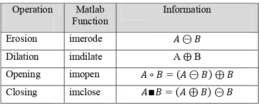
Image Processing morphology is an important tool in digital image processing based on human intuition and perception. Morphology based on the geometry, which emphasizes the geometry of the image. Morphology of the process is mainly used to remove the imperfections that exist in the form of an image. No exception in the field of medicine / medical, often obtained results rontgent or scanning the resulting images do not have the accuracy of the expected image quality. This is because factors of body movement or instrument (not focusing) so that the resulting image blur and distorted. One method is to enhance this image by using morphology method. With operations erosion and dilation as well as a combination of both in the process of opening and closing, the morphology of high level / complex projects could be implemented. The key to success lies in the selection process of the morphology of mathematical operations and the choice of structured elements. Even the selection of filters and methods of transformation in this process is often not used. In a study obtained optimal results for the distorted image with SNR 19.891 dB, reduction bits 2.206, and Gain 13.27 dB.
Keywords: morphology, enhanced image, erosion, dilation, structured elements
1. Introduction
Modern digital technology has made it possible to manipulate multi-dimensional signals with systems that range from simple digital circuits to advanced parallel computers. An image defined in the ‘real world’ is considered to be a function of two real variables, for example: a(x,y) with a as the amplitude (e.g. brightness) of the image at the real coordinate position (x,y).
An image may be considered to contain sub-images sometimes referred to as regions of interest (ROI) or simply regions. An image is digitized to convert it to a form which can be stored in a computer’s memory or on some form of storage media such as a hard disk or DVD-ROM. This digitization procedure can be done by a scanner or by a video camera connected to a frame grabber board in a computer. Once the image has been digitized, it can be operated upon by various image processing operations. Image processing operations can be roughly divided into three major categories:
Image compression, involves reducing the amount of memory needed to store a digital image Image enhancement , Image defects which could be caused by the digitization process or by faults
in the imaging set up
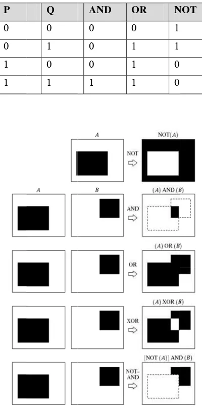
Morphol and zero bina
perations rela ed in the pro 0 or by chang
ckground to ings: the im red element)
ns AND, OR
Some operat
chnique or p hology [1]. I inal image [2 process is alw
ary values [3 ated to the s ocess of morp
ging certain the foregrou
process used In image pr 2]. Meanwhi ways closely 3]. Furtherm shape or mor phology, nam
parts of the und area. Cha morphology o here are thre ab 1. Morpho
Q
for image p rocessing, th le, Chris Sol y related to n more, the mor rphology of mely the bit 1
foreground i ange foregro of the type ee basic ope ology of the b
of binary log ber 1 represe
processing (i he expected lomon and T neighborhood
rphology of an image [4 1 or known a into the back ound and bac
of operation erations in m
basic operati
gic. Binary b ents 0.
image) based result is ba Toby Breckon
ds which are an image is 4]. In practice
as the foregro kground and ckground are
n, and arran morphology ion
NOT
black and wh
d on the pri ased on the n further said e formed from s a collection
e, binary sys ound and bac vice versa t ea is closely r
ngement of operations,
hite represent
inciple of shape or related to elements namely:
In binary morphology, an image was viewed as a subset of Euclidean space Rd or grid Zd integer to the value of the dimension d. Benefits of using the morphology of which is to eliminate the existing noise. Getting to know the characters form an image, and used to improve image quality. In the form of 2D, the morphology of the process used for the extraction of the characters in the image. As for 3D, this process is used in the medical field. One is to get objects from a collection of objects together in the cardiac surgical, neuro surgical and functional MRI for mind [5]. The process is also used in the morphology of the police to fingerprint identification in order to clarify the flow pattern existing hand lines.
2. Study References
According to Luc Vincent [7] stated that combination between morphological gray scale and sequential technique result in a hybrid grayscale reconstruction algorithms which is an order of magnitude faster than any previously known algorithm.
Marian M. Choy and Jesse S. Jin [8] stated that assessment of cardiac function using imaging techniques requires accurate identification of borders. Combination between morphological images with a second derivative operator that is Laplican of Gaussian can reduce noise and increase contrast of the image.
Karol Mikula, Tobias Preuber, and Martin Rumpf [9] stated that using morphological multi-scale method for image sequence processing will give result denoise the whole sequence while retaining geometric features such s spatial edges and highly accelerated motions.
L. Vincent [10] gives an efficient algorithm for the implementation of morphological operations with random structure elements, assuming a chain code encoding of the binary objects.
The types of operations to transform an input image a[m,n] into an output image b[m,n] can be classified into three categories:
Point: the output value at a specific coordinate is dependent only on the input value at that same coordinate
Local: the output value is dependent on the input values in the neighborhood of that same coordinate
Global: the output value is dependent on all the values in the input image
To obtain the third form, the image on the sample is can be classified two forms of structured element or neighborhoods pixels:
Rectangular sampling Hexagonal sampling
Local operations produce an output pixel value b[m=mo,n=no] based upon the pixel values in the
neighborhood of a[m=mo ,n=no]. Some of the most common neighborhoods are the 4-connected
neighborhood and the 8-connected neighborhood. Operations that are fundamental to digital image processing can be divided into four categories:
Operations based on the image histogram On simple mathematics
On convolution
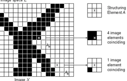
An arran
ngement of e umn or block
F
igure 2, the on (e.g., 3 x eft side clos ng element i
Fig 3. Stru
ing element often used in mage, each p
ation and Er process in th mage, by plac el neighborho n for dilation
cess of erosio ng element ackground p n for erosion
elements (str k.
is a common n linear filter
pixel by pixel
rosion he morpholo cing one by ood foregrou
is expressed
on is the pro one by one ixels (value
is expressed
ructuring ele
les of structu element pix
cturing elem As for the b middle of the
ments that is f
n form used rs process an l interlaced i
ogy of identi one after the und pixel (va d as follows.
ocess of remo in the foreg 0), then the d as follows.
ement) is a b
ure elements xel are drawn
ment is in the block dimens e block (e.g
fill the area o
d in the morp n image [4]. image neighb
cal image is e central stru alue 1), the b
oving pixels ground pixel
e value is in
block array i
and the cent n in the gray.
e middle of sion is even, ., block of 4
of origin in a
phology of th When the s borhood of o
can do by a ucturing elem
background c
within the ob s (value 1). n the foregro
s 0 or 1 or r
tral structurin
the block w the central s 4 x 3 and 4 ment for each changed to th
bject image If there is a ound to the
rectangular, t
ng
when the bloc structuring e
x 4 then th
fter another.
a convolutio lement is pla
s in the origin h pixel backg
he foregroun
by putting th a neighborho background
the block
ck is odd lement is he central
on kernel he central ood pixel d change.
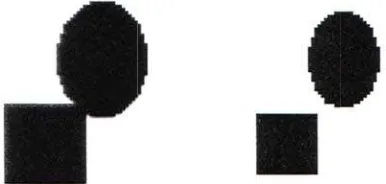
F
The pixe case the the origi neighbor shrinking to each o the objec
Fig 4. The ori
el value will value of cro in pixel has rhood turn g the size of other. While ct to flatten t
Fig 5. Th
F
iginal image
l only be wo oss -1) with s a value eq into such a f the image o e the dilation
the edge of th
he process of
Fig 6. Dilatio
is covered b
orth it as the values other qual to the b
block struc object so tha n will increas
he object is l
f erosion can
n process is
by a block arr
e central stru r than the cen
block arrang cturing elem at it can be u
se its size so lost or damag
be used to s
used to conn
rangement o
ucturing elem nter to zero. gement of e ment. With t used to separ o it can thick
ged.
eparate objec
nect the fragm
of the elemen
ment of appr While in th lements of t the erosion rate objects t ken the objec
cts that bond
mented objec
nts one by on
ropriate valu he process of the pixel va process res that mutually ct image and
d together.
ct.
ne.
2.2 Ope Opening using str
Opening original
While cl operation
Closing
ening and C g the morpho ructuring elem
g widely use shape.
Fig 7. Ope
losing is the n using the s
method is us
F Closing
ology is the p ments the sa
ed for the p
ening the pro
e process of same structur
Fig 8. Exam is us
sed when you
Fig 9. Closing
process using ame, which c
rocess of re
ocess used to
morphology ring element
mple of a 3x3 sed for the pr
u want to clo
g process is u
g erosion and an be expres
emoving sma
separate obj
y by perform . Written wit
3 structuring rocess of ero
ose the gap w
used to fill th
d then the pr ssed by the fo
all objects i
jects as well
ming a dilatio th the follow
element wit sion and dila
with the objec
he empty par
rocess contin ollowing not
n an image
as to smooth
on operation wing notation
th a binary 1 ation.
ct retains its
rt of an objec
nued with the tation.
but still ret
h the image
followed by n:
original shap
ct.
e dilation
(3) tain their
y erosion
(4)
pe.
2.3 Bou
by first doi reduced by t
11. Using a s
orphology in me of the bas athWorks Inc
action he boundary ing the proc the image ori
structuring e
n Matlab sic process o
c. (www.mat cess of erosio
igin. Bounda Matlab func Matlab
objects or k on with a sm
known by the mall block s n Notation is
ss extraction
e boundary e structuring e s written as f
n
cess of bound y
with the help g table. element, and
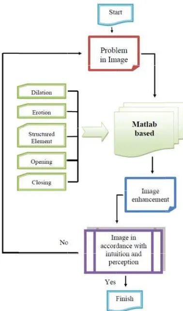
Fig 12. F
Flow chart o
study using ed with: bon e been distor
rence. g a collection of structured
ined image w g program fo
dilation, eros ing all of the mming in orde ults of the me ting of perfec
of research pr
a variety of nes, joints, sk rted because
n based algo elements wi with the optim or each type sion, opening e algorithms er to facilitat edical image ct image and
rocess with t
f medical im kull, heart (g
of movemen orithm in Ma ith different v mal shape in
of morphol g and closing
and program te the researc e that has bee d then made a
the morpholo medical field
mage or coll gray scale). B
nt by lens or atlab program variations th
terms of eye ogy which i g as well as m ms in the mo
ch of several en improved a conclusion.
ogy of algori d
lecting medi Both images
by patients a mming, prim
sources of m d with the me
.
thms to impr
ical imagery are obtained and then the marily determ
o compare w y used to im een the four
a whole proj medical imag ethod morph
rove image q
y, especially d in perfect c perfect imag mines the ch with each oth mprove image
components oject based M
ges.
hology compa
quality in
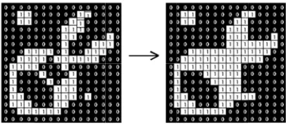
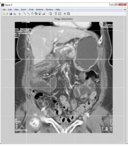
Fig 13. One o
Fig 14. R by using s
of the origina
econstruction structured ele
al Colon.jpg i
n Colon.jpg ement in Mat
image is use
with the pro tlab [0 1 0; 1
d for researc
cess dilate 1 1 1;0 1 0]
ch.
Fig 16. 0 1 0
Reconstruct 0 0 1; 0
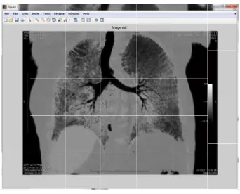
Fig 15. R by using st
tion Lung.jpg 1 0 1 0
Reconstructio tructured ele
g with the pr 1 0; 1 1
on Lung.jpg w ement in Mat
rocess erode 1 1 1 1
1]
with the proc tlab [0 1 0; 1
by using stru 1; 0 1 0
cess erode 1 1; 0 1 0]
uctured elem 0 1 1 0 0
ment in Matla 0; 1 0 0
ab [1 0 1 0 0
Fig 17. I
Fig 18. I 0 1 0
Image Pediat
Image Pediat 0 0 1; 0
tric.jpg with
tric.jpg with 1 0 1 0
mix process 0; 1
mix process 1 0; 1 1
s between dil 1 1 1; 0
s between dil 1 1 1 1
1]
late and erod 1 0]
late and erod 1; 0 1 0
de using struc
de using struc 0 1 1 0 0
ctured eleme
ctured eleme 0; 1 0 0
ent[0 1
Tab 3. The amount of Gain, number bits on reduction process, and SNR Image Gain (dB) Reduction Bit (bits) SNR (dB)
Adrenal.jpg 13.47 2.24 18.88
Cardiac.jpg 14.28 2.37 20.18
Chest.jpg 12.32 2.05 17.02
Colon.jpg 11.27 1.87 17.08
Esophagus.jpg 10.43 1.773 16.56
Gastrointestinal.jpg 15.63 2.60 22.90
Genitourinary.jpg 16.52 2.74 26.27
Kidney.jpg 7.74 1.28 14.58
Liver.jpg 15.94 2.65 29.48
Lung.png 14.22 2.36 21.60
Musculoskeletal.jpg 18.21 3.02 22.09
Pancreas.jpg 9.88 1.64 16.46
Pediatric.jpg 11.65 1.94 17.03
Spleen.jpg 14.22 2.36 18.35
∑ / µ 185.78 / 13.27 30.893 / 2.2066 278.48 / 19.891
3. References
[1] http://www.wordiq.com/definition/Morphological_image_processing [2] yzgrafik.ege.edu.tr/~aybars/ip/.../Morphological%20Operations.ppt
[3] Fundamentals of Digital Image Processing, Chris Solomon and Toby Breckon, Wiley-blackwell Press.
[4] http://www.cs.auckland.ac.nz/courses/compsci773s1c/lectures/ImageProcessing-html/topic4.htm
[5] Morphological segmentation for Image Processing and visualization, Lixu Gu, J. Robarts Research Institute London.
[6] Morphology Lecture on the Image, Thomas Moeslund, Computer Vision and Media Technology Lab. Aalborg University.
[7] Luc Vincent, Morphological Grayscale Reconstruction: Definition, Efficient Algorithm and Application in Image Analysis, Proc. IEEE on Comp. Vision and Pattern Recog pp 633-635 June 1992.
[8] Marian M. Choy and Jesse S. Jin, Morphological image analysis of left-ventricular endocardial borders in 2D echocardiograms, School of Computer Science and Engineering University of New Sauth Wales Sydney Australis.
[9] Karol Mikula, Tobias Preusser, and Martin Rumpf, Morphological image sequence processing, Journal Computing and Visualization in Science Volume 6 Issue 4, April 2004.
![Fig 17. IImage Pediattric.jpg with mix process0; 1s between dil1 1 1; 0 late and erod1 0] de using strucctured elemeent [0 1](https://thumb-ap.123doks.com/thumbv2/123dok/757179.577567/12.612.151.461.391.660/fig-iimage-pediattric-process-late-using-strucctured-elemeent.webp)
