Kikuchi-Fujimoto Disease
Case Report
Soepardi Soedibyo
Abstrak
Penyakit Kikuchi-Fujimoto pertama kali dilaporkan oleh dua orang ahli patologi secara terpisah di Jepang. Penyakit ini termasuk idiopatik, merupakan self limited necrotizing lymphadenitis. Manifestasi klinik berupa pembesaran kelenjar limfe leher yang multipel, yang disertai gejala demam, nyeri otot, leukopeni dan kemerahan pada kulit. Makalah ini melaporkan satu kasus penyakit Kikuchi-Fujimoto yang pertama terdiagnosis pada seorang anak perempuan usia 12 tahun di RS.Dr.Cipto Mangunkusumo Jakarta. (Med J Indones 2005; 14: 107-12)
Abstract
Kikuchi-Fujimoto disease (KFD) was first reported by 2 Japanese pathologists, Kikuchi and Fujimoto, independently in 1972. KFD is an idiopathic, self-limited necrotizing lymphadenitis. The most common clinical manifestation is cervical lymphadenopathy accompanied by fever, myalgia, leukopenia, and skin rash. The purpose of this paper is to report the first case of Kikuchi-Fujimoto disease in a twelve year old girl in Dr.Cipto Mangunkusumo Hospital, Jakarta. (Med J Indones 2005; 14: 107-12)
Keywords : Cervical limphadenopathy, self-limited necrotizing lymphadenitis
Kikuchi-Fujimoto disease (KDF) was first reported by two Japanese pathologist Kikuchi and Fujimoto independently in 1972.
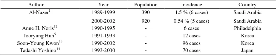
It is a rare disorder with incidence ranging from 0.54% to 1.5% in Saudi Arabia and 5.7 % in Taiwan.1,2
Epidemiology
The incidence of Kikuchi Fujimoto Disease (KDF), also called histiocytic necrotizing lymphadenitis is rare.1,2,4,6,7 KFD is more prevalent among Asians and is a relatively common disorder among Koreans.8,9 It remains a poorly recognized entity,4,11 is still frequently confused with malignant lymphoma and systemic lupus erythematosus (SLE)4,9 and has been under-diagnosed and therefore underreported.6
The age range is 19 month to 75 year old,8 but it typically affects in young adults, mean age 30-year
old1,3,4,6,7,10 and only 5-10% of cases are younger
than 21 years of age.11
Females are more commonly affected than males, with female-to-male ratio of 3 to 4:1,4,6,7,10 but now most series report an equal distribution between females and males or slightly females predominance (1.1:1 to 2.75:1).1,12
Clinical manifestation
Cervical lymphadenopathy accompanied by fever, myalgia, skin rash and leucopenia is a clinical manifestation of KFD. Cervical nodes are affected in about 80% - 98% of cases.6,12 Frequently posterior cervical nodes are involved (65-88.5%).2,6,8 Cervical lymphadenopathy is isolated to a single (83 %) location, but multiple chains may be involved. Generalized adenopathy occurs in 1-22% of cases,8,15 pain and tenderness of the lymph nodes in 50-59% of cases,2,8,9 nodes tend to be 0.5 - 4 cm in diameter in Departement of Child Health, Faculty of Medicine University of
93.4% of cases, usually unilateral (88.5%),8 firm, mobile, and they are not fluctuant.6
A flulike prodrome with fever is present in 30-50% of cases.6,8,16 The following are less common symptoms: headache, nausea, vomiting, malaise, fatigue, weight loss (10%),12 night sweats, chill (4%),12 abdominal or chest pain6 and fever of unknown origin.17 Systemic symptoms are found more frequently when extranodal involvement is present.7,8
Cutaneous involvement of KFD has been observed in 5 - 40%.2,4,6,7 Finding are varied and nonspecific and include maculopapular lesions, papules, plaques, nodules or ulcers mainly affecting the upper part of the body, such as trunk, the upper extremities and the facial skin.2,4,6 These cutaneous lesions have been reported to develop simultaneously with or after KFD4 and resolve in few weeks to months, similarly to the lymph adenopathy.3,6
Hepatosplenomegaly2,4,6 and neurologic involvement are rare but has included conditions such as aseptic meningitis,2,6,10 acute cerebellar ataxia, and encephalitis.6
Rarely involved bone marrow and myocardium.6 The ocular involvement has been reported as a possible association with KFD include bilateral panuveitis developed 2 years after the onset of lymph adenopathy.18 The manifestation of KFD as extensive small and large joint synovitis has been reported by Graham.19
The range of symptoms and abnormalities is wide.2,6 Most cases resolve within several weeks to 6 months or more.3,6,7,10 The time from onset of symptoms to diagnosis ranges from 1-24 months.4
Pathophysiology
The etiology of KFD is not yet well known.2,5-9 Currently, the most favored theory proposes that
KFD results when an as-yet-unidentified infectious agent triggers a self-limited autoimmune process.6,10 A preceding fever, leukopenia, occasional skin rashes, and no response to antibiotics suggest a viral etiology.8,9 Several viral candidates have been proposed, including cytomegalovirus (CMV), Epstein-Barr virus (EBV), herpes virus 6 and 8, varicela zoster virus, parainfluenza virus, parvovirus B 19, myxovirus,6 rubella,10 Toxoplasma and Yersinia also have been reported as possible causative agents.6,8,15,16,20 However, serologic and molecular studies have failed to link KFD to a specific pathogen6,9 because several other studies that, by using the same and other molecular pathology procedures to localize the virus genome, have concluded that neither EBV nor herpes virus 6 or 8 has a putative role in the pathogenesis of KFD.8,21 Chan et al22 reported that, there is probably no one single cause for this lesion, and it is probably a hyperimmune reaction to different etiological agents, be it microbial, chemical, physical or neoplastic. No reports have been made documenting possible transmission from one person to another.23 It is possible that KFD might represent an extuberant T cell-mediated immune response in genetically susceptible people to a variety of nonspecific stimuli.8
Several authors have reported an association between KFD and Systemic Lupus Erythematosus (SLE).6,9,21,24 KFD has been diagnosed before, during, and after a diagnosis of SLE was made in the same patient. Some authors have suggested that KFD be an attenuated form of SLE,6,9or might be the initial diagnosis in patients who go on to develop SLE,7 but this theory has not been substantiated, and the association of KFD with SLE, if any, remains unclear.6
Diagnosis
The diagnosis of KFD is confirmed by characteristic pathologic findings of involved nodes.6,10
Table 1. The incidence of Kikuchi-Fujimoto Disease
Author Year Population Incidence Country Al-Nazer1 1989-1999 390 1.5 % (6 cases) Saudi Arabia
2000-2002 920 0.54 % (5 cases) Saudi Arabia Anne H. Noris12 1990-1995 - 6 cases Philadelphia
Jooryung Huh9 1991-1993 - 12 cases Korea
Laboratory abnormalities include leucopenia that occurs in 25-50% of cases,2,6,10,16 leucocytosis in 2.9-5% of cases, mild granulocytopenia in 20-50% of cases, and atypical lymphocytosis in 25% of cases. Elevated ESR occurs in 70 % of cases (30-60 mm/hour).10,12,13 C-reactive protein (CRP) and LDH might be increased in some patients.2,6 Bacterial, fungal, and acid-fast stains and cultures of body fluids and biopsy material are usually negative.10 Serology for viruses, toxoplasma, autoimmune studies are also usually negative.1,23
Chest radiograph in KFD is normal. The CT and MRI appearance of KFD can be variable, and can mimic not only lymphoma, but various nodal
diseases with nodal necrosis, including metastasis and tuberculosis.13,25,26
The usefulness of fine-needle aspiration (FNA) cytology to establish a cytologic diagnosis of KFD has been limited, most authors recommend confirmation by excisional biopsy. FNA had overall accuracy of 56.75%.6,8
The histology of the excised lymph nodes characterized by necrotic areas and surrounding histiocytic infiltrate in the lymph node paracortex, the occurrence of variable numbers of immunoblasts, and the notable absence of polymorphonuclear leukocytes.12,27 The extent of necrosis is variable ranging from 5 to 95% of the node.1
Figure 1. The characteristic histopathologig finding of KFD12
A. Lymph node with Kikuchi-Fujimoto disease shows geographic foci of necrosis.
B. Necrotic foci contain numerous kayorrhectic nuclear fragments and eosinophilic granular debris phagocytized by crescentic histiocytes
The histomorphological changes in KFD pass through three stages, a proliferative stage, a necrotic stage, and a xanthomatous stage.3,6,9 Since the diagnosis of KFD is usually made in the xanthomatous stage when the lesion is full blown, the chance of detecting
most common in the late postnecrotic stage with few B and NK cells (natural killer).3
Differential Diagnosis
About 40% of patients with KFD were initially misdiagnosed as having lymphoma and were consequently over treated with chemotherapy.6,12 This can be differentiated by incomplete architectural effacement with patent sinuses, the presence of numerous reactive histiocytes containing phagocytic debris, the paucity of mitotic activity and absence of
“starry-sky” pattern or Reed-Sternberg cell.3,6 Systemic lupus erythematosus can sometimes pose a diagnostic
problem, but the clinical setting, “hematoxylin bodies”
with infiltration by granulocytes, and karyorrhectic debris within vessels differentiates KFD from
SLE.3,10 Kawasaki’s disease can be differentiated by
its younger age of presentation, mucosal involvement, abnormal endothelial-lined vessels containing micro-thrombi and irregular areas of necrosis involving the follicles.3
Treatment
Nonsteroidal anti-inflammatory drugs (NSAIDs) may be used to alleviate lymph node tenderness and fever. The use of corticosteroids, such as prednisone, has been recommended in severe extra nodal or generalized KFD.6 Immunosuppressants have been given as an adjunct to corticosteroids in severe, life-threatening cases.6,7
Prognosis
KFD is a self-limited condition that can recur in about 3 – 4 % of cases. Therefore, the prognosis is excellent.2,6-8,12 recurrence has been recorded over a period of 2 to 12 years after initial presentation.28
CASE REPORT
A 12-year-old girl presented to Out Patient Department of Child Health Dr. Cipto Mangunkusumo, Jakarta on October 18, 2004 with chief complain of there is a mass located at the left posterior cervical area since 19 days prior came to the hospital. There was no
improvement after several cycles of antibiotics given by other medical doctor.
On physical examination, there were two lymph nodes 1 x 1 x 1 cm3 and 2 x 2 x 1 cm3 in size, firm, mobile, and tender in palpation in the left posterior cervical region. This complaint accompanied by fever, intermittent with the highest temperature of 40oC, myalgia, and decreased of appetite. There were no other symptoms like cough, cold, vomiting or diarrhea. She had no pets and denied history of cat scratches. A diagnosis of lymphadenitis colli was made. She was given antibiotic cephradine, mefenamic acid and tinoridine Hcl. She was given paracetamol, which afforded temporary relief. On October 21st, due to persisted symptoms, she was brought to pediatric surgery outpatient department for consultation. On October 29th, symptoms persisted. The lymph nodes became more tender and maculopapular rashes appeared starting from the chest and spread to all over the body. Within 2 days the facial rashes disappeared.
On October 30th, still with persisted of symptoms, there was a new lymph nodes enlargement on the right posterior cervical area, 1.5 x 1 x 0.5 cm3 in size with the same characteristic as previously. Fever on and of, usually at night. Bowel movement was decreased. Her tongue was coated. Laboratory evaluation showed a hemoglobin (Hb) levels of 11.2 g/dL, a hematocrit (Ht) of 35 vol %, a white blood cell count of 3900 /L with a differential of (%): 8 bands, 53 neutrophils, 39 lymphocytes and a platelet count of 221.000 /L. ESR was 48 mm/hour, bleeding time and clotting time were normal. A diagnosis of lymphadenitis TB superimposed with typhoid fever was made.. She was afebrile and was advised for chest x-ray, PPD skin test, complete blood count and ESR.
On November 7th, the 19th day of illness, she was underwent lymph nodes excisional biopsy on the right posterior cervical region.
Pathologic assessment (PA no. 0405652)
A skin test with a purified protein derivative of tuberculin (PPD) was nonreactive. Chest x-ray showed minimal pulmonary infiltrate with hillar lymph node enlargement. She was consulted to a pulmonologist, which pulmonary TB was negative. Complete blood count revealed 10.7 g/dL, Ht 32 vol %, trombocytes 321.000 /L, white blood cell count of 4000 /L with a differential of (%): 2 eosinophils, 5 bands, 55 neutrophils, 33 lymphocytes, 5 monocytes. ESR was 50 mm/hour. Acute antibody titers against cytomegalovirus, toxoplasma gondii were negative. The serum C3, C4 and IgG were normal. Scar post excisional biopsy was noted on the right posterior cervical region. The rest of the head and neck lymphnode on examination were unremarkable.
DISCUSSION
A girl, 12 years old came to Department of Child Health Dr.Ciptomangunkusumo, Jakarta, Indonesia with the main complain of multiple cervical lymphadenopathy.The lymph nodes are enlarged,
with pain and tenderness, accompanied by fever, myalgia and decrease of appetite. Skin rashes appeared starting from the chest and spread to all over the body. An excisional biopsy of right posterior cervical lymph nodes was performed and histophatologic finding are hyperplastic of lymphoid follicle, there were plenty of macrophages which it cytoplasma containing of cellular debris compatible with chronic lymphadenitis and identified as Kikuchi-Fujimoto disease.
She was managed as outpatient and was given symptomatic treatment with ibuprofen.
Since this is a self-limiting disorder, requires no specific management, long-term closely follow-up is needed because frequently confused with systemic lupus erythematosus (SLE).
CONCLUSIONS
Health Dr. Cipto Mangunkusumo and was reported in this journal.
2. KFD should be considered in the differential diagnosis of any persistent pediatric cervical lymph nodes enlargement, may be distinguished from more serious infectious, neoplastic or autoimmune conditions.
3. Base on clinical manifestation, blood laboratory findings and tissue pathologic assessment a diagnosis of Kikuchi-Fujimoto disease was established. She was managed as outpatient and was given symptomatic treatment.
Acknowledgement
The author highly appreciates Prof. Dr. I Made Nasar in Pathologic Laboratory Department who performed the tissue specimen examination.
REFERENCES
1. Nazer M, Hadad A, Aithan S, Salem A, Al-Faraj A, Al-Saeed H. Kikuchi-Fujimoto disease. Saudi Med J 2002; 23(4):405-8.
2. Scheinfeld NS. Cutaneous Kikuchi disease. Updated at November 25, 2003. From
http://www.eMedicine.com/ped.htm. Downloaded at October 13, 2004.
3. Mohanty SK, Arora R, Saha M. Kikuchi-Fujimoto disease: an overview. J Dermatol 2002; 29:10-14. 4. Kato A, Kono T, Ishii M, Wakasa K, Taniguchi S. Spontaneous
clearance of psoriasis during the course of Kikuchi-Fujimoto disease. J Am Acad Dermatol 2002; 47:S287-8.
5. Kawai H, Hasegawa M, Hagiwara S, Hosomura Y.
Kikuchi’s disease with leukocytoclastic vasculitis in a
10-year-old girl. Pediatr Int 1999; 41:323-6.
6. Boone JL, Kuzma C. Kikuchi disease. Updated at Augustus 19, 2004. From http://www.eMedicine.com/ped.htm. Downloaded at October 13, 2004.
7. Chiang YC, Chen RMY, Chao PZ, Yang TH, Lee FP. Pediatric Kikuchi - Fujimoto disease masquerading as a submandibular gland tumor. Int J Pediatr Otorhino-laryngol 2004; 68:971-4.
8. Bosch X, Guilabert A, Miquel R, Campo E. Enigmatic Kikuchi-Fujimoto disease: a comprehensive review. Am J Clin Pathol 2004; 122(1): 141-152.
9. Huh J, Hyun SC, Sung SK, Gong G. A study of the viral etiology of histiocytic necrotizing lymphadenitis (Kikuchi-Fujimoto disease). J Korean Med Sci 1998; 13:27-30. 10. Debley JS, Rozansky DJ, Miller ML, Katz BZ, Green
ME. Histiocytic necrotizing lymphadenitis with autoimmune phenomena and meningitis in a 14-year-old girl. Pediatrics 1996; 98:130-3.
11. Murga Sierra ML, Vegas E, Blanco-Gonzalez J, Martinez P, Gonzales A, Calero MA. Kikuchi disease with multisystemic
involvement and adverse reaction to drugs. Pediatrics 1999; 104(2):1-4.
12. Norris AH, Krasinskas AM, Salhany KE, Gluckman SJ. Kikuchi-Fujimoto disease: a benign cause of fever and lymphadenopathy. Am J Med. 1996; 101:401-05. 13. Kwon SY, Kim TK, Kim YK, Ki YL, Nam JL, Hae YS.
CT Findings in Kikuchi disease: analysis of 96 cases. Am J Neuroradiol 2004;25:1099-1102.
14. Yoshino T, Mannami T, Ichimura K, Takenaka K, Nose S, Yamadori I, et al. Two cases of histiocytic necrotizing lymphadenitis (Kikuchi-Fujimoto’s disease) following diffuse large B-cell lymphoma. Human Pathology 2000; 31 (10):1328-31.
15. Naddaf H, Al-Balla SR, Al-Sami H, Hafeez MA. Kikuchi disease associated with overlap syndrome responding to steroids. From the Department of Medicine, Rheumatology Division, King Khalid University Hospital, Riyadh, Saudi Arabia. September 1994. Didapat dari http://www.kfshrc.edu.sa/annals/145/cr9316.htm. Downloaded at October 13, 2004.
16. Piccirillo JF, Lanza DC, Stasio EA, Moloy PJ. Histiocytic
necrotizing lymphadenitis (Kikuchi’s disease). Arch Otolaryngol Head Neck Surg 1991;117:800-2.
17. Parappil A, Rifaath AA, Doi SAR, Pathan E, Surrun SK. Pyrexia of unknown origin: Kikuchi-Fujimoto disease. Clin Infect Dis 2004; 39:138-43.
18. Taguri AH, Mcllwaine GG. Bilateral panuveitis: a possible association with Kikuchi-Fujimoto disease. Am J Ophthalmol 2001; 132:419-21.
19. Graham LE. Kikuchi-Fujimoto disease and peripheral arthritis : a first!. Ann Rheum Dis 2002; 61:475.
20. Correa H. Kikuchi-Fujimoto Disease: An exuberant localized T cell activation arrested by histiocytes? Medscape General Medicine 1999; 1(1):section 1-7. 21. Tsaousis G, Kolevris N, Vaidakis E, Mintzias D,
Rondogianni D, Chalevelakis G. Kikuchi’s disease and systemic lupus erythematosus: case report and review. Europ J of Int Med 1999; 10:53-55.
22. Chan JKC, Wong KC, Ng CS. Fatal case of multicentric
Kikuchi’s histiocytic necrotizing lymphadenitis. Cancer
1989;63:1856-62.
23. Gleeson MJ, Siodlak MZ, Barbatis C, Salama NY.
Kikuchi’s – a new cause of cervical lymphadenopathy. J Laryngol Otol 1985;99:935-9.
24. Martinez-Vazques C, Hughes G, Bordon J, Alonso-Alonso J, Anibarro-Garcia A, Redondo-Martinez E, et al. Histiocytic necrotizing lymphadenitis,
Kikuchi-Fujimoto’s disease, associated with systemic lupus
erythematosus. Q J Med 1997;90:531-3.
25. Dong GN, Chung TS, Hong SB, Hong DK, Young HK, Jeong HY. Kikuchi disease: CT and MR findings. Am J Neuroradiol 1997:18:1729- 32.
26. Bennie MJ, Bowles KM, Rankin SC. Necrotizing cervical lymphadenopathy caused by Kikuchi-Fujimoto disease. The Br J Radiol 2003; 76:656-8.
27. Facchetti F, de Wolf-Peeters C, van den Oord JJ, de Vos R, Desmet VJ. Plasmacytoid monocytes (so-called plasmacytoid T-cells) in Kikuchi’s lympadenitis: an immunohistologic study. Am J Clin Pathol 1989; 92:42-50. 28. Blewitt RW, Kumar SN, Abraham JS. Recurrence of Kikuchi’s